I would use zebrafish and combine a physical record of divisions, an inherited DNA record, and independent tests of predicted progenitor outputs. The intended result is a set of lineage trees for individual animals, registered to brain regions and mature cell identities, with confidence limits. Expression-based developmental pseudotime orders molecular states; neither it nor a patch of cells sharing a colour is sufficient on its own to establish ancestry.
I would begin by defining the sampled interval and outputs: neural-plate progenitors, their expansion into forebrain, midbrain and hindbrain, and later neuronal and glial descendants. Sampling only neurons would miss persistent progenitors and glia; sampling one brain region would make an apparent restriction difficult to interpret. Nuclear and membrane fluorescent reporters, anatomical landmarks and a staging series would anchor the map. I would establish a molecular reference with single-cell RNA sequencing, then verify selected cell identities by spatial transcript detection and morphology. Expression identifies a state at the observation time; a neural progenitor can switch off its original marker while all its descendants remain in the lineage.
Direct imaging would provide the earliest ground truth. Multiview light-sheet fluorescence microscopy would image labelled nuclei and membranes from neural specification through the tractable early period. The time interval must resolve divisions and rapid cell rearrangements; multiple views reduce missed cells behind opaque regions. Automated segmentation and tracking would be manually checked at division events, crowded boundaries, apoptosis and apparent long jumps. I would maintain alternative branch assignments where the images do not resolve a mother–daughter relation. Unimaged siblings and lower-light controls would test whether mounting or illumination changes proliferation, survival or brain morphology. Whole-embryo light-sheet tracking has been demonstrated in early zebrafish development, but that does not guarantee an error-free record through all later brain growth.[ref]
Reconstruction of zebrafish early embryonic development by scanned light sheet microscopy by Philipp J. Keller and others. 2008, pp. 1065–1069, multiview nuclear tracking and digital embryo reconstruction
For a small set of progenitors, I would photoconvert a stable nuclear fluorescent protein in a single verified cell and follow its daughters continuously. Imaging immediately before and after conversion establishes whether neighbouring cells were also labelled. Repeated independent single-cell conversions provide a spatially interpretable test of the large-scale molecular map. Subsequent protein turnover and dilution eventually limit this record, so a permanent inherited label is preferable for long chase times. Sparse induction of a validated recombination reporter is another option, but the actual induction window and the chance of multiple independently labelled founders must be measured. Labelling based on a promoter reports the descendants of cells that expressed that promoter during the pulse, not necessarily one unique anatomical progenitor.
An inducible CRISPR recording system would extend the map to later stages. A neutral genomic barcode array would receive early edits, followed by separately timed editing pulses during neural development. Descendants inherit an ancestor's edits and can acquire additional scars. Recovering barcode sequences together with single-cell transcriptomes then links the inherited record to cell identity. A constitutive founder label or an independent array would help distinguish unrelated founders with recurrent common edits. The feasibility of coupling temporally induced scars to cell-type classification in the zebrafish brain was demonstrated by scGESTALT.[ref]
Simultaneous single-cell profiling of lineages and cell types in the vertebrate brain by Bushra Raj and others. 2018, Figures 1 and 3–6, inducible editing, lineage reconstruction and transcriptome readout
I would validate the operating parameters in the chosen line rather than assume the published recovery rate or editing spectrum transfers unchanged.Each pulse requires no-inducer, no-guide or no-Cas9 controls as appropriate, a measurement of editing duration, and tests that the recorder does not alter cell-cycle progression, survival or cell-type composition. Too little editing yields unresolved branches; too much editing saturates sites and can delete earlier information. I would sequence genomic DNA from a subset of cells to estimate how often a barcode is present but absent from the transcriptome readout: this estimates lineage-tracing dropout. Independent target sites and longer sequence tags reduce, but do not eliminate, recurrent-edit homoplasy. The same small deletion arising twice is not strong evidence for a common progenitor. Editing probabilities should be measured in each recording interval, because a large nominal space can contain only a few common outcomes.
An explicit barcode collision calculation would guide labelling density. If independently labelled founders receive barcode with probability , the pairwise match probability is . Among founders the expected number of matching pairs isFor a uniform library this becomes for possible barcodes. These are design calculations, not measurements from the paper. They explain why sparse labelling and empirically measured diversity matter more than calling a recorder “high complexity”. Shared common scars, missing earlier scars and sequencing errors would be represented as uncertain branches rather than forced into a completely resolved tree. The root and temporal order of new edits must agree with the actual induction schedule.
I would reconstruct ancestry from the inherited sequences independently of cell-type assignment, then ask which molecularly defined outputs occur on each branch. Otherwise using expression similarity to construct the tree and then declaring related cell types to share ancestry would be circular. Closely related cells may differentiate into different types, while the same type may arise from multiple progenitor pools. Spatial recovery by in situ barcode readout or matched tissue sections would test whether a sequence-defined clone occupies the predicted regions. A pooled brain atlas can compare homologous populations across fish, but independently generated barcode trees from different animals cannot simply be merged as though identical scars named the same historical cell.
A concrete prediction might be that one early progenitor population yields both a particular neuronal class and a glial class, before later progenitors become restricted. I would sparsely label one cell in that population, confirm the single founder by imaging, and score all its descendants for the two identities using several markers and anatomy. A true mixed-output clone supports clonal multipotency. Several cells labelled by the same promoter do not. Earlier and later pulses would test when the mixed output disappears. Apparent restriction after a late pulse would be compared with the probability of missing rare outputs: if each of descendants of a class is recovered independently with probability , the probability of recovering none is . Nonindependent dropout would need a more realistic estimate. Complete imaging of selected clones would therefore be especially valuable.
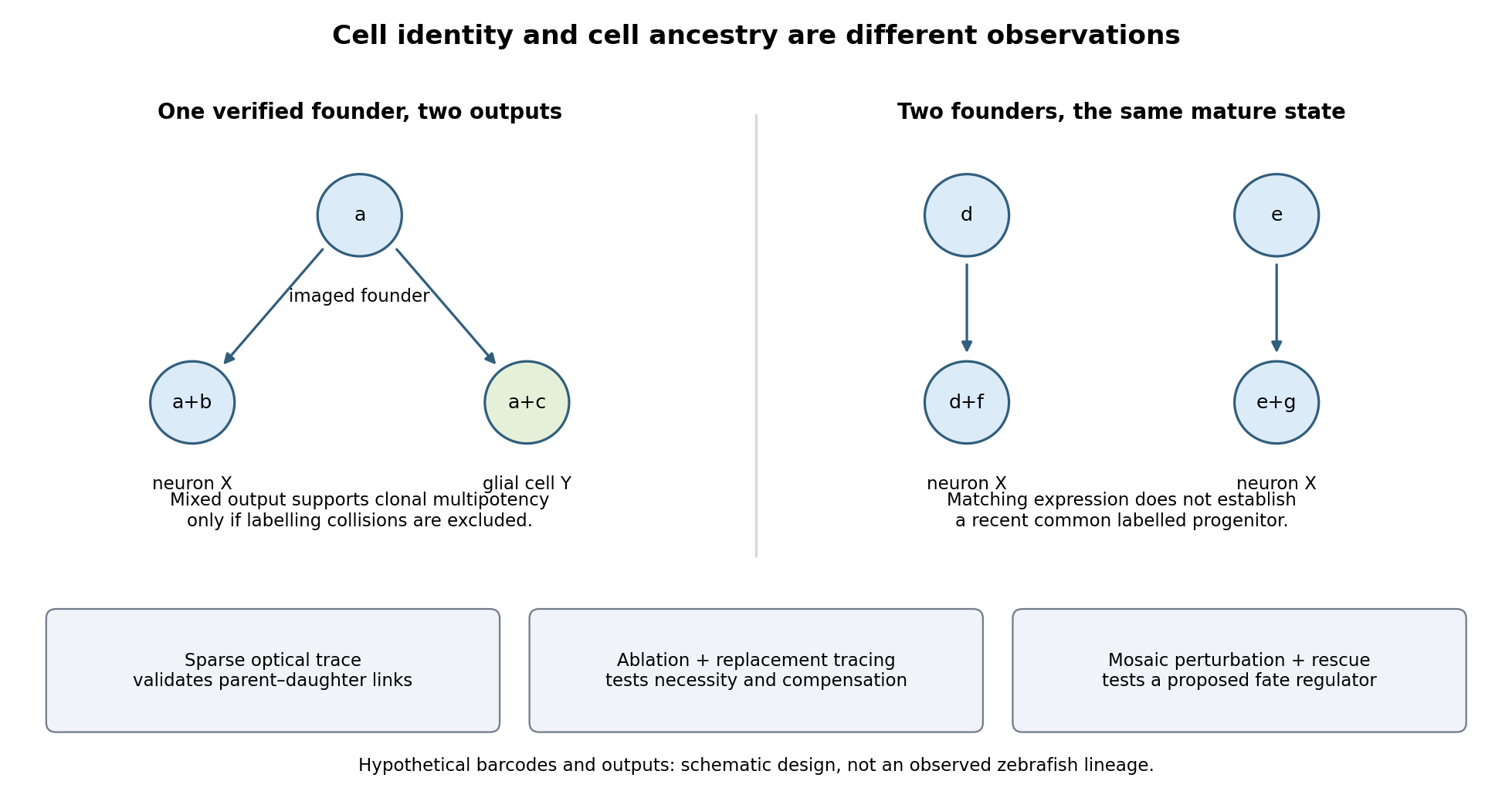

Functional tests would be designed to distinguish ancestry, necessity and developmental potential. Selective laser ablation of the labelled founder before its predicted bifurcation should remove or reduce both outputs if there is no compensation. A sham exposure, ablation of an adjacent equally sized population, and immediate assessment of collateral damage are essential controls. If outputs recover, I would trace the replacing cells: recovery can reveal regulative development rather than refute the original ancestry. Conversely, loss of a neuronal population after ablation could reflect loss of a required signal from the targeted cells, so it cannot alone prove that they were its ancestors.
I would then transplant labelled progenitors between matched hosts, including orthotopic controls and a heterotopic or temporally altered environment. Retention of both outputs under a viable alternative environment would support commitment; adoption of the host's outputs would show environmental instruction. Failure to differentiate after poor engraftment is not evidence of restriction. To test a proposed branch regulator, I would perturb it in sparse, permanently labelled clones and measure descendant identities, clone size, proliferation, apoptosis and position in parallel. A change in fate proportions at comparable survival and clone size is more persuasive than disappearance of the entire clone. Rescue by a perturbation-resistant allele, independent perturbations and comparison with neighbouring unperturbed clones would test specificity and cell autonomy. The lineage label should persist even if the cell-state marker used to select the progenitor is later lost.
Replication would be at the level of independently treated embryos and parents as well as clones; thousands of cells from one fish are not thousands of independent developmental replicates. I would report label specificity, collision and dropout estimates, alternative tree resolutions, and the sampled developmental window. A robust map is one in which sequence-based ancestry predicts independently observed divisions and clone outputs, while perturbations reveal which developmental choices are causal and which remain plastic.
Articles by others on the same topic
There are currently no matching articles.