
An active site is the region of an enzyme where a substrate binds and undergoes a chemical reaction. In a protein enzyme, it includes the amino acid residues that contact the substrate and those that directly assist catalysis. These roles can overlap: an amino acid side chain can both orient a substrate and participate in chemistry. The active site occupies only part of the folded protein; the remaining amino acid residues help establish its protein structure, stability and movement. An active site need not be a consecutive stretch of protein primary structure: protein folding can bring distant amino acid residues together. A ribozyme instead supplies an RNA active site. See Alberts et al., Molecular Biology of the Cell, Protein Function: binding sites and catalytic sites.
Binding and catalysis answer different questions. A protein binding site recognizes shape and chemistry through such interactions as hydrogen bonds, electrostatics and the hydrophobic effect. At an active site, appropriate positioning also helps a substrate reach the transition state of a chemical reaction. Some amino acid side chains transfer a proton; others participate transiently in a covalent bond. The enzyme-substrate complex is the associated enzyme and substrate, before conversion to a reaction product. Merely accumulating this complex does not establish rapid catalysis: stabilizing the bound substrate without sufficiently stabilizing the transition state can leave a large barrier. The important complementarity is chemical as well as geometric. See Geoffrey M. Cooper, The Cell, The Central Role of Enzymes as Biological Catalysts: substrate binding and transition-state stabilization.
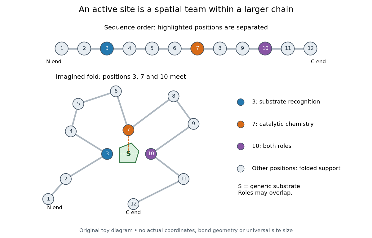
Distant amino acid residues can cooperate at an active site
. Original twelve-residue polypeptide chain cartoon, with the same order and eleven connections in both drawings. Positions 3, 7 and 10 approach a generic substrate after an imagined fold. Blue means recognition, orange means catalytic chemistry, and purple means both; these assignments are invented for explanation. Dashed contacts represent cooperation, not specified hydrogen bonds or actual atomic coordinates. The other nine positions support the imagined protein structure; three highlighted positions are not a universal active site size. Details.The cartoon makes a useful experimental distinction. Changing a contacting amino acid residue might weaken substrate binding, change the reaction rate, or do both. Changing a distant amino acid residue can also disrupt the active site by changing protein folding. Therefore a loss of activity after a mutation alone does not locate a catalytic group: compare binding, protein structure and enzyme kinetics as well. This is an interpretation guide, not a report of a particular mutation experiment.
A concrete example is hexokinase: it transfers the terminal phosphate group of ATP to the -position hydroxyl group of a sugar such as glucose. Specificity need not mean only one acceptable substrate: the hexokinase reaction class includes several possible acceptor sugars. Which are used, and at what reaction rates, depends on the particular enzyme and conditions. See Swiss Institute of Bioinformatics, ENZYME entry EC 2.7.1.1: hexokinase reaction and acceptor range.
Bennett and Steitz compared yeast hexokinase A crystallized with glucose against hexokinase B crystallized without glucose. X-rays scattered by the crystals supplied diffraction measurements from which the authors reconstructed protein structure; comparing coordinates located movement, rather than measuring a reaction rate. The glucose-containing cleft was narrower: one lobe was rotated by about relative to the other, with some protein backbone displacements reaching . This makes the active site a moving arrangement of interacting groups, rather than a permanent hole. See William S. Bennett Jr. and Thomas A. Steitz, Glucose-induced conformational change in yeast hexokinase, PNAS 75 (1978), 4848–4852.
The structural comparison has two explicit limitations: different forms of hexokinase and different crystal environments were compared, and static coordinates do not record the order of binding and movement. Closure is consistent with induced fit, but those structures alone cannot exclude conformational selection. The solution experiment under induced fit addresses whether compaction also occurs outside a crystal.


New to topics? Read the docs here!