
Language is not necessary for the basic capacity to discriminate or organize colors, but it can influence color judgments, attention and memory in particular tasks. The evidence supports neither the claim that words create all color experience nor the claim that language is irrelevant whenever the stimuli are visible.
The starting distinctions are essential. A receptor sensitivity, a color-matching relation, a discrimination threshold, a category boundary and a color name are different measurements. Molecular differences can change available cone signals, while linguistic experience can change how available information is grouped or used. Finding different words for two samples does not show that one group cannot discriminate them. Finding the same matching relation does not establish identical subjective appearance or identical naming.
The biological basis imposes constraints independent of a person's vocabulary. Three cone classes provide overlapping spectral measurements, and later circuits compare those measurements. Altered receptor signals or adaptation can change performance without requiring a new word. Conversely, changing a response strategy can change performance while leaving receptor input intact. The central empirical question is therefore which stage and task language influences, not whether one can name a color without knowing its name.
Prelinguistic evidence rules out a strict requirement for color words. Alice E. Skelton and colleagues used infants' recognition-memory responses to sample relations among colors across the hue circle. Infants' categorical distinctions showed organization related to common divisions in adult color lexicons and to early visual mechanisms.[ref]
Biological origins of color categorization by Alice E. Skelton and others. PNAS 114, 5545–5550; infant recognition-memory method and category structure
Infants did not need to produce the relevant color terms to show those distinctions. The inference is not that newborns possess an immutable adult lexicon: the infants already had visual experience, and looking-time measures require controls for discriminability, novelty and attention. The title's biological interpretation should not be strengthened into proof that experience plays no role.Cross-language studies also challenge a rigid universal partition. Debi Roberson and colleagues compared English speakers with Berinmo speakers using naming, recognition and category-learning tasks. Effects of linguistic category boundaries appeared for the categories used by each group, and response-bias controls weakened a simple claim that universally focal colors always enjoy a memory advantage.[ref]
Color categories are not universal: Replications and new evidence from a stone-age culture by Debi Roberson and others. J Exp Psychol Gen 129, 369–398; recognition, response-bias controls and boundary comparisons
The article's historical title is retained as a citation, not adopted as a description of people. The comparison shows that category use and memory cannot be reduced to one vocabulary-independent set of preferred labels. Because language group covaries with cultural and visual experience, it does not isolate vocabulary as the sole cause.A more direct manipulation was used by Jonathan Winawer and colleagues. Russian has conventional basic terms distinguishing lighter and darker blues that English usually groups under “blue.” Participants saw a target square and two alternatives together and selected the matching alternative. Russian speakers showed a speed advantage for comparisons crossing their linguistic boundary. A concurrent verbal task removed that advantage, while a spatial interference task did not produce the same pattern.[ref]
Russian blues reveal effects of language on color discrimination by Jonathan Winawer and others. PNAS 104, 7780–7785; simultaneous matching, category boundary and interference conditions
The simultaneous display reduces the long-term color-memory explanation, and the interference manipulation implicates access to language-related processes during performance. It does not establish a new receptor limit, a complete inability to discriminate within a category, or a necessary change in private color qualia. Concurrent tasks can affect more than one processing stage.These results motivate separating sensitivity from response facilitation. A faster match can reflect attentional selection, category-assisted comparison or decision formation. To claim changed sensory resolution, an experiment should measure psychometric functions over controlled stimulus separations, fit location, slope and lapse parameters, and test whether any effect persists with nonverbal responses and reversed response mappings. Equal physical distance in a standard color space does not automatically ensure equal discriminability for every participant; individual thresholds and adaptation should be measured where they matter.
A stronger causal program can combine within-person interference, language-learning interventions and longitudinal or bilingual comparisons, while matching exposure and task difficulty. Testing transfer to new samples distinguishes memorized stimulus labels from a changed category rule. Comparing immediate discrimination, delayed recognition, naming and appearance matches identifies whether an effect generalizes across measurement levels. These are proposed strategies rather than invented experimental outcomes. No test based only on verbal descriptions can directly establish equality or inequality of another observer's subjective experience.
The seemingly opposing findings can therefore coexist. Biology and early experience can produce color organization before fluent naming, while learned labels subsequently guide the use of that organization. Color vision does not depend on language for its existence; linguistic resources can nevertheless alter how people attend to, classify, remember and rapidly compare colors under specified conditions.
L, M and S describe the relative spectral sensitivities of cone classes. Calling them red, green and blue confuses receptor sampling with the color appearance constructed from comparisons and context. The shorthand also wrongly suggests three narrow, nonoverlapping detectors or the primaries of a display.
First, each class has a broad sensitivity curve. A long-wavelength-sensitive cone responds to many wavelengths, and its sensitivity maximum is not a unique red stimulus. L- and M-cone sensitivities overlap extensively, while S cone cells are relatively more sensitive at shorter wavelengths. Light at one wavelength can therefore excite more than one class. Andrew Stockman and colleagues derived L- and M-cone sensitivity estimates using color-matching data and measurements from observers of known pigment genotype. Their analysis emphasizes broad overlap and the need for isolation procedures to measure one class without contamination from others.[ref]
The spectral sensitivities of the middle- and long-wavelength-sensitive cones derived from measurements in observers of known genotype by Andrew Stockman and others. Vision Res 40, 1711–1737; spectral-sensitivity measurements and cone fundamentals
Exact curves depend on pigment variant and optical filtering, so the letters denote relative classes rather than three invariant color labels.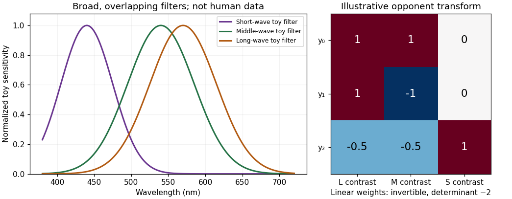
Original Gaussian toy receptor filters and an invertible opponent transform; the filters illustrate broad overlap and are not measured human cone fundamentals or a calibrated color chart
. Details. 
Second, the principle of univariance prevents one cone from identifying hue. For a monochromatic stimulus, let mean absorption be . Whenever and are positive, intensities satisfying give the same . The single receptor cannot distinguish those two inputs on absorption count alone. Calling that receptor a red detector would assign a color judgment to a signal that cannot even uniquely distinguish the two wavelengths. It remains true that spectral sensitivity constrains the comparison; univariance does not mean all receptor types are identical.
Third, perceptual color requires combinations. Comparing L and M signals and comparing S with L+M supplies chromatic information; summing appropriately weighted signals contributes to luminance-related processing. An L-cone increment can be part of several stimulus changes depending on what happens to M and S and on the adapted background. Neither an L response nor a raw L:M ratio by itself specifies a context-independent color word. The retinal and cortical opponent circuitry demonstrated by Dennis M. Dacey and Bevil R. Conway provides mechanisms for comparisons, rather than a relabelling of three cone outputs as three sensations.[ref]
The blue-on opponent pathway in primate retina originates from a distinct bistratified ganglion cell type by Dennis M. Dacey and others. Nature 367, 731–735
[ref]Fourth, “blue” or “red” need not identify a single wavelength. Spectrally different lights can be metamers, and a purple appearance can arise from a mixture rather than a monochromatic wavelength. Conversely, the same physical spectrum can look different against another surround or after adaptation. Cone labels cannot replace the distinction between emitted or reflected spectrum, receptor catch, opponent representation and reported hue.
Fifth, the display analogy is backwards. An RGB screen emits mixtures from three selected primary spectra. The cone cells receive and integrate the resulting spectrum; their response curves are not identical to those primary spectra. If primary intensities are and cone sensitivities to each primary form matrix , then . Off-diagonal terms are generally nonzero because each primary can excite several cone classes. If is nonsingular, an intended catch change can be computed through within the display's feasible gamut. It does not follow that every requested catch vector is realizable with nonnegative bounded primary intensities. Calibration and observer differences therefore matter in cone-isolating experiments.
Finally, color terms themselves vary across languages and tasks, whereas receptor class is a biological classification. Receptor genes, color matches, discrimination and linguistic categorization are related but distinct measurements. The labels L, M and S prevent an unwarranted assumption that one anatomical cell class directly instantiates one learned word or unique subjective hue.
The useful statement is that trichromatic vision starts with three broad, overlapping receptor classes and extracts chromatic information by comparison. “Red, green and blue cone cells” conceals both the information loss at each receptor and the transformations needed to see color.
Photoreceptors determine which optical differences are available for vision, with what noise and spatial–temporal precision. They strongly constrain light, color and material perception without uniquely determining the eventual percept. Later processing can exploit context and action, but cannot recover arbitrary information already lost in receptor sampling.
For cone class , a simplified expected quantum catch over one sampling interval iswhere is the incident photon-number spectrum at the receptor, including ocular filtering, and effective absorption probability. An energy spectrum requires conversion by photon energy before this counting interpretation. The integral is many-to-one: equal catches do not imply equal spectra. The principle of univariance means that an individual receptor does not independently report photon wavelength and number.
Multiple receptor classes provide comparisons, but ordinary cone-mediated trichromacy still samples a spectral distribution with only three class responses at a specified location and time. In a discretized model, with three rows and more than three spectral bins. The matrix has a nonzero kernel. Around a strictly positive spectrum, a sufficiently small perturbation in that kernel preserves nonnegative intensities while leaving all three catches unchanged. This derives the possibility of metamers; it does not mean all real-world spectral differences are unobservable under every condition. Altering illumination, observer or spatial–temporal context can break a particular match.
Sensitivity also has a photon budget. In an ideal Poisson model, mean catch has variance , so relative counting uncertainty is . Gathering more photons reduces this uncertainty but requires a larger effective collecting area or more integration time. Temporal averaging can obscure rapid change, while spatial pooling can sacrifice fine detail. Receptor and downstream noise add limitations beyond shot noise. Rods support low-light detection with different sensitivity and pooling from cone cells; if only one receptor class provides usable signals, wavelength–intensity ambiguity cannot be resolved by comparisons within that class. Mesopic vision, where rods and cone cells can both contribute, is not captured by declaring all dim-light vision achromatic.
The receptor array is also uneven. Christine A. Curcio and colleagues mapped rods and cone cells in whole-mounted human retinas using microscopic counts. Cone density was highest centrally and fell strongly with eccentricity, while the rod distribution differed and the central foveal region was rod-free.[ref]
Human photoreceptor topography by Christine A. Curcio and others. J Comp Neurol 292, 497–523; methods and photoreceptor density maps
Thus acuity and sensitivity should not be treated as uniform over the visual field. Optical blur and downstream sampling constrain performance alongside receptor spacing. The S-cone mosaic is sparser than the combined L/M mosaic, so equal spatial detail cannot simply be assumed for all chromatic signals.A regular one-dimensional model of photoreceptor sampling makes one limitation explicit. Sampling a sinusoid at spacing gives identical samples for frequencies and , because the added phase at sample is . Restricting a real signal to frequencies below avoids this basic aliasing ambiguity. Real retinal mosaics are two-dimensional, irregular and optically filtered, so this is a benchmark rather than a literal universal retinal cutoff. Fine material texture can consequently disappear or alias even when coarser color and shape remain visible.
Individual variation changes the sampling system itself. Heidi Hofer and colleagues' adaptive-optics measurements showed large L:M cone-ratio variation among observers passing normal color tests.[ref]
Organization of the Human Trichromatic Cone Mosaic by Heidi Hofer and others. J Neurosci 25, 9669–9679; retinal densitometry and corrected abstract
Gabriele Jordan and colleagues found behavioral evidence consistent with an extra chromatic dimension in one of a selected set of carriers with potential extra pigment diversity, rather than in every carrier.[ref]The dimensionality of color vision in carriers of anomalous trichromacy by Gabriele Jordan and others. J Vis 10(8):12; behavioral and genetic comparisons
These findings separate receptor availability from downstream use: receptor counts and pigment variants constrain possibilities without directly specifying appearance or functional dimensionality.Material perception adds an inverse problem. For a matte surface under a single simplified illuminant, the reflected photon spectrum is proportional to , so catches mix illumination and surface reflectance. The same catches can therefore arise from different surfaces, even before considering three-channel spectral compression. Specular reflection, subsurface scattering, translucency and fluorescence introduce further dependencies on direction, geometry or wavelength conversion. Three cone values cannot independently label every physical material property.
Nevertheless, humans can exploit cues across space, movement and time. Highlights change with viewpoint; texture and shading constrain shape; surrounding surfaces provide illumination evidence. Such information can reduce ambiguity under useful assumptions, while metamerism and sampling limits remain. Receptor properties define an information bottleneck and a set of trade-offs. Successful material recognition depends on combining what survives that bottleneck with contextual constraints, not on receptors directly measuring “wood,” “metal” or intrinsic surface color.
Sensitivity adjustments occur at several stages and over several timescales. They reveal adaptive gain control and flexible reference points, but no single aftereffect uniquely identifies one anatomical site. Changes in threshold, apparent hue, an achromatic setting and a receptor current must be distinguished before explaining them with one adaptation mechanism.
At the receptor level, a finite photocurrent range has to represent environments with very different mean light levels. Julie L. Schnapf and colleagues measured membrane currents from individual macaque cone outer segments. Responses to a maintained light step had an initial component followed by a smaller steady response, and a steady background reduced sensitivity to an added weak flash. This desensitization was distinguishable from loss of pigment through bleaching.[ref]
Visual transduction in cones of the monkey Macaca fascicularis by Julie L. Schnapf and others. J Physiol 427, 681–713; outer-segment recording, steps, backgrounds and bleaching comparisons
Direct recording establishes that some gain adjustment occurs before any color name, decision or cortical object representation. The preparation is macaque tissue, and a measured cone current is not identical to a human appearance judgment.An idealized normalization model illustrates the benefit. If a mechanism's incremental gain is proportional to , maintaining a fixed response increment requires . At high backgrounds this gives approximately constant fractional contrast , a Weber-like regime. It should not be confused with a universal law across all intensities. Under a known-background Poisson counting model with mean , the noise standard deviation is ; a small increment has signal-to-noise ratio approximately . A fixed signal-to-noise criterion then gives fractional threshold proportional to instead. The two predictions derive from different assumptions about what limits performance.
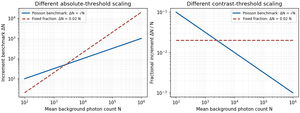
Original Poisson-counting and fixed-fraction threshold benchmarks; arbitrary criterion constants, not measured human adaptation data
. Details. 
Mean adaptation and contrast adaptation also differ. A diagonal cone-gain model replaces by , with gains related to recent stimulation. It can reduce the effect of a persistent chromatic shift in the illuminant. If the signal changes approximately as , choosing cancels that change. This is exact only for the assumed channelwise scaling; real spectral illuminant changes need not transform every surface's catches by the same three factors.
To test adjustments beyond mean cone excitation, experiments can modulate color around an unchanged time average. John Krauskopf and colleagues found direction-selective threshold elevation after temporal chromatic or luminance modulation, supporting separable adaptable mechanisms for particular directions near threshold.[ref]
Cardinal directions of color space by John Krauskopf and others. Vision Res 22, 1123–1131; selective threshold elevation for chromatic and luminance directions
Michael A. Webster and colleagues then measured appearance after adaptation to modulation along different color-space directions. A color monitor, a modulated adapting field and asymmetric appearance matches allowed testing effects beyond detection thresholds. Appearance shifted away from the adapting direction, including directions intermediate to the simplest cardinal color directions.[ref]Changes in colour appearance following post-receptoral adaptation by Michael A. Webster and others. Nature 349, 235–238; constant-mean modulation and asymmetric matching
Thus a three-independent-channel account sufficient for some threshold results was not sufficient for all suprathreshold appearance changes. Constant mean catches constrain a simple mean-receptor explanation, but do not prove that all receptor nonlinearities have been removed.Longer experience changes reference points too. Jay Neitz and colleagues altered chromatic experience with colored filters over days and measured unique yellow settings. Shifts persisted for one to two weeks after filter use ended.[ref]
Color perception is mediated by a plastic neural mechanism that is adjustable in adults by Jay Neitz and others. Neuron 35, 783–792; altered chromatic experience and persistent unique-yellow changes
Persistence after removing the immediate adapting stimulus demonstrates more than an instantaneous receptor response. It supports experience-dependent normalization without showing that a color category or verbal label has been rewritten. Any interocular transfer must also be measured explicitly; duration alone does not identify cortex.A natural perturbation extends the range to months. Peter B. Delahunt and colleagues followed achromatic settings before and after removal of cataractous lenses. Removal greatly changed short-wavelength input; settings continued to readjust over roughly three months, whereas chromatic discrimination recovered much faster.[ref]
Long-term renormalization of chromatic mechanisms following cataract surgery by Peter B. Delahunt and others. Vis Neurosci 21, 301–307; longitudinal achromatic settings and comparison with discrimination
An achromatic setting compensates for a perceived tint, so its physical shift should not be mistaken for a same-direction tint in appearance. The findings indicate distinct adjustment timescales. They are observational evidence with a changing optical input, not a randomized isolation of one neural site or a general treatment recommendation.These studies support a multilevel experimental strategy. Measure receptor responses, detection thresholds and appearance separately; manipulate mean and contrast independently; test spatial specificity, interocular transfer and recovery. Control exposure, eye position, stimulus calibration, response criterion and learning. A loss of sensitivity can reflect adaptation that preserves dynamic range rather than damage, and stable appearance can coexist with changed receptor inputs. Conversely, perfect compensation is not guaranteed.
Adaptation is a family of adjustments serving different constraints. Its most informative use is to distinguish which signal statistics, stages and reference points change, rather than treating every aftereffect as evidence for one fatigued color channel.
Post-receptoral processing compares cone signals, organizes those comparisons across space and time, and uses them to represent surfaces and objects. Three receptor classes do not imply three neurons labelled with three color names. A complete answer must distinguish the information available at the receptors from its neural transformation and its perceptual interpretation.
The principle of univariance supplies the starting constraint. For one cone class, an absorption-dependent response does not identify wavelength independently of intensity. Comparing differently sensitive classes makes chromatic information available. Relative to an adapted background, cone contrasts can be written , where is the background catch. This representation separates a change relative to the prevailing input from the raw number of absorbed photons. It is a useful coordinate system, not a claim that every neuron implements this exact formula.
Retinal circuitry immediately transforms the signals. Light hyperpolarizes cone cells and changes their transmitter release. ON and OFF bipolar pathways respond differently to changes in cone output, while horizontal and amacrine circuitry contribute spatial and temporal interactions. Retinal ganglion cells transmit spike trains rather than simply forwarding a cone's voltage. Their center–surround organization compares regions, so chromatic and spatial structure are already intertwined. Near the central retina, the midget pathway can oppose an L- or M-dominated center to a surround containing other cone input. The same circuitry can carry information useful for fine spatial structure and for chromatic contrast; these are not necessarily separate wires.
A distinct S-cone route illustrates the anatomical specificity. Dennis M. Dacey and colleagues combined physiological characterization with identification of ganglion-cell morphology in primate retina, finding that the S-ON opponent response belonged to a small bistratified ganglion-cell type rather than simply another midget cell.[ref]
The blue-on opponent pathway in primate retina originates from a distinct bistratified ganglion cell type by Dennis M. Dacey and others. Nature 367, 731–735; physiological and morphological identification
This supports parallel circuits with different cone combinations. Calling the pathway “blue-on” is shorthand for its stimulus response, not proof that the cell itself produces a unique blue experience.At a simplified computational level, an achromatic-like combination and two chromatic combinations can be represented asThis invertible three-channel opponent transform reorganizes three coordinates rather than creating additional independent spectral information. Their matrix has determinant , so the transform is invertible: , and . Actual weights depend on cell, location, adaptation and stimulus conditions. This algebra is an illustrative opponent transform, not a fitted retinal wiring diagram or a complete perceptual color space.
The lateral geniculate nucleus preserves several parallel retinal pathways while also participating in cortical interactions. A. M. Derrington and colleagues studied macaque geniculate cells with gratings whose luminance and chromatic modulation could be varied independently on a calibrated color television display. Finding directions that changed cone stimulation without driving a cell constrained its linear cone weights.[ref]
Chromatic mechanisms in lateral geniculate nucleus of macaque by A. M. Derrington and others. J Physiol 357, 241–265; null-plane method and chromatic stimulus space
For a nonzero linear weight vector, the zero-response contrasts obey , defining a chromatic null plane. A null response therefore tests a relation between inputs, rather than merely assigning the cell a preferred color name. The resulting opponent directions need not coincide with human unique hues.Cortical processing adds more complex spatial comparisons. Bevil R. Conway mapped cone inputs to color-responsive neurons in alert macaque V1 using cone-isolating spots and eye-position-corrected reverse correlation. Some cells combined chromatic opponency with opposite tuning in adjacent spatial subregions.[ref]
Spatial Structure of Cone Inputs to Color Cells in Alert Macaque Primary Visual Cortex by Bevil R. Conway. J Neurosci 21, 2768–2783; receptive-field mapping and double opponency
Such double-opponent cells can respond strongly to a chromatic boundary while responding less to a spatially uniform change. This offers a mechanism useful for separating surface differences from shared illumination changes. It does not establish that one V1 population alone solves color constancy under arbitrary scenes.Further processing must relate color to form, illumination, material and the current task. The same cone contrasts can be interpreted differently in a shadow, highlight or translucent object. Surface color judgments, discrimination thresholds and color naming therefore constrain different computations. Feedback and attention can influence which information is selected, while learning can alter how a signal is interpreted or normalized. A lesion or imaging response can constrain contributions to those tasks, but an area responsive to color is not automatically an isolated “color center.”
The strongest picture is consequently a hierarchy of interacting computations: cone-specific signals, opponent and spatial filtering, combinations tuned to richer image structure, and object-level judgments. Trichromatic sampling limits spectral information; later processing determines how that information supports boundaries, stable surface appearance and behavior.
There are unlisted articles, also show them or only show them.