Microtomy prepares thin physical sections with a microtome, exposing interior structure for microscopy. The knife separates a layer from a supported specimen while a controlled feed determines the nominal depth increment. Physical section thickness, section orientation and cutting-induced deformation all enter the interpretation of an image. Ultramicrotomy is the specialized thin-section technique used in applications such as transmission electron microscopy.
The preparation method also serves materials research. Yale's instrument description lists a Leica Ultra UC7 with a trimming mill and glass knife maker, and identifies polymers, material fibres and soft metals among suitable specimen materials.[ref]
Microtome at the Yale Institute for Nanoscience and Quantum Engineering. About the instrument, accessories and material list
Manufacturer guidance discusses polymer blends and composite materials, along with the possibility of section compression and cutting artifacts.[ref]Ultramicrotomy Techniques for Materials Sectioning by Leica Microsystems. Opening materials discussion; How to prepare high-quality sections of polymers
A useful teaching experiment would compare cuts through a straight, known circular material fibre at different section orientations. A normal vector to the cut aligned with the fibre axis gives a circular profile. An oblique cut gives an ellipse even when the fibre has suffered no deformation. Image the recovered sections with an optical microscope, measure profile dimensions with an established image stack axis calibration, and record the angle between the section's normal vector and the fibre axis. This tests the geometry of the section rather than assuming that every measured elongation is a physical distortion.
For an original ideal calculation, model the fibre boundary as an infinite circular cylinder of radius , with axis along the unit vector , where . Parallel section planes have equation . The squared Euclidean distance to the axis is , obtained by subtracting the squared axial orthogonal projection from . At the section plane, the boundary therefore satisfiesThis oblique section of a circular cylinder is an ellipse with semi-major and semi-minor axes and areaThe area of an ellipse gives the last expression. For chosen and , the semi-major and semi-minor axes are approximately and . The profile area is times the perpendicular-profile area, an increase of about 41.4 percent entirely explained by section orientation.
The center of an ellipse in this model has coordinate . In an ordered image stack, two sampled section planes separated by consequently have lateral centre displacement . At the chosen and , this displacement is . Here is the spacing between sampled planes, which can differ from the physical section thickness if some sections are not imaged.
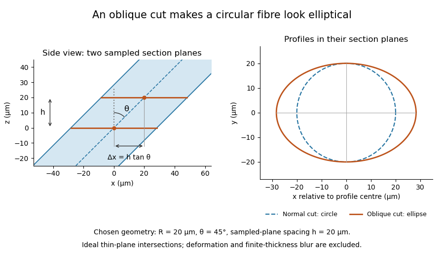

The calculation assumes a circular, straight fibre, known section geometry and negligible physical section thickness relative to resolved features. One elliptical profile alone cannot distinguish an oblique circular fibre from a differently shaped fibre or an unknown deformation. Comparing known reference features and following their positions through an image stack provides additional evidence. Image registration can align images, but an inferred shift may contain real specimen geometry as well as instrument motion; removing every shift would erase the tilt information in this example.
Articles by others on the same topic
There are currently no matching articles.