An optical microscope uses light and optical lens assemblies to reveal small structures, such as the boundaries of biological cells or defects in a manufactured surface. In a compound optical microscope, the microscope objective supplies an enlarged real image; the microscope eyepiece makes that intermediate microscope image subtend a larger viewing angle. The transverse magnification of the first stage and the angular magnification of the second answer different questions.[ref]
2.8 Microscopes and Telescopes. University Physics Volume 3, §2.8 Microscopes, image formation and Eqs. 2.35–2.37
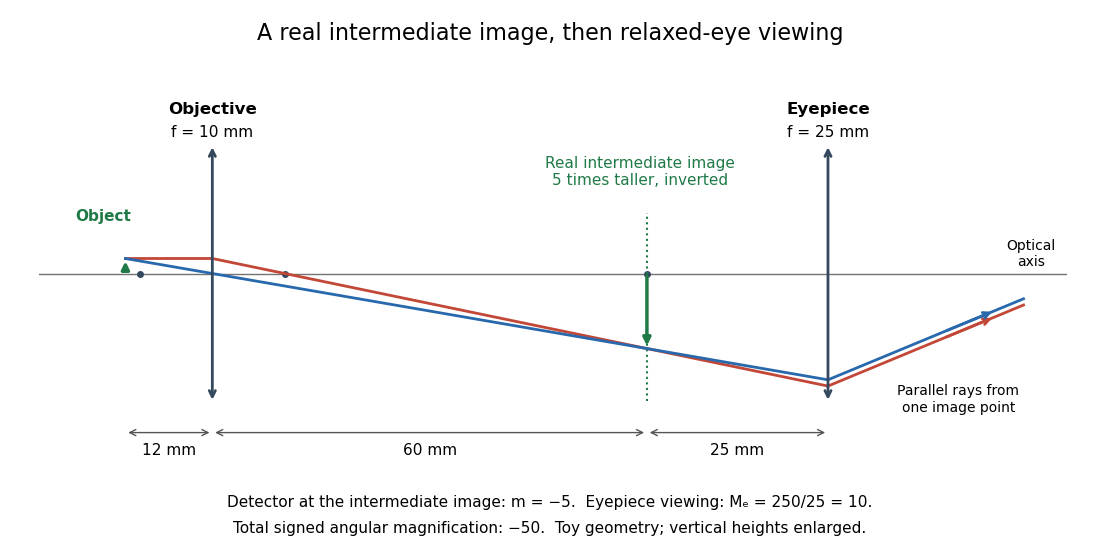
Consider an original ideal finite-conjugate microscope in air, with both optical lens assemblies treated as converging thin lens elements in the paraxial approximation. Choose objective focal length and object distance . The thin lens equation and signed transverse magnification givePlace a microscope eyepiece of focal length a further beyond the intermediate microscope image. The objective-to-eyepiece separation is therefore . Each object point then supplies a parallel outgoing bundle of light rays, appropriate to a relaxed eye focused at infinity. Define the unaided reference as the same object viewed at distance ; this is a rating convention, not a universal property of eyesight. The eyepiece angular magnification and total signed angular magnification areFor instance, a hypothetical object height becomes at the intermediate microscope image. The signed viewing angles are approximately with the instrument and unaided. Their ratio is : enlarged and inverted. These are stipulated model dimensions, not measurements or specifications of a modern commercial optical microscope.
The attraction of an optical microscope is that seeing can become measuring: a stage micrometer supplies a known object-space length, and microscope scale calibration converts an image readout into a specimen length. This also separates a large-looking image from a trustworthy size estimate. Meanwhile, diffraction-limited resolution depends on numerical aperture and wavelength; empty magnification enlarges an existing blur without revealing additional structure.