Vascular surgery is the specialty concerned with assessing and treating disorders of arteries and veins, including reconstructive operations and minimally invasive endovascular surgery. Its usual clinical scope includes major vessels outside the heart and brain. The specialty also includes nonoperative management and long-term assessment, rather than requiring an operation for every vascular condition.[ref]
What is a Vascular Surgeon? by the Society for Vascular Surgery. Specialty scope and open, endovascular and nonoperative care
The scientific problems involve both vessel structure and blood flow. A narrowed passage can impede flow, while an aneurysm concerns a different structural failure: abnormal dilation of the vessel. A bypass graft creates an alternative flow path; a repair must also account for its connections, material behavior and biological interface. Geometry, fluid dynamics and biomechanics therefore enter the same clinical engineering problem.
An idealized calculation shows why lumen geometry matters. For steady laminar flow of a Newtonian fluid through a straight rigid circular tube, the Hagen-Poiseuille equation givesIf only the radius is halved, predicted flow falls to of its original value at fixed pressure difference. This is an explanatory tube model, not a rule for predicting the flow through an actual short narrowing. Real vessels are compliant and branching, blood flow is pulsatile, and local geometry can alter the flow regime.
Assessment can combine anatomical imaging with measurements of flow. Computed tomography angiography describes vessel geometry, while Doppler ultrasound provides information about motion and velocity. A structural image and a functional measurement answer related but distinct questions. Outcomes likewise need a stated follow-up period: a technically completed reconstruction, an open flow path and sustained tissue function are different observations.
The specialty’s connection to tissue engineering is particularly direct when evaluating vessel substitutes. Mechanical strength, attachment at a connection, interaction with blood and continued function must be investigated together. An ideal flow calculation can clarify a mechanism, while clinical evidence is needed to assess a treatment in its intended setting.
A vascular graft is a conduit used to replace or reconstruct a blood vessel or to create a new flow route. A bypass graft is one application: the conduit directs blood flow around a diseased segment. The graft can use biological tissue or a manufactured material; a tissue engineering approach attempts to build a vessel substitute with living cells and supporting matrix.
The engineering requirements are simultaneous. The lumen must permit flow, the wall must withstand loading, the connections must remain intact, and the blood-contacting surface must interact appropriately with circulating blood. Graft patency records whether the route remains open; it is a different endpoint from the wall's strength or its appearance under microscopy.
A simplified thin-walled circular tube illustrates one mechanical demand. Balancing the pressure force on a lengthwise half-cylinder against circumferential wall tension gives hoop stresswhere is lumen radius, wall thickness and the pressure difference across the wall. For stipulated , and , this gives approximately . The approximation assumes a thin wall and simple geometry; it does not predict the strength of a suture connection or a real multilayer graft.
A primary engineered-vessel study by Laura E. Niklason and colleagues measured static and dynamic compliance on a bench using controlled pressures and video-based tracking of vessel diameter. Separate tests assessed rupture strength. These apparatus measurements distinguished resistance to expansion, response during pulsatile loading and failure pressure.[ref]
Morphologic and mechanical characteristics of engineered bovine arteries by Laura E. Niklason and others. Compliance measurements, printed pp.631–632; Figure9, p.636
That distinction motivates vascular graft compliance mismatch and vascular graft burst pressure as separate assessments. A conduit can be strong at rupture yet differ mechanically from the vessel it joins. Conversely, surviving an initial pressure test does not establish long-term graft patency or biological remodeling. The measurement, tested material, loading history and follow-up interval must accompany claims about graft function.
Vascular graft burst pressure is the internal-to-external pressure difference at which a vascular graft fails by rupture in a specified pressurization test. It is a destructive strength measurement. The vascular graft burst-test conditions, including mounting, surrounding pressure, pressure ramp and failure location, are needed to interpret the reported number. Leakage at a connector and rupture of the graft wall are different observed events.
An elementary thin-walled pressure vessel calculation explains the role of geometry. Cut a cylindrical tube of radius , wall thickness and length along its axis. The resultant pressure force on a half-cylinder is , balanced by two cut walls carrying . Thus the hoop stress isIf a stipulated wall failure stress is , and , this model predictsThis is an illustrative membrane-force balance, not a measured graft specification. It assumes a thin, uniform wall and ignores local defects, changing radius, nonlinear material response and attachment effects. The measured rupture pressure need not equal this estimate.
The primary engineered-bovine-artery study measured rupture strength as well as static and dynamic mechanical response within 24 hours of harvesting vessels. Those were distinct endpoints, rather than interchangeable definitions of graft quality.[ref]
Morphologic and mechanical characteristics of engineered bovine arteries by Laura E. Niklason and others. Printed pp. 631–632: bench-top mechanical measurements; mechanical-strength Results
A graft that survives a particular pressure test has not thereby demonstrated a specified graft patency or repeated-loading lifetime. Conversely, a burst-pressure comparison alone does not measure vascular graft compliance mismatch. A useful report separates the observed failure event, its pressure difference and the test conditions from predictions about subsequent biological performance.
Vascular graft burst-test conditions specify the specimen state, loading and failure observation used to assign a vascular graft burst pressure. Relevant quantities include specimen dimensions, mounting and end constraints, test fluid and temperature, surrounding pressure, pressure-measurement location and loading history. A reported peak pressure is interpretable only together with the event classified as failure.
For example, a pressure trace can fall because the graft wall ruptures, a connection leaks or the specimen slips from its mounting. Those are different observations. A rupture comparison should identify the failure location and distinguish wall failure from apparatus failure, rather than silently combining them into one material-strength number.
The measured load is the internal-to-external pressure difference. If stipulated absolute lumen pressure at failure is and bath pressure is , the difference is . Reporting 150 as the wall load would confuse an absolute pressure with the distending pressure.
Constant fluid delivery also does not guarantee a constant pressure ramp. In a simple leak-free quasistatic model, define the compliance of a burst-test apparatus as . ThenStipulated delivery and total compliance give . Doubling that compliance halves the ramp, even at unchanged pump delivery. The total includes the specimen and any compliant tubing or trapped gas; the calculation is an instrument model, not a test prescription.
The primary engineered-bovine-artery study describes bench-top mechanical measurements after harvest and separately reports static, dynamic and rupture properties.[ref]
Morphologic and mechanical characteristics of engineered bovine arteries by Laura E. Niklason and others. Printed pp. 631–632: mechanical apparatus and measured properties
Specifying conditions makes such endpoints comparable; it does not turn destructive strength into a direct measurement of graft patency or long-term biological performance.Vascular graft compliance mismatch is a difference between the pressure-dependent deformation of a vascular graft and that of the adjoining blood vessel, compared under specified loading conditions. Equal unloaded diameters do not imply equal diameter changes during a pressure pulse. The comparison must specify both the geometric observable and the pressure interval.
For the diameter compliance of a vascular graft, one useful definition isBoth have inverse-pressure units; the second is an interval estimate referenced to . This differs from the volume-based vascular compliance . For a cylindrical segment of fixed length, , so differentiation gives . A diameter convention and a volume convention cannot be compared merely because both are called compliance.
Suppose two stipulated segments each have diameter at . At their diameters are and . The fractional diameter increases are 10 percent and 2.5 percent, givingOne interval compliance is four times the other despite equal initial diameters. Attaching these segments constrains their deformation near the join; this calculation alone does not determine the resulting local stress or biological response.
In a primary experiment on engineered bovine arteries, a video motion analyzer tracked vessel edges while a pressure transducer recorded loading. A bellows pump supplied pulsatile loading; static loading was assessed separately. The authors observed lower dynamic than static compliance overall, illustrating that the loading history and analysis method belong in the measured property.[ref]
Morphologic and mechanical characteristics of engineered bovine arteries by Laura E. Niklason and others. Printed pp. 631–632: video, transducer and pump apparatus; Figure 9 and accompanying Results: static versus dynamic response
Their diameter-based values expressed as percent per 100 millimeters of mercury require a factor of relative to fractional change per millimeter of mercury.A mismatch assessment should therefore compare like observables at comparable pressure, frequency and reference diameter. It is distinct from vascular graft burst pressure, which concerns rupture, and from graft patency, which concerns whether the conduit remains open.
Diameter compliance of a vascular graft quantifies how the measured diameter of a vascular graft changes with distending pressure. A fractional differential convention isThis logarithmic diameter compliance has inverse-pressure units. State whether is an internal or external diameter and use the same convention throughout a comparison. The distending pressure is lumen pressure minus surrounding pressure.
An interval estimate often uses the lower-pressure diameter as reference:For constructed external diameters and , this gives . Expressed as percent diameter change per 100 millimeters of mercury, its numerical value is . The factor combines percent conversion and the 100-millimeter pressure interval; it is not a change in the measured deformation.
A logarithmic interval average instead givesIt differs slightly because its normalization follows the changing diameter. Neither interval value is automatically the local derivative at every pressure.
A primary apparatus measured external vessel-edge motion with video and paired it with transducer measurements of static or pulsatile pressure. The published compliance equation uses systolic and diastolic diameters and pressures, expressed as percent per 100 millimeters of mercury.[ref]
Morphologic and mechanical characteristics of engineered bovine arteries by Laura E. Niklason and others. Printed pp. 631–632: video motion analyzer, pressure transducer and compliance equation; Figure 9: measured response
Thus loading frequency, pressure interval and the chosen diameter observable belong to the reported result.This fractional diameter response differs from volume-based vascular compliance . For a cylindrical segment with fixed length, implies . Comparisons of vascular graft compliance mismatch should therefore identify both the observable and normalization before comparing numbers.
Vascular stenosis is narrowing of the lumen of a blood vessel. The location, length, shape and cause of the narrowing matter, as does the reference used to express its severity. Narrowing can leave an open lumen; it is not synonymous with complete occlusion. A vascular stent supports an existing route, while a bypass graft creates an alternative route around a diseased region.
Diameter versus area stenosis illustrates an important measurement distinction. If is the narrowest measured diameter and is the stated reference diameter, define the illustrative diameter reductionFor circular lumens the associated area reduction isA narrowing from to is therefore by diameter and by area. These are geometric calculations using a specified reference, not interchangeable clinical grading conventions. Different reference segments or reconstruction methods can give different reported percentages.
An original ideal-flow example connects geometry to an observation. For incompressible flow with the same instantaneous volume flow through both sections, conservation of mass gives . Reducing circular diameter by half reduces area to one quarter and raises mean velocity fourfold at that maintained . The real circulation can change , so this ratio is conditional, not a universal diagnostic cutoff or prediction of tissue perfusion.
Ultrasound can examine both vessel appearance and moving blood. The Society for Vascular Surgery’s carotid-duplex description identifies a hand-held sound-wave probe and coupling gel, used to look for narrowing and examine flow. Such an apparatus makes anatomy and flow-related observations available together; a velocity estimate is not itself a direct measurement of every lumen dimension.[ref]
Characterizing vascular stenosis therefore combines the geometric convention, imaging method and physiological question. A percentage alone does not establish the pressure loss, the cause of symptoms or the best intervention. Persistent patency and clinical consequences require separate follow-up observations.
Diameter versus area stenosis distinguishes two geometric ways to express vascular stenosis relative to a stated reference lumen. A percentage reduction in diameter is generally not the same percentage reduction in area.
For a circular narrowed lumen with diameter and a circular reference with diameter , define the fractional reductionsSince , the conversion isMultiply a fraction by 100 to report a percentage. A diameter change from to gives and . Conversely, a area reduction corresponds to about diameter reduction in this circular model. Converting a geometric definition is not the same thing as converting clinical grading systems.
Eccentric lumen stenosis shows why the shape assumption matters. Suppose the reference is a circle of diameter , while the narrowed lumen is an ellipse with full axes and . Its area is , half the reference area. A single view measuring the axis would show a diameter reduction, but treating that view as a circular lumen would incorrectly infer a area reduction.
The reference also matters. For the same measured narrow diameter , references and give diameter reductions and . A report should therefore identify the measurement plane or projection, the reference segment, the geometric convention and the imaging method.
For a small uncertainty in , the circular-model sensitivity is . Uncertainty in the reference and narrow diameters, including shared measurement errors, must be propagated through their ratio rather than treating the percentage as a directly measured primitive quantity.
Ultrasound can supply vessel images and flow-related observations using a probe and coupling gel.[ref] A geometric stenosis estimate and a flow observation remain distinct measurements. Even a correctly converted lumen area does not determine the pressure loss or tissue perfusion without an appropriate model of the vessel and circulation.
Eccentric lumen stenosis concerns a narrowed vessel lumen whose geometry breaks the symmetry of a circular, concentric reference model. Noncircular shape and displacement away from the reference centre are distinct geometric features; neither alone identifies the composition of the surrounding tissue. The practical issue is that one observed diameter may not characterize the remaining lumen area.
For an original noncircular model, take an elliptical lumen with semiaxes and . Its area and area-equivalent lumen diameter areIf full principal diameters are and , then . This circle has the same area, not necessarily the same flow resistance, as the ellipse.
The width of the ellipse projected onto an axis at angle to its major axis isWith and , principal projected widths range from to . Relative to a circular reference, those two widths would suggest diameter reductions and , although the model's true area reduction is fixed at . The area-equivalent diameter is about .
This ideal projection calculation explains why observation direction matters. It is not a complete model of a contrast-filled, curved, moving vessel. An observed image also depends on viewing geometry, overlap, resolution and the boundary-reconstruction method. Judkins' primary coronary-arteriography work used selective catheterization with serial and cine recording, providing an apparatus for observing the contrast-filled arterial route.[ref]
Selective Coronary Arteriography: Part I: A Percutaneous Transfemoral Technic by Melvin P. Judkins. 1967, Material and Technic, selective catheterization and serial/cine recording
Diameter versus area stenosis conversions that square one diameter ratio assume a circular model. They should not be applied automatically to a single width of an eccentric or noncircular lumen. Two principal diameters can determine the area of the stated ellipse, but a general irregular outline requires more shape information. Shape, percentage narrowing and the physiological consequence of vascular stenosis remain separate questions.
The area-equivalent lumen diameter is the diameter of a circle having the same area as a measured lumen. For positive area ,It converts an area observation into a length scale without asserting that the actual lumen is circular. The definition applies to an irregular outline as well as to an ideal geometric shape.
For an ellipse with full principal diameters , area is , soAn original ellipse with diameters and has area and area-equivalent diameter . The arithmetic mean of its two diameters is , which would define a circle of larger area.
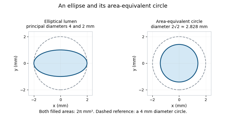

Using an equivalent diameter in a circular area-ratio formula is valid by construction:Using an arbitrary single projected width instead is not. Eccentric lumen stenosis can show different widths in different views while retaining the same area. Primary selective coronary arteriography used serial and cine recording to observe the contrast-filled arterial route; interpreting such images still requires the chosen projection and reconstruction model.[ref]
Selective Coronary Arteriography: Part I: A Percutaneous Transfemoral Technic by Melvin P. Judkins. Material and Technic, serial/cine recording
The equivalent diameter also differs from hydraulic diameter, , which includes wetted perimeter . Two equal-area outlines can have different perimeters and flow resistance. An area-equivalent circle is therefore not automatically an equivalent flow model.
For small area uncertainty, differentiation gives . A relative standard uncertainty in area gives approximately in the derived diameter if other inputs are fixed. This propagates area uncertainty; it does not correct an incorrectly segmented outline or a mismatched reference region.
A bypass graft is a conduit that creates an alternative route for blood flow around a diseased or obstructed vessel segment. It can use biological vessel tissue or a synthetic material. Creating the route does not itself remove the original obstruction or eliminate its underlying disease.[ref]
Surgical Bypass by the Society for Vascular Surgery. What is Surgical Bypass?
A bypass connects an inflow region to an outflow region through a surgical anastomosis at each end. The graft’s geometry, material response, connections and interaction with blood all affect performance. Graft patency describes whether the conduit remains open; an open lumen, sufficient flow and useful downstream tissue function are related but distinct observations.
An idealized resistance model clarifies the new flow path. If a remaining native segment has hydraulic resistance and a graft connects the same two junctions with resistance , the two paths are in parallel:For illustrative and in consistent resistance units, . The local flow would increase sixfold at fixed pressure difference across those junctions. But if other vessels contribute a series resistance , the total resistance changes from to , giving a flow ratio at fixed overall pressure difference. The example shows why a local geometric improvement does not determine the whole circulation’s response.
The model assumes steady linear resistance and fixed driving conditions. Real grafts and vessels are compliant, branching biological structures with pulsatile flow. Bench measurements of pressure and flow, mechanical tests of the conduit and its connections, and imaging follow-up address different parts of the problem. The Society’s primary description includes ultrasound follow-up for graft monitoring.[ref]
Surgical Bypass by the Society for Vascular Surgery. Follow-up discussion
A bypass graft is therefore both a fluid pathway and a material–tissue interface. Its scientific evaluation connects vascular surgery, biomechanics, biocompatibility and, for engineered substitutes, tissue engineering.
Graft patency is the openness of a graft’s lumen to blood flow. For a bypass graft, an open route is a necessary anatomical observation, but it does not by itself establish adequate tissue perfusion or a favorable overall clinical outcome. A conduit can remain open while narrowing progressively.
Reporting must also specify the history of that openness. In the 1997 vascular reporting standard, primary graft patency describes uninterrupted patency without a qualifying intervention on the graft or its anastomosis. Assisted primary graft patency allows intervention before an occlusion; secondary graft patency allows restoration after occlusion under the stated reconstruction rules. Procedures confined to disease beyond the graft and its anastomoses have specific exceptions. The exact endpoint definition must accompany the result.[ref]
Recommended standards for reports dealing with lower extremity ischemia: Revised version by Robert B. Rutherford and others. Printed pp. 524–525, Patency status: primary vs secondary patency
An original complete-follow-up example shows why one percentage is insufficient. Ten grafts are followed for the same interval: six need no procedure, two remain open after preventive graft interventions, one occludes and is successfully reopened, and one remains occluded. The corresponding endpoint proportions are primary, assisted primary and secondary patency. They describe the same cohort, not three competing estimates of one quantity. The last figure includes grafts that never required rescue.
With unequal observation times, the percentage of grafts open at their last visit is not a fixed-time patency estimate. Survival analysis, including the Kaplan–Meier estimator, tracks time to the chosen endpoint and handles right censoring under stated assumptions. Loss to follow-up and death need explicit treatment; simply deleting them can bias a comparison.
Observation is also method-dependent. Ultrasound follow-up can document graft status and detect developing abnormalities. Measurements of narrowing or flow velocity, the occurrence of occlusion and the need for intervention are related but different records. Specifying which was measured makes graft patency useful for comparing graft materials, surveillance strategies and vascular surgery outcomes.[ref]
Surgical Bypass by the Society for Vascular Surgery. Follow-up ultrasound discussion
A graft patency risk set contains the observed grafts still eligible to experience the specified patency endpoint immediately before a given time. It is the denominator for a time-specific failure calculation, not simply the original number of operations or the number of open grafts at the last recorded visit.
The endpoint must be chosen first. Primary graft patency, assisted primary graft patency and secondary graft patency use different intervention and occlusion histories. An intervention that ends one endpoint need not end another. The vascular reporting standards define these categories and discuss life-table and Kaplan–Meier estimator methods for unequal follow-up.[ref]
Recommended standards for reports dealing with lower extremity ischemia: Revised version by Robert B. Rutherford and others. Printed pp. 524–525, patency definitions; p. 525 onward, Estimating patency rates
For a single time-to-first-failure endpoint, let be graft 's failure time and its last observable follow-up time. With a specified common time origin, defineAt an observed failure time , the risk set size is evaluated just before the failures. If qualifying failures occur then, the Kaplan–Meier estimator of patency isTies between failure and censoring times require a consistent observation convention.
In an original example, ten grafts enter follow-up. Two meet the chosen endpoint at day 10, three of the remaining grafts are last observed without failure at day 15, and one of the five still followed fails at day 20. ThenThe three censored grafts do not cause a survival-curve drop at day 15; they reduce the later denominator. The crude expression answers neither the same follow-up-adjusted question nor the question of what happened after censoring.
The censoring assumptions are substantive. If grafts are preferentially lost when failure is imminent, observed risk sets may not represent those no longer followed. Death also prevents later graft-failure observation and can be a competing risks, rather than merely an ordinary missed visit. A cumulative incidence question with death as a competing event differs from a net-patency analysis that censors deaths. Endpoint definition, time origin, observation process and treatment of death must accompany a patency estimate.
Secondary graft patency credits continued function of the original graft reconstruction, including qualifying restoration after an occlusion. It differs from assisted primary graft patency, which requires that patency never have been lost. The 1997 reporting standard limits what counts as salvaging the original reconstruction: a replacement that does not preserve the required original graft continuity is not simply a continuation of its secondary patency.[ref]
Recommended standards for reports dealing with lower extremity ischemia: Revised version by Robert B. Rutherford and others. Printed p. 525, secondary patency and redo-reconstruction rules
Secondary patency is a durability endpoint that can credit successful salvage. It does not assert that blood flowed continuously during an interval in which the graft was occluded. An instantaneous graft patency assessment and a reconstructed follow-up history therefore need not be the same data object.
An original example follows five grafts through six months. One occludes at month 2 but is successfully reopened at month 3 with a qualifying salvage of the original reconstruction. Another occludes at month 4 and is not restored. The remaining three stay patent without procedures. At the end of this complete observation period, retain secondary patency while only have uninterrupted patency without a qualifying intervention. A newly created replacement bypass should be recorded under the stated reconstruction rules, rather than silently resetting the old graft’s clock.
A time-dependent analysis must define when secondary patency is considered irretrievably lost, how salvage episodes are adjudicated and what happens when follow-up ends before that status is resolved. Death can prevent later observation of graft failure; treating it as ordinary right censoring estimates a particular hypothetical durability quantity and requires assumptions. A real-world probability of failure before death instead calls for a competing risks analysis.
Reporting primary graft patency, assisted primary graft patency and secondary graft patency together makes the role of preventive and rescue interventions visible. The associated procedure counts, observation times and clinical outcomes remain necessary to distinguish prolonged graft function from the wider burden and consequences of maintaining it.
Assisted primary graft patency credits maintenance of an open graft by interventions performed before it occludes. The distinction is temporal: assistance preserves uninterrupted patency, whereas successful reopening after an occlusion belongs to secondary graft patency. The 1997 reporting standard introduced this distinction explicitly when revising how preventive graft procedures were counted.[ref]
Recommended standards for reports dealing with lower extremity ischemia: Revised version by Robert B. Rutherford and others. Printed p. 525, assisted-primary-patency discussion
A qualifying preventive intervention can end primary graft patency without ending assisted primary graft patency. Thus the two endpoints measure different aspects of durability: the former concerns performance without that assistance, while the latter also credits preservation work. They should share a stated time origin and compatible rules before their curves are compared.
Consider an original four-graft cohort. At month 2, one graft undergoes a preventive procedure while still open. At month 3, a different graft occludes, then is successfully reopened at month 4. The other two remain open without procedures. With complete follow-up to month 4, primary patency is , assisted primary patency is , and secondary patency is , assuming the restoration qualifies for the original reconstruction’s secondary endpoint. The reopened graft does not regain an uninterrupted assisted-primary history.
Surveillance influences what is observed and when assistance is undertaken. A narrowing may be detected before occlusion, so reports need their detection and intervention rules. A high assisted-primary proportion can coexist with many preventive procedures; it is not a measure of low procedural burden. Conversely, two cohorts with different surveillance intensity are not made directly comparable merely by using the same endpoint name.
With incomplete follow-up, survival analysis uses a graft patency risk set rather than treating every graft as observed to the final study date. Clinical interpretation also requires the separately measured consequences for tissue function and the patient. Preserving a conduit is an important intermediate outcome, not a complete description of benefit.
Primary graft patency measures how long a graft remains open without an intervention that terminates the primary-patency endpoint. It concerns the history since the original reconstruction, not merely whether a bypass graft is open on today’s image. Under the 1997 reporting standard, work confined to disease beyond the graft and its anastomosis can be an exception; an intervention on the graft to prevent its failure is not.[ref]
Recommended standards for reports dealing with lower extremity ischemia: Revised version by Robert B. Rutherford and others. Printed p. 525, primary-patency definition and native-vessel exceptions
For an explicit time-to-event model, define as the first qualifying loss of primary patency. Its survival function isOpening a narrowed but still patent graft by a qualifying preventive procedure can end primary graft patency even though blood never stopped flowing. Later rescue does not put that same graft back into the original primary-patency history. Assisted primary graft patency and secondary graft patency answer different questions about preserving or restoring function.
An original five-graft example demonstrates the denominator. All enter follow-up together. One has a qualifying intervention at month 2, another is last observed event-free at month 3, and a third occludes at month 4; the other two remain event-free through month 4. The Kaplan–Meier estimator givesThe graft patency risk set falls after both the first endpoint and the later right censoring. The censored graft is neither declared a failure nor assumed patent indefinitely. Interpreting this estimate requires an appropriate independent-censoring assumption.
A report should identify the original procedure date, qualifying interventions, documented occlusions and observation limits. A status image alone cannot reconstruct every earlier event. Primary graft patency is therefore most informative alongside the intervention history and separately defined clinical outcomes, rather than as an unexplained percentage.
Endovascular surgery comprises interventions performed from within a blood vessel, commonly using catheters and imaging to reach and treat a vascular region. It differs in access and apparatus from an operation that directly exposes the entire treated segment. The field includes methods such as balloon angioplasty and placement of a vascular stent; the appropriate intervention depends on the clinical problem.[ref]
What is a Vascular Surgeon? by the Society for Vascular Surgery. Open and endovascular procedure scope
An angiographic system combines vascular access with images of a contrast-filled vessel. The Society for Vascular Surgery describes an angiogram as potentially diagnostic or therapeutic, with balloon angioplasty and stent placement among the possible accompanying interventions.[ref]
Angiogram by the Society for Vascular Surgery. Procedure overview and possible therapies
The image supplies information about the lumen and device position, but an image of an apparently open region is not by itself a complete measurement of downstream tissue function.A simple imaging example shows why geometry matters. In a bench phantom, suppose a marker of known length spans 25 image pixels. At that marker’s plane, the scale is per pixel, so an 18-pixel feature would represent under the same projection conditions. Applying that scale to an object at a different depth can be wrong because magnification changes. These chosen values illustrate measurement calibration, not device sizing for a patient.
Fluoroscopy provides time-dependent projection images that help relate device motion to anatomy. Projection, motion, overlapping structures and image resolution limit what can be inferred from one view. A sound technical account therefore distinguishes the device’s mechanical action, the image measurement and the biological outcome.
The scientific questions extend beyond immediate access: how the intervention changes the flow pathway, how a device interacts with the vessel wall and whether the intended function persists. Those questions connect endovascular surgery to fluid dynamics, biomaterials, imaging and the measurement of clinical outcomes.
A vascular stent is a tubular implant providing radial support inside a blood vessel or vascular graft. Unlike a bypass graft, it supports the existing route. Support is a mechanical function; sustained patency is an outcome to measure.[ref]
Non-Clinical Engineering Tests and Recommended Labeling for Intravascular Stents and Associated Delivery Systems by the FDA. Section I, Intravascular Stent
A balloon-expandable vascular stent retains its enlarged shape after balloon deflation. A self-expanding vascular stent enlarges after release from its constrained delivery configuration, through material properties, geometry or both.[ref]
Non-Clinical Engineering Tests and Recommended Labeling for Intravascular Stents and Associated Delivery Systems by the FDA. Section I, Balloon Expandable Stent and Self-expanding Stent
Stent recoil gives a simple worked measurement. If bench imaging measures diameter during expansion and after balloon deflation, defineFor illustrative diameters and , recoil is about . The lumen area falls by about if both sections are circular, since area scales with diameter squared. Neither percentage alone predicts the change in pulsatile blood flow.
Bench radial-compression tests record diameter against external pressure. Loading and unloading distinguish reversible response from permanent damage; cycling probes mechanical fatigue. The FDA engineering guidance separately considers microscopy and durability tests. These measurements connect vascular stents to biomechanics and biomaterials; bench performance does not establish clinical effectiveness.[ref]
Non-Clinical Engineering Tests and Recommended Labeling for Intravascular Stents and Associated Delivery Systems by the FDA. Sections IV.B.4–6 and IV.B.9–11, recoil, integrity, radial response and durability
Stent recoil is a reduction in the diameter of an expanded vascular stent after the expanding load is removed. For a balloon-expandable device, the relevant acute comparison is commonly the diameter during balloon expansion against the diameter after balloon deflation. It is distinct from later biological tissue growth inside the treated segment.[ref]
Non-Clinical Engineering Tests and Recommended Labeling for Intravascular Stents and Associated Delivery Systems by the FDA. Section IV.B.4, Recoil for Balloon Expandable Stents
Using matched diameter measurements during expansion and after release, defineThe denominator is the expanded diameter. An original bench example with and gives . If the sections are circular, the corresponding area loss isDiameter and area percentages should not be interchanged, and neither alone specifies the resulting change in blood flow.
The mechanical distinction is between permanent expansion and recoverable deformation. A loading–unloading test relates stent recoil to the device’s material response and geometry. Imaging should compare the same location along the device; averages can hide differences near its ends. The primary FDA engineering guidance consequently asks for diameter-specific measurements and attention to variation with length and position.[ref]
The ratio also has a useful measurement property. Multiplying both diameters by the same scale factor leaves unchanged, while an equal additive edge-location offset generally does not. For independent diameter standard uncertainties , first-order uncertainty propagation givesWith in the example, percentage points. Shared image-scale or fitting errors require a covariance term instead of the independent-error formula.[ref]
Evaluation of measurement data — Guide to the expression of uncertainty in measurement by JCGM. Section 5.2, correlated input quantities
Balloon angioplasty enlarges a narrowed vessel segment by inflating a balloon carried on a catheter within the lumen. It is a form of endovascular surgery. The balloon applies outward pressure to the narrowed region and is subsequently deflated and removed. A vascular stent, if used, is a separate retained support; balloon dilation by itself does not leave that scaffold behind.[ref]
The mechanical target is vascular stenosis. Increasing lumen radius can strongly reduce resistance, but geometry is only part of the response. Under the ideal assumptions of Hagen-Poiseuille flow through a uniform rigid circular tube,An illustrative radius increase from to would reduce that segment’s model resistance to of its former value. This is not a predicted patient flow increase: a short irregular narrowing, compliant walls, pulsatility and the rest of the circulation depart from the ideal tube model.
The historical primary report by A. R. Grüntzig, A. Senning and W. E. Siegenthaler describes a catheter system with controlled balloon inflation in a 50-patient coronary series. In the 32 reported successful cases, mean percentage stenosis changed from to , while mean pressure gradient changed from to . Measuring both anatomy and pressure made the proposed mechanical effect observable. The report also records failures and emergency bypasses and calls for randomized comparison, so the successful-case averages are not a complete treatment-effect estimate.[ref]
Nonoperative dilatation of coronary-artery stenosis: percutaneous transluminal coronary angioplasty by A. R. Grüntzig and others. 1979 abstract, apparatus, successful-case measurements and limitations
A modern scientific evaluation similarly separates immediate lumen enlargement, the pressure gradient, persistent patency and later clinical outcomes. A larger lumen on an angiographic image answers an anatomical question at that observation time; it does not establish that narrowing cannot recur or that every downstream tissue problem has been resolved.
Coronary angioplasty enlarges a narrowed segment of a coronary artery using an intravascular intervention. In balloon angioplasty, a balloon catheter applies outward pressure within the affected segment and is then deflated. A vascular stent, when used, is a separate retained support rather than the balloon itself.
The historical primary report by A. R. Grüntzig, A. Senning and W. E. Siegenthaler describes a catheter system introduced through a systemic artery under local anaesthesia, with controlled balloon inflation. Its 50-patient series reported 32 technical successes and measured both the stenosis and the coronary pressure difference.[ref]
Nonoperative dilatation of coronary-artery stenosis: percutaneous transluminal coronary angioplasty by A. R. Grüntzig and others. 1979 abstract, apparatus, treated series and paired measurements
This connects a mechanical intervention to observations of anatomy and a physiological consequence.An original series-resistance model explains why improving one segment does not determine the whole circulation's response. Let a narrowed segment have vascular resistance , the rest of a represented pathway resistance , and assume a fixed total driving pressure difference . ThenIn consistent illustrative units, take , and , giving . If intervention reduces the local resistance to while the other assumptions remain fixed, flow becomes . A fourfold increase in the local segment's conductance has increased total flow only by a factor , not fourfold. The pressure loss across that segment changes from to about in the same chosen units.
Real coronary blood flow also involves pulsatility, deformable vessels and responses of smaller vessels, so this is an explanatory network model rather than a patient prediction. Diameter versus area stenosis adds another caution: a geometric narrowing percentage requires its measurement convention and reference segment before it is converted into an area or a flow-model parameter.
Evaluation separates immediate technical success, persistent openness, symptoms, later interventions and other clinical outcomes. The original authors reported failures and emergency bypasses and called for prospective randomized comparison with surgical and medical management.[ref]
Nonoperative dilatation of coronary-artery stenosis: percutaneous transluminal coronary angioplasty by A. R. Grüntzig and others. Abstract, adverse observations and final comparative-evaluation statement
A selected successful-case measurement does not supply the outcome distribution for every attempted procedure or establish comparative benefit by itself. Articles by others on the same topic
There are currently no matching articles.