In signal transduction, inositol trisphosphate usually means inositol 1,4,5-trisphosphate: a soluble intracellular second messenger. An extracellular ligand, its cell-surface receptor, IP3 and the released calcium ions are distinct components. IP3 binds an IP3 receptor on the endoplasmic reticulum, promoting stored release into the cytosol.[ref]
Pathways of Intracellular Signal Transduction. Phospholipids and Ca2+, Figure 13.27: soluble IP3 and intracellular calcium release
The useful separation is spatial: an extracellular message can control an internal store without entering it. The existing intracellular-injection experiment under metabotropic receptor tests bypassing reception; the Pancreatic IP3 store fractionation experiment below tests which internal compartment supplies the released calcium. Neither question is answered merely by observing a downstream membrane electric current.
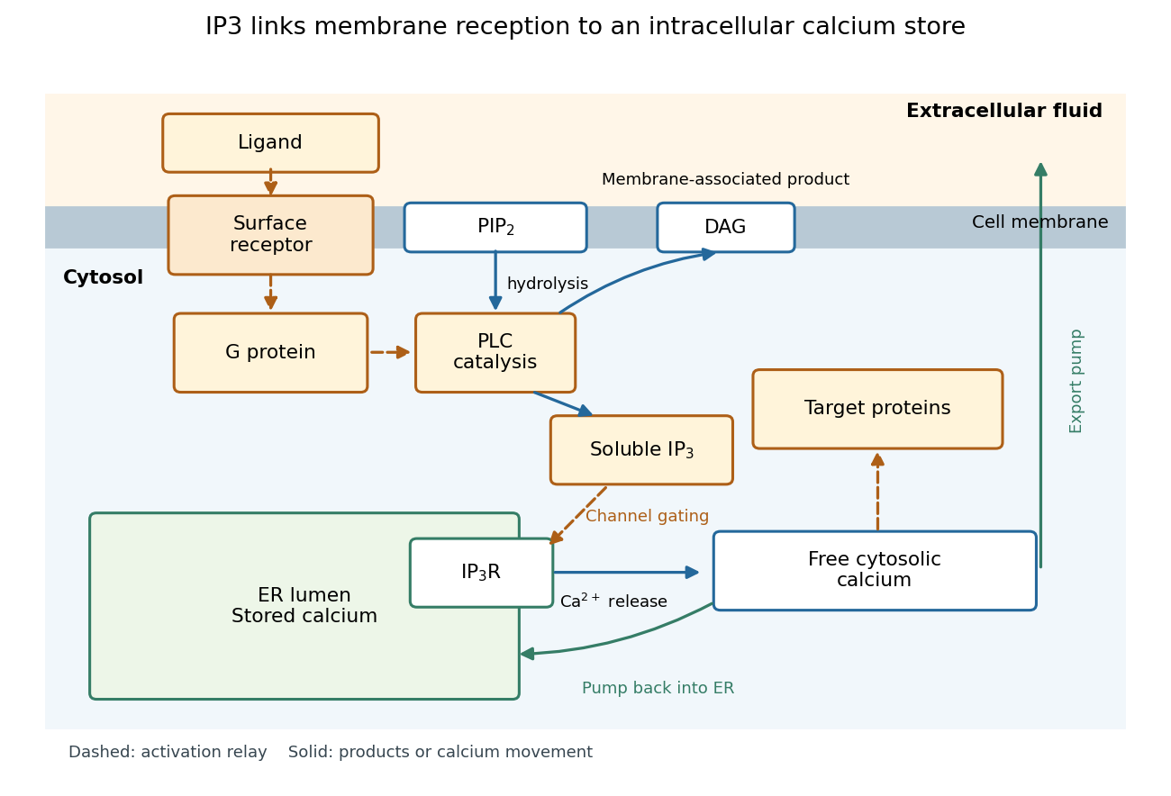

Phospholipase C catalyzes hydrolysis of membrane PIP2, producing soluble IP3 and membrane-associated diacylglycerol. This consumes a phospholipid substrate, rather than manufacturing calcium. G protein-regulated and receptor tyrosine kinase-regulated forms of PLC provide different routes to this reaction.[ref]
Pathways of Intracellular Signal Transduction. Phospholipids and Ca2+, Figures 13.24–13.25: PIP2 hydrolysis and distinct PLC activation routes
An IP3 receptor is an intracellular ligand-gated ion channel, rather than the extracellular signal's cell-surface receptor. Its opening is regulated by both IP3 and cytosolic calcium: IP3 binding is not a command that fixes one constant release rate. Mammalian IP3 receptors have three subtypes, with possible mixed assemblies and splice variants; these need not have identical regulation or cellular roles. The 2010 authored account distinguishes established channel behavior from then-unresolved structural explanations.[ref]
IP3 Receptors: Toward Understanding Their Activation by Colin W. Taylor and Stephen C. Tovey. Taylor and Tovey, IP3 Receptors: Toward Understanding Their Activation, Abstract; regulation by cytosolic Ca2+ and Structural Determinants of IP3R Activation: three subtypes and tetrameric assemblies
An IP3-sensitive calcium store contains releasable calcium behind a membrane bearing functional IP3 receptors. The endoplasmic reticulum is an important example. Pumping calcium into it or exporting calcium across the cell membrane lowers free cytosolic calcium; intracellular redistribution and loss from the whole biological cell are different balances.[ref]
Pathways of Intracellular Signal Transduction. Phospholipids and Ca2+, paragraph accompanying Figure 13.27: calcium pumps across plasma and ER membranes
Finite availability matters: a release pathway cannot deliver more calcium than its accessible store contains unless replenishment occurs. A falling cytosolic calcium response can reflect reduced release, increased removal, or both. Store content, release rate and free cytosolic concentration therefore answer different questions. Binding to proteins can also change the free fraction without changing the total number of calcium ions.
A 1984 primary experiment localized IP3-evoked release using pancreatic acinar cells isolated from rat pancreas with collagenase. A homogenized preparation was separated by differential centrifugation, then by a sucrose step density gradient or precipitation with magnesium chloride. The readout was IP3-induced calcium release per mass of protein across fractions. Release tracked two endoplasmic reticulum markers, RNA content and an enzyme activity, across several separation methods. Marker activities for mitochondria and the cell membrane supplied contrasting comparisons: release correlated negatively with them.[ref]
Effect of inositol-1,4,5-trisphosphate on isolated subcellular fractions of rat pancreas by H. Streb and others. Streb, Bayerdörffer, Haase, Irvine and Schulz, Effect of inositol-1,4,5-trisphosphate on isolated subcellular fractions of rat pancreas (1984), abstract: preparation, separation methods, protein-normalized release and marker correlations
Agreement across distinct fractionations supports an endoplasmic reticulum-associated source in this preparation. “Nonmitochondrial” alone only excludes a candidate; positive compartment markers add localization evidence. Marker comparisons control the localization inference, rather than replacing a no-IP3 release comparison. Fractionation may mix or damage compartments, and correlation does not itself identify the IP3 receptor protein. This calcium release readout also does not directly measure secretion or establish identical responses in every biological cell.
For an original mathematical model, imagine a sustained, imposed release pathway, with no store refill during the plotted interval. Let be releasable store amount of substance, excess freely dissolved cytosolic calcium amount of substance, and the cumulative amount removed from into a separate sink. Choose , , cytosolic volume , release rate constant and clearance rate constant . These are stipulated values, not physiological measurements or a model of IP3 receptor gating.
Assume fixed volume, uniform mixing, no binding, no reverse transfer, no outside entry and first-order loss from each donating compartment. ThenThe last equality is conservation of mass for the tracked calcium across the store, mobile excess and sink together; it does not assert conservation inside a biological cell if the sink represents export. A constant background is held separate from this excess bookkeeping. Since , the response is .
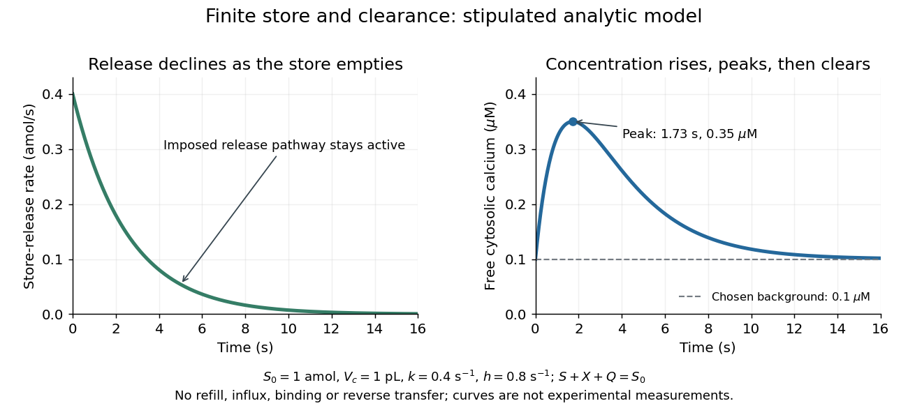

Solving these ordinary differential equations givesRelease declines from the start, yet initially rises because delivery exceeds clearance. Its peak occurs when , at . There and . Ultimately and tend to zero, tends to and returns to . The delayed peak is a consequence of material balance, even though this model has no delayed channel activation.
This distinction guides interpretation: a large released amount need not mean a large simultaneous free cytosolic concentration. Clearance and volume intervene. Real biological cells add binding, store refilling, IP3 turnover and IP3 receptor regulation; the two plotted curves isolate one finite-store effect rather than explaining every calcium response.
Articles by others on the same topic
There are currently no matching articles.