A receptor tyrosine kinase combines extracellular ligand recognition with an intracellular protein kinase protein domain. A membrane-spanning segment connects these parts across the cell membrane: the signal changes the intracellular machinery without requiring the ligand to enter the cytosol. The protein kinase transfers a phosphate group from ATP to selected tyrosine residues on proteins, including the receptor itself.[ref]
Signaling through Enzyme-Linked Cell-Surface Receptors. “Activated Receptor Tyrosine Kinases Phosphorylate Themselves”: extracellular binding, intracellular catalytic domains and ATP-dependent phosphate transfer
This architecture makes a reception event useful in two ways: it can change catalytic activity and assemble a local group of signaling proteins. Trans-autophosphorylation can create phosphotyrosine residues recognized by SH2 domains. Recruitment positions a downstream enzyme or relay protein near its partners. Neither every modified site nor every recruited protein has the same effect; association is a specific molecular connection, rather than a universal command to activate everything nearby.[ref]
Signaling through Enzyme-Linked Cell-Surface Receptors. “Phosphorylated Tyrosines Serve as Docking Sites For Proteins With SH2 Domains”, Figure 15-53: signaling enzymes, adaptors and negative regulators
Compare the epidermal growth factor receptor with the insulin receptor. Their common topology does not impose one activation geometry or make assembly equivalent to activity. The receptor phosphorylation assay consequently needs more than evidence that two protein chains associate.
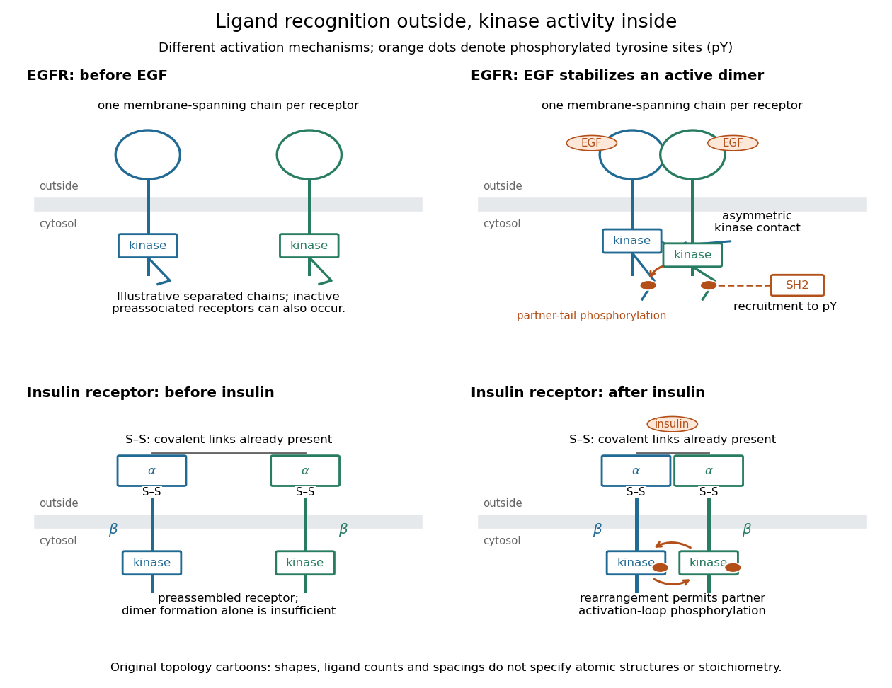
Receptor tyrosine kinase architecture and distinct activation mechanisms
. Original topology cartoons contrasting EGFR with the preassembled insulin receptor. Orange dots mark phosphotyrosine residues; S–S marks protein disulfide bonds. The EGFR panel separates asymmetric catalytic contact from partner-tail phosphorylation. Shapes, distances and drawn ligand counts are explanatory, rather than atomic structures or binding stoichiometry. Details.
In an illustrative EGFR activation route, EGF stabilizes an associated pair of receptor chains. Asymmetric contact between their protein kinase protein domains activates one catalytic partner without requiring its activation-loop phosphorylation. Subsequent phosphorylation of cytoplasmic tails creates recruitment sites. Preassociated inactive EGFR populations can also occur: the cartoon's separated starting chains are not a universal initial condition.[ref]
Cell signaling by receptor-tyrosine kinases by Mark A Lemmon and Joseph Schlessinger. Lemmon and Schlessinger, “Mechanisms of Receptor Activation” and “Allosteric activation of TKDs”, Figure 2F
The mature insulin receptor already contains two extracellular chains and two membrane-spanning chains, joined through protein disulfide bonds, before insulin binds. Insulin induces rearrangement of this assembly. Partner-mediated phosphorylation of the inhibitory activation loop permits increased protein kinase activity. Ligand-induced creation of a previously absent dimer is therefore an unsuitable universal explanation.[ref]
Cell signaling by receptor-tyrosine kinases by Mark A Lemmon and Joseph Schlessinger. “Mechanisms of Receptor Activation” and “TKD autoinhibition by the activation loop: insulin and FGF receptors”, Figure 2E
An insulin receptor substrate is a docking protein phosphorylated by the activated insulin receptor. Its phosphotyrosine residues recruit downstream signaling proteins, including SH2 domain-containing components of phosphoinositide 3-kinase. This connects reception to the pathway described under insulin, rather than making the insulin receptor substrate another extracellular ligand.[ref]
Cell signaling by receptor-tyrosine kinases by Mark A Lemmon and Joseph Schlessinger. “Linking RTK activation to cell signaling”: IRS docking proteins and Figure 3A
A receptor phosphorylation assay asks whether, and how much, a specified receptor is modified by phosphorylation under defined conditions. It is distinct from a binding assay for receptor binding occupancy, a test of protein kinase activity on a supplied substrate, and a test of downstream signal transduction. Those measurements answer related but different questions.
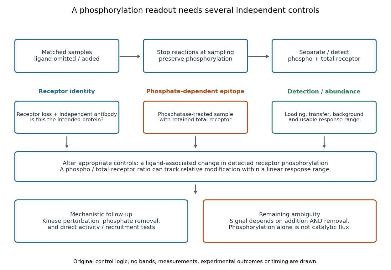
For a Western blot implementation, compare matched samples with ligand omitted or added, detect a specified phosphorylated epitope, and separately detect total receptor abundance. Preserve the sampled state by stopping further enzyme reactions during sample preparation; otherwise protein kinase or protein phosphatase activity after sampling can change the result. A general anti-phosphotyrosine residue antibody recognizes many modified proteins; a site-selective antibody gives a narrower readout, but still needs Western blot antibody specificity controls.
Reason through the controls before interpreting the difference. Loss of the proposed signal after receptor-specific gene deletion supports protein identity. Treatment with a protein phosphatase, alongside preservation of the total-receptor signal, tests phosphate-dependent recognition. Western blot loading controls and Western blot transfer controls address sample recovery; a usable detection range addresses Western blot response saturation. Lower signal after perturbing protein kinase activity supports dependence on that perturbation, but alternative targets and altered receptor abundance can complicate the inference. These are proposed assay comparisons, not outcomes attributed to the historical membrane experiment.

An original idealized measurement model explains the normalization. Let be the retained number of receptor molecules, the fraction carrying the selected modified epitope, and fixed detection gains. After subtracting their respective backgrounds, suppose the phosphorylated and total-receptor signals lie in a linear response range:Dividing by total receptor cancels under these assumptions. It differs from total-protein Western blot normalization, whose denominator includes many other proteins. For invented values , samples with and give and , but both give . Twice the phospho signal here means twice the retained receptor amount, with no change in fractional modification. Unequal recovery, changed epitope accessibility, nonzero uncorrected background or Western blot response saturation invalidates the cancellation. Unknown relative gains prevent reading an absolute modified fraction directly from the ratio.
A motivating experiment used isolated membranes from A-431 biological cells, which supplied abundant EGFR. Separate membrane aliquots were incubated at with ATP carrying radioactive phosphorus in its terminal phosphate group, plus magnesium or manganese ions. Radioactivity in material recovered by chemical acid-induced precipitation supplied the readout. Adding EGF beforehand enhanced incorporation relative to the unstimulated preparation; this placed a ligand-dependent biochemical response in a cell-free membrane system.[ref]
Epidermal Growth Factor by Stanley Cohen. Cohen, Epidermal Growth Factor, printed p. 339: A-431 membrane aliquots, terminally labeled ATP, divalent ions and trichloroacetic acid-insoluble incorporation
The follow-up comparisons separated increased addition from suppressed removal: phosphate removal was rapid but unaffected by EGF, and anti-EGF antibody reversed activation by removing the ligand. These observations support reversible regulation of membrane-associated protein kinase activity.[ref]
Epidermal Growth Factor by Stanley Cohen. printed p. 341, numbered observations 1–4: rapid activation, dephosphorylation, lack of soluble kinase release and anti-EGF reversal
Total incorporation alone cannot identify which protein or amino acid residue received the label, prove intrinsic receptor catalysis, or predict an intact biological cell's response.The same account records an instructive identification error: receptor phosphorylation was initially assigned to the wrong amino acid residue because released modified amino acids migrated together in the electrophoretic electric field. Reanalysis identified phosphotyrosine residues. Retained radioactivity is evidence about the assay's separation and detection, not unique chemical identity.[ref]
Epidermal Growth Factor by Stanley Cohen. printed p. 341: co-migration in the electrophoretic system and subsequent identification of tyrosine phosphorylation
Protein kinase activity adds modification, while protein tyrosine phosphatase activity can remove it. An original one-site model shows why a receptor phosphorylation assay cannot determine either process from a single modified fraction. Let be the fraction of a fixed receptor population carrying one selected phosphotyrosine residue. Stipulate constant effective first-order rate constants , with ATP availability absorbed into , and no receptor synthesis, degradation or trafficking:Here is the steady state fraction and the time constant. The ordinary differential equation has the exponential function solutionThe balance equation and a single relaxation step give this result; it is an illustrative model, rather than a fit or a claim about every RTK.
With stipulated rate constants , and . Doubling both rate constants leaves but changes to . The balanced phosphate turnover per site,doubles from to . Identical steady state modification therefore permits different catalytic fluxes. Removing the addition term instead gives ; this illustrates why a perturbation that stops protein kinase activity can expose removal dynamics, subject to the model's unchanged-removal assumption. A maintained steady state with ongoing ATP consumption is not chemical equilibrium.
Articles by others on the same topic
There are currently no matching articles.