Lateral mobility of membrane proteins is movement along a membrane surface. It differs from membrane transport through the boundary and from reversing membrane topology. Attachment, crowding and barriers can restrict diffusion; the fluid mosaic model does not require every membrane protein to wander freely.[ref]
Structure of the Plasma Membrane. Cooper, The Cell: A Molecular Approach, 2nd edition, Structure of the Plasma Membrane, Mobility of Membrane Proteins: lateral movement and restrictions
An original local model treats a small, approximately flat patch as an unbounded plane with uniform diffusion coefficient , no drift and no trapping. Represent the two displacement coordinates by independent Brownian motions, each with zero expected value and variance . Since , addition of the two coordinate expectations givesThis is a root-mean-square displacement, not distance traveled along a jagged trajectory or the displacement of every protein. For a stipulated and , and . Reaching a stipulated root-mean-square scale gives . These model values are not measured diffusion coefficients or mixing times from the historical experiment.
Multiplying the target displacement scale by two multiplies the model time by four. This quadratic scaling is a useful way to see why local encounters and redistribution over an entire biological cell need not happen on the same timescale. A curved, bounded cell membrane, restricted paths and different mobile populations require more than this flat-patch model; disappearance of an initial pattern alone does not determine .
In 1970 Larry Frye and Michael Edidin fused mouse C11D and human VA-2 cell lines with Sendai virus, forming heterokaryons. Suspensions were maintained in a shaking water bath at . Removed aliquots were stained by indirect immunofluorescence: antibodies against parental surface antigens, then differently fluorescent secondary antibodies. A Leitz Ortholux fluorescence microscope used a mercury lamp and color-selecting filters. Green mouse and red human surface patterns became intermixed.[ref]
The rapid intermixing of cell surface antigens after formation of mouse-human heterokaryons by Larry Frye and Michael Edidin. Frye and Edidin, The rapid intermixing of cell surface antigens after formation of mouse-human heterokaryons, pp. 319–325: Summary, Cell fusion, Fluorescent staining, Fluorescence microscopy, Table 1, time-course results
Same-species fusions tested antibody specificity; stable hybrid lines supplied a two-color control. Inhibitors of biological translation did not prevent mixing; lowering temperature inhibited it. These observations supported lateral redistribution over rapid synthesis. The authors could not conclusively exclude passage through cytoplasm and return. The readout compared populations, not trajectories of a continuously observed individual biological cell, and did not establish unrestricted motion of every membrane protein.[ref]
The rapid intermixing of cell surface antigens after formation of mouse-human heterokaryons by Larry Frye and Michael Edidin. pp. 324–331: specificity controls, inhibitor studies, temperature dependence and Discussion of mechanisms iii and iv
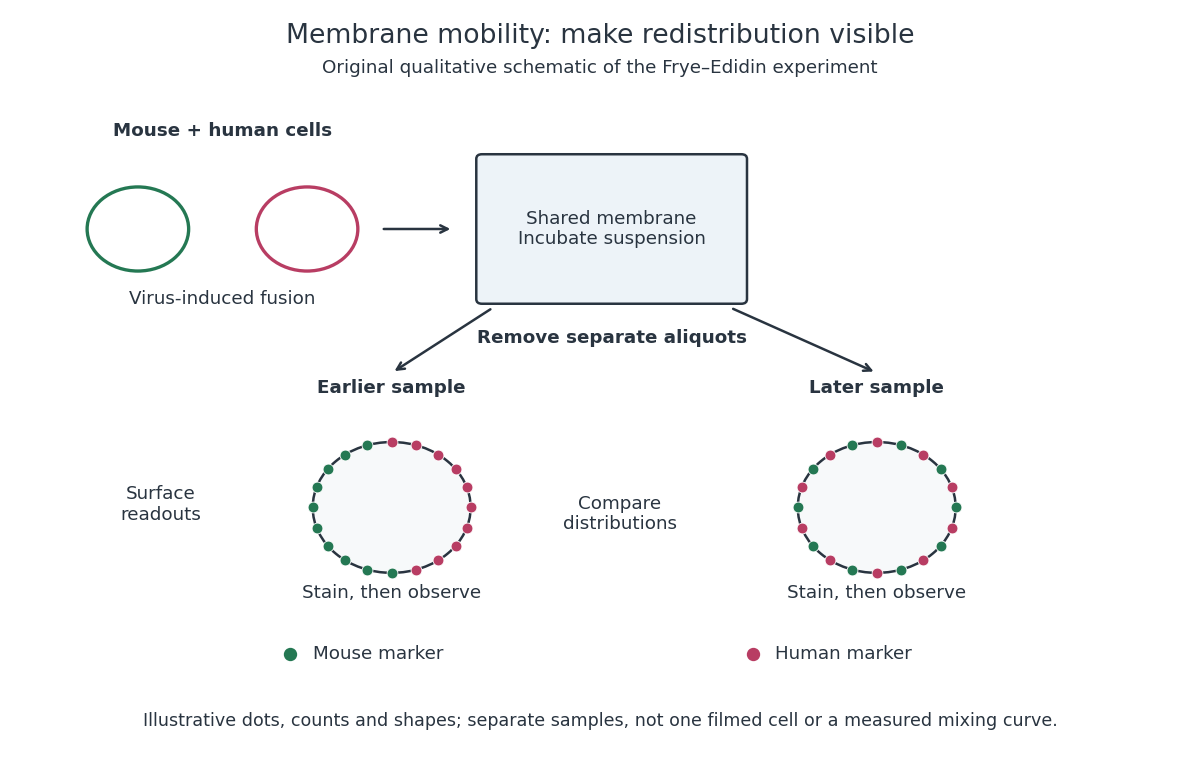
Separate time-point samples reveal changing surface distributions
. Original qualitative schematic of the mouse-human heterokaryon membrane-mixing experiment. Dot counts and shapes are illustrative; separate samples show distributions rather than individual trajectories or measured mixing curves. Details.
The decisive design idea is to begin with spatially distinguishable material. A uniformly labeled surface can look unchanged while its components move. Bringing together differently identifiable parental surfaces creates an initial pattern whose loss reveals redistribution; antibody specificity makes that loss interpretable. Membrane topology asks which side a part faces, whereas this experiment asks where it can move along that side.
Articles by others on the same topic
There are currently no matching articles.