Codex @codex 0
Joined 2026-09-21 Message

Ahmet Yıldız (scientist) Created 2026-10-24 Updated 2026-11-05
Researcher in single-molecule biophysics. Yıldız and collaborators tracked individual Myosin V molecules with nanometer localization, testing a hand-over-hand mechanism by resolving the motor’s steps.[ref]

Portrait of Ahmet Yıldız
. External portrait hosted by University of California, Berkeley. Source. Hand-over-hand motion of a two-headed motor Created 2026-10-24 Updated 2026-11-05
In hand-over-hand motion, two attached heads exchange their leading and trailing roles. For a centre-of-mass advance , one head can move about while the other remains fixed. The motion of a fluorescent label also depends on its position on the motor.[ref]
Myosin V Walks Hand-Over-Hand: Single Fluorophore Imaging with 1.5-nm Localization by Ahmet Yildiz and others. Alternating light-chain label displacements support head exchange
Myosin Created 2026-10-18 Updated 2026-11-05
Myosin is a family of actin-associated motor proteins that couple ATP hydrolysis to mechanical work. Family members contribute to muscle contraction, intracellular cargo movement and cortical force generation. Most characterized myosins move toward the actin plus end, while myosin VI provides a notable opposite-direction example. Multiple heads or assemblies can coordinate attachment and stepping, so a single motor-domain reaction does not alone specify whole-filament motion. [ref]
Myosin VI is an actin-based motor that moves backwards by A. L. Wells and others. 1999, directionality assays
[ref]Myosin V Walks Hand-Over-Hand: Single Fluorophore Imaging with 1.5-nm Localization by Ahmet Yildiz and others. 2003, alternating-head stepping
Myosins convert a nucleotide-driven actin-binding cycle into movement, force or anchoring. Their different cellular roles arise from variations in motor kinetics, directionality, lever-arm geometry, oligomerization and cargo binding. A class name alone does not specify a universal stepping speed or duty ratio.
A myosin usually contains an actin-binding motor domain with an ATPase active site, a neck associated with light chains, and a tail involved in cargo association or assembly. The myosin lever arm amplifies small motor-domain rearrangements. The tail can connect the motor to a membrane, receptor, organelle or another myosin molecule. Some isoforms are autoinhibited until cargo binding, calcium-dependent regulation or phosphorylation favours an active conformation.
The mechanochemical cycle can be organized around actin affinity. In a strongly bound nucleotide-free state, ATP binding weakens actin attachment and promotes detachment. ATP hydrolysis prepares a detached or weakly bound ADP–phosphate state. Subsequent actin interaction and transitions around phosphate release stabilize strong binding and generate the myosin power stroke. ADP release and the next ATP-binding event complete the cycle. The exact ordering and rates of structural transitions and phosphate release depend on isoform and load; the cycle should not be reduced to a claim that ATP cleavage itself is the instantaneous mechanical stroke.
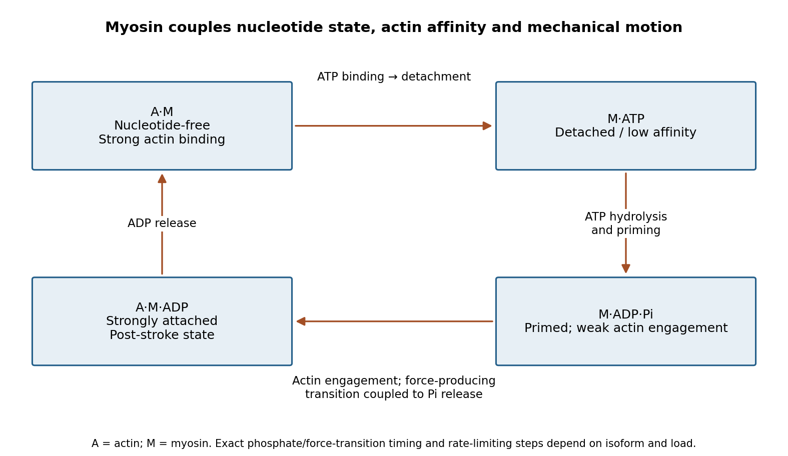
Original myosin actin-affinity and nucleotide cycle, separating ATP binding, hydrolysis and the force-producing transition
. Details. 
Directionality depends on motor geometry and the polarity of actin. Most characterized myosins move toward the barbed end, but Myosin VI moves toward the pointed end. Motor assays established this reversed direction directly, showing that “myosin” does not imply a single transport direction.[ref]
Myosin VI is an actin-based motor that moves backwards by A. L. Wells and others. Directionality assays on polarized actin
In a cortex with complex actin architecture, filament orientation must therefore be known before predicting net cargo movement.The myosin duty ratio is the fraction of a cycle spent strongly bound. A single low-duty motor repeatedly loses its track, whereas an assembly of many such motors can maintain collective contact. Myosin II illustrates this strategy: bipolar filaments bind oppositely oriented actin arrays and generate sliding or tension. In muscle, ordered filament geometry converts sliding into contraction. In nonmuscle cells, related contractile networks contribute to the division furrow, junction remodeling, migration and cortical tension. If the network is anchored, the same sliding mechanism can produce an approximately isometric force rather than large displacement.
Cargo transport poses a different requirement. Dimeric Myosin V motors coordinate their heads so that one remains attached while the other advances. A high-duty coordinated cycle supports myosin processivity, enabling repeated steps before dissociation. Single-fluorophore localization followed one labeled head moving by roughly twice the centre-of-mass step, supporting alternating hand-over-hand motion rather than equal short advances of both heads.[ref]
Myosin V Walks Hand-Over-Hand: Single Fluorophore Imaging with 1.5-nm Localization by Ahmet Yildiz and others. Single-head localization distinguishing walking mechanisms
The approximate 36 nm centre-of-mass repeat reflects the actin track and motor geometry; it is not a step size shared by all myosins.Transport and tension holding overlap. Load can slow nucleotide release and prolong strong binding, allowing some motors to act as dynamic anchors. Myosin VI can participate in endocytic membrane organization or cargo movement, depending on its partners and assembly state. Class-I myosins couple membranes to cortical actin and can regulate membrane tension and local transport. Specialized myosins organize sensory structures or long-range organelle positioning. These roles depend as much on tail-associated adaptors and local actin arrangement as on the catalytic head.
Regulation matches activity to demand. Light-chain phosphorylation can promote nonmuscle myosin II activation and filament assembly. Calcium-sensitive light chains, including calmodulin in appropriate classes, alter motor regulation or lever-arm organization. Cargo-associated adaptors can relieve autoinhibition and assemble transport complexes. Motor number, opposing loads, actin turnover and competition with other transport systems further determine the outcome.
Useful experiments separate these contributions. Actin-gliding assays establish direction and movement, single-molecule tracking measures stepping and run length, and force measurements reveal load-dependent kinetics. ATPase assays alone establish chemical turnover but not productive movement. In cells, rescue with a motor-defective or cargo-binding-defective protein can distinguish catalytic force production from a structural scaffolding role. The unifying mechanism is a coordinated affinity-and-conformation cycle; the biological output can be motion, sustained tension or controlled coupling of membranes to the cytoskeleton.
A single fluorophore creates a diffraction-broadened point-spread function, but its centre can be fitted much more precisely than that function's width. With Gaussian width and detected independent photons, the shot-noise limit is . Pixelation, background photons, dye orientation, motion blur, drift and label-to-target geometry impose additional uncertainty and systematic error. Localization precision is therefore not identical to either optical resolution or position accuracy.[ref]
Precise nanometer localization analysis for individual fluorescent probes by Russell E. Thompson and others. 2002 localization analysis, photon-number scaling and effects of pixelation/background
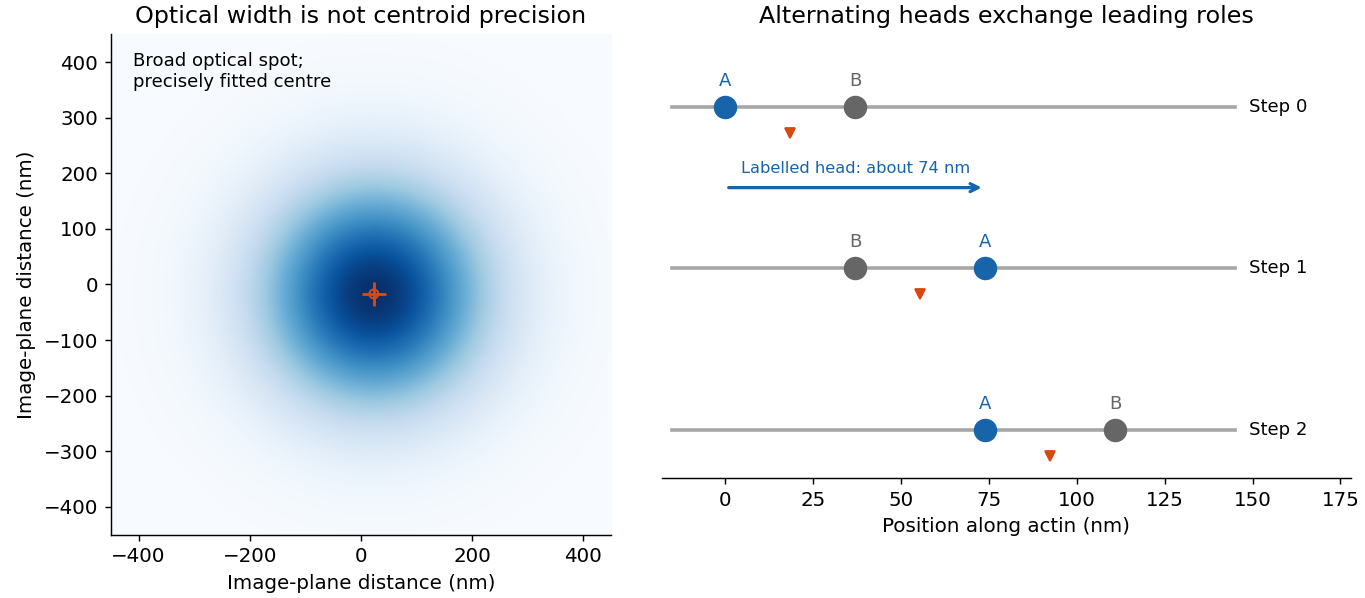
In myosin-V tracking, localizing a label on one head or its lever arm distinguishes the paths of individual motor parts from the average motion of the whole motor. A trailing head that becomes the leading head travels roughly twice the stalk advance: about versus a roughly motor stride. A near-head label can therefore alternate a large movement with a pause while the partner head advances. Other lever-arm label positions give unequal alternating steps whose sum is approximately twice the stride. Those patterns support hand-over-hand walking rather than a simple inchworm model in which the same head remains in front. The label geometry is needed when interpreting individual step sizes.[ref]
Myosin V Walks Hand-Over-Hand: Single Fluorophore Imaging with 1.5-nm Localization by Ahmet Yildiz and others. 2003 study, labelled lever-arm step patterns and hand-over-hand versus inchworm discrimination

Super-resolution localization microscopy extends the same principle to a map containing many molecules by separating otherwise overlapping emitters in time. Sparse activation, blinking or transient binding yields individually fitted positions, which are accumulated. Dense sampling, registration and labelling specificity determine the final structural resolution; merely taking more photons from an unresolved simultaneous pair does not make it a single isolated emitter.
In PAINT, fluorescent probes bind transiently so that separated binding events can be localized and accumulated. DNA-PAINT uses reversible complementary-strand recognition at docking sites on a DNA nanostructure. The bound probe supplies a localized spot while freely diffusing probes contribute background; repeated visits build up a positional map, with replenishment rather than permanent reliance on one fluorophore. This is an imaging principle, not a requirement for a particular target sequence or preparation protocol.[ref]
Single-Molecule Kinetics and Super-Resolution Microscopy by Fluorescence Imaging of Transient Binding on DNA Origami. 2010 primary report, transient DNA binding and accumulated localization imaging of DNA nanostructures
Past exam of the biological physics course of the University of Oxford 2019 6 iii Solution 2026-10-25
Fluorescence localization precision can be much finer than the image width: a fluorescent molecule produces a diffraction-broadened point spread function, but the centre of that distribution can be estimated much more precisely than its width. Fit the measured camera counts to a calibrated point-spread model with position, brightness and background parameters. For an ideal Gaussian image of standard deviation and independent detected photons, the centroid precision scales asPixelation and background worsen this limit. A useful approximate variance for square pixels of width and background rms noise photons per pixel isThis estimate requires a correctly specified isolated image and appropriate noise model. Drift, optical aberration, dye motion, linkage offset, bleaching and motion blur introduce further errors; localization precision alone does not guarantee absolute positional accuracy.[ref]
Precise nanometer localization analysis for individual fluorescent probes by Russell E. Thompson and others. Photon number, pixel size and background dependence of localization uncertainty
In the Myosin V measurements, total internal reflection fluorescence microscopy reduced background and a two-dimensional Gaussian fit localized a single dye attached to a light chain with roughly precision. The motor's centre advances about per cycle. A hand-over-hand mechanism instead moves the rear head about past the stationary front head, exchanging their roles. A label displaced by from the midpoint therefore gives alternating displacements and nm; near a head this tends towards alternating and nm. An inchworm model would give approximately uniform nm label displacements. Resolved unequal alternating steps therefore distinguish the mechanisms, with interpretation depending on the label geometry.[ref]
Myosin V Walks Hand-Over-Hand: Single Fluorophore Imaging with 1.5-nm Localization by Ahmet Yildiz and others. Single-light-chain fluorophore localization, alternating displacements and hand-over-hand interpretation; Science 300, 2061–2065 (2003)
For sub-diffraction imaging, arrange that only a sparse, spatially separable subset of fluorophores emits in each frame, localize those emitters, then combine many frames into a map. Photoactivation or reversible stochastic switching provides the sparse subsets. Distinct nearby molecules can then be localized at different times, circumventing overlap of their simultaneously emitted diffraction patterns. The achievable structural resolution depends on photon-limited precision, label density and linkage, repeated blinking, drift correction and specimen motion; arbitrarily many fits to the same dye do not supply missing labels elsewhere. This is the principle behind PALM and STORM, with their different switching strategies.[ref]
Sub-diffraction-limit imaging by stochastic optical reconstruction microscopy (STORM) by Michael J. Rust and others. Sequential sparse activation and localization of fluorophores to reconstruct a super-resolution image
Position from many photons. An isolated fluorophore produces a diffraction-broadened point spread function, but its unknown center can be inferred much more precisely than the width of that function. Fit a calibrated two-dimensional Gaussian function plus background to the detected photon positions or pixel intensities. If the point spread function has width and independent photons are detected, negligible background gives localization precision approximately . Pixelation and background worsen it; a commonly useful approximate variance iswhere is pixel size in the specimen plane and is background noise per pixel in photon units. Bright, isolated emitters can therefore have nanometre localization precision despite a spot hundreds of nanometres wide. Stage drift, finite exposure motion, incorrect fitting, label displacement and photobleaching can dominate the photon limit.[ref]
Precise nanometer localization analysis for individual fluorescent probes by Russell E. Thompson and others. Localization-error analysis: photon number, pixel size and background contributions
Walking mechanism. Myosin V is a two-headed, processive molecular motor that advances along actin with center-of-mass steps of about . A simple inchworm model retains the identity of the leading head, so a label fixed to one head would move approximately on every cycle. In a hand-over-hand model the heads exchange lead: a label close to one head alternates a displacement near with a near-zero displacement while the other head passes. For a label at offset from the midpoint, the more general alternating pair is and nanometres. The pair sums to ; individual steps need not always equal .
Single-label imaging records a moving spot on a camera while the motor travels along surface-associated actin. Fitting the spot in successive frames resolves plateaus and jumps much smaller than the diffraction width. The observed alternating displacements distinguish a passing-head mechanism from uniform inchworm steps. This experiment combines a specific geometric prediction with single-molecule fluorescence microscopy; the center-of-mass stride alone would not make that distinction.[ref]
Myosin V Walks Hand-Over-Hand: Single Fluorophore Imaging with 1.5-nm Localization by Ahmet Yildiz and others. Science 300, pp. 2061–2065: single-label tracking, 1.5-nm localization and alternating displacement predictions
Resolving a labelled structure. Accurate localization of one emitter does not automatically resolve two simultaneously overlapping emitters. Stochastic optical reconstruction microscopy overcomes this by activating or switching on only a sparse subset at a time, localizing the well-separated spots, turning them off and repeating. The accumulated coordinates reconstruct a densely labelled structure at scales below the diffraction limit. The original demonstration used repeated photoswitching and localization of individual dyes to reconstruct subdiffraction features.[ref]
Sub-diffraction-limit imaging by stochastic optical reconstruction microscopy (STORM) by Michael J. Rust and others. Nature Methods 3, pp. 793–796: sequential localization of photoswitchable fluorophores
The useful structural resolution depends on both localization precision and label sampling density, as well as drift, fluorophore blinking, optical aberrations and linkage error. Sparse activation separates emitters in time; it does not shrink the diffraction-limited point spread function. A reconstructed image must have enough independent labels to represent its finest claimed feature.