The common ones.
The rare ones. Notably present in peptidoglycan.
The weird one, not directly coded in the genetic code.
The study of the proteome.
Ciro Santilli defines a "model protein" as a protein which has been significantly used in the history of protein science, in analogy to the term model organism.
Key characteristics of model proteins include:
- they are easy to obtain and are stable
- they are important to medical applications
- they are small and easier to understand for early studies
Important model proteins include:
- insulin: as a peptide hormone, this was small. Also it was useful and widely available even at pharmacies, The Eighth Day of Creation says you could get it a Boots, a major British pharmacy chain, and as such was a natural choice for the first sequencing by Frederick Sanger published in 1951
- hemoglobin
- keratin
For an initial concrete example, consider e. Coli K-12 MG1655 gene thrA.
How Enzymes Work by RCSBProteinDataBank (2017)
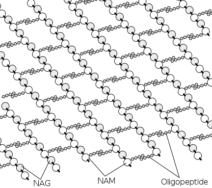
Source. Shows in detail how aconitase catalyses the citrate to isocitrate reaction in the citric acid cycle.Forms the bacterial cell wall.
From the Wikipedia image we can see clearly the polymer structure formed: it is a mesh with:
- sugar covalent bond chains in one direction. These have two types of monosaccharide, NAM and NAG
- peptide chains on the other, and only coming off from NAM
Peptidoglycan polymer structure
. Source. Responsible for muscle contraction.
Kinesin protein walking on microtubule by XVIVO
. Source. Clip from Video "The Inner Life of the Cell by XVIVO Scientific Animation (2011)" with captions added in.Kinesin Motor Protein 3D Animation by Art of the cell
. Source. Binds an amino acid to the correct corresponding tRNA sequence. Wikipedia mentions that humans have 20 of them, one for each proteinogenic amino acid.
The second protein to have its structure determined, after myoglobin, by X-ray crystallography, in 1965.
Breaks up peptidoglycan present in the bacterial cell wall, which is thicker in Gram-positive bacteria, which is what this enzyme seems to target.
Part of the inate immune system.
It is present on basically everything that mammals and birds excrete, and it kills bacteria, both of which are reasons why it was discovered relatively early on.
With X-ray crystallography by David Chilton Phillips. The second protein to be resolved fter after myoglobin, and the first enzyme.
Published at: Structure of Hen Egg-White Lysozyme: A Three-dimensional Fourier Synthesis at 2 Å Resolution (1965). The work was done while at the Davy Faraday Research Laboratory of the Royal Institution.
Phillips also published a lower resolution (6angstrom) of the enzyme-inhibitor complexes at about the same time: Structure of Some Crystalline Lysozyme-Inhibitor Complexes Determined by X-Ray Analysis At 6 Å Resolution (1965). The point of doing this is that it points out the active site of the enzyme.
www.nature.com/articles/206757a0 on Nature 181, 662-666. Paywalled as of 2022. Has some nice pictures in it.
www.nature.com/articles/206761a0 on Nature 206, 761-763. Paywalled as of 2022. Has some nice pictures in it.
Published at: a Three-Dimensional Model of the Myoglobin Molecule Obtained by X-Ray Analysis (1958). The work was done at the Cavendish Laboratory.
Articles by others on the same topic
Protein is a macromolecule that is essential for the structure, function, and regulation of the body's tissues and organs. It is made up of long chains of amino acids, which are organic compounds composed of carbon, hydrogen, oxygen, nitrogen, and sometimes sulfur. There are 20 different amino acids that combine in various sequences to form proteins, each of which has a specific function in the body.


