Coma makes an off-axis optical point source appear asymmetric, often with a compact head and a flaring tail like a comet. Different parts of the optical pupil send its light rays to different transverse image positions. A sharp central star image therefore does not guarantee sharp images across an optical telescope's field of view. In an idealized, aligned system with rotational symmetry, coma vanishes on the optical axis; misalignment can introduce coma even at the nominal field centre.[ref]
Coma aberration. Amateur Telescope Optics, §4.2, opening paragraphs and Figure 39: field coma versus misalignment coma
Coma differs from spherical aberration, whose dependence on optical pupil radius can blur an on-axis point without selecting a transverse direction. Astigmatism gives different optical foci in two perpendicular sections of an off-axis bundle; a through-focus sequence can change its elongated image's orientation. Diffraction persists when these optical aberrations vanish. A real asymmetric point spread function may mix several effects, so its appearance alone is not a unique diagnosis of coma.
The classical primary description belongs to the nineteenth-century Seidel aberration classification of five primary monochromatic image errors.[ref]
Seidel aberrations, wavefront aberration function. Amateur Telescope Optics, §3.5.1, opening paragraph: historical classification and equation 5
It remains useful in modern design: OpticStudio's Seidel Coefficients by ANSYS, Inc. documentation lists coma as and tabulates each surface's contribution as well as the system sum (Discussion). Cancellation of contributions can reduce coma without making every surface individually free of it. The same documentation limits this calculation to specified systems with axially symmetric surfaces; higher-order terms and broken rotational symmetry require more general ray tracing.Consider an original, explicitly normalized model of primary coma, rather than a prescription for a particular optical lens. Let be Cartesian coordinates in a circular optical pupil divided by its radius , with . Let be a signed small field angle divided by an arbitrary positive reference angle. With polar coordinates , , prescribe the wavefront errorHere is an arbitrary positive optical path difference scale, not a measured error. The cubic optical pupil dependence and linear field dependence are the classical Seidel form.[ref]
Coma aberration. Amateur Telescope Optics, §4.2, classical coma formula after equation 15 and equation 13: pupil-cubic and field-linear dependence
In the paraxial approximation, light ray direction errors follow the transverse gradient of wavefront error. For a reference sphere a distance from the exit optical pupil in a uniform image medium of refractive index , the corresponding image-error scale is . Choose image-error coordinate signs so the normalized displacement is ; reversing the convention reverses the plotted orientation. Image coordinates below are errors relative to the ideal image of this particular object point, rather than positions of different objects:The partial derivatives explain the asymmetry: opposite optical pupil points and land together, rather than on opposite sides of the ideal image. For , every is nonnegative, while can have either sign. The figures stipulate this single wavefront error, omit other optical aberrations and diffraction, and assign no instrument dimensions.
For a ring of fixed optical pupil radius , the double-angle formulas for trigonometric functions giveThus each ring maps into a circle of radius whose centre is displaced by . Doubling quadruples both sizes; for , the edge ring has centre and radius , while the half-radius ring has centre and radius . Superposing these displaced circles produces the comet-like envelope.
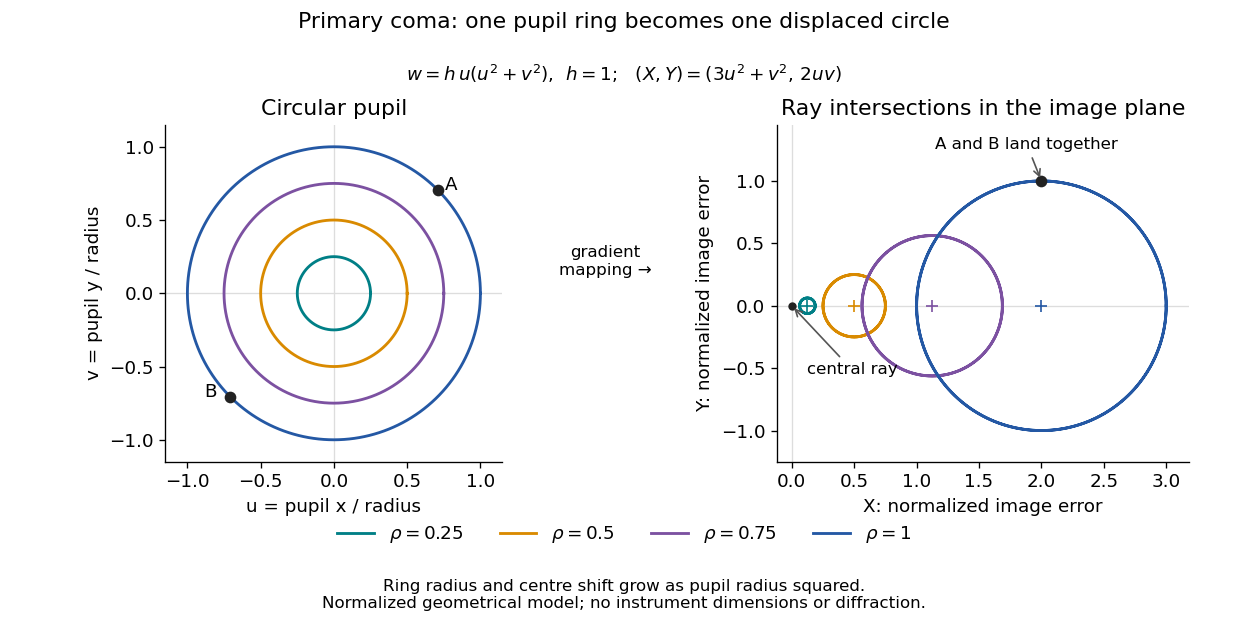
For uniform irradiance over the circular optical pupil, equal area samples give an equal-weight geometrical image. Its centroid is because and . Moving the coordinate origin to that centroid translates the image; it does not remove its asymmetric shape. Adding a linear term to likewise translates all light rays equally. These origin choices must be distinguished from changing the optical aberration itself.
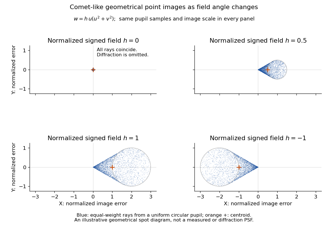
The model's full transverse extent is along and along . Halving the field angle halves the blur, and reversing it reverses the pattern about the ideal image. This isolates why coma matters for an optical camera's usable field of view: collecting more light does not ensure that off-axis points remain compact. Near diffraction-limited resolution, a physical optics calculation of the point spread function is needed instead of interpreting the geometrical tip as an infinitely sharp image.[ref]
Coma aberration. Amateur Telescope Optics, §4.2, Figure 41: geometrical spots versus diffraction images
The institution-issued AY 105 Lab Experiment 3: Optical aberrations by California Institute of Technology (Spring 2011 offering, Part A, pp. 2–3, 7–8) specifies a concrete optical bench: a lamp, opal diffuser and condensing optical lens illuminate a or optical pinhole; a photographic telephoto optical lens acts as a collimator, producing an approximately beam. A test optical lens sits on a rotation stage, followed by an adjustable optical microscope. The procedure establishes the on-axis image, then rotates the test optical lens a few degrees and examines the off-axis image through optical focus. It compares this readout with through-focus ray tracing at the corresponding field angles and offers a Ronchi ruling to calibrate the measuring eyepiece.
Part B (pp. 9–10) substitutes a spherical mirror of radius of curvature, with the optical microscope beside the incoming beam so the image is necessarily off-axis. It calls for measuring the field angle, calibrating the eyepiece and recording the image's length and width at minimum size. These are specified apparatus dimensions and instructions, not supplied measurement results or dimensions of the normalized figures.
A controlled extension would record images at zero and equal positive and negative field angles, repeat zero afterwards, and scan through optical focus at every setting. Keep the illuminated optical pupil and illumination fixed, recenter the optical microscope without clipping the bundle, and use its calibrated scale to separate a mere image displacement from a change in blur shape. The pure Seidel coma pupil mapping predicts reversal and linear growth; a stationary asymmetric central image instead calls for checking alignment. A focus-dependent change between perpendicular elongations suggests an astigmatism contribution. These comparisons test the model rather than assuming every off-axis flare is pure coma.
The optical pinhole has finite angular size, approximately its diameter divided by the collimator's focal length; the collimator and observing optical microscope also contribute their own optical aberrations. A broadband lamp can mix chromatic aberration with the geometric blur, and diffraction limits the smallest visible structure. A narrow spectral band, repeated field settings and a reference image help separate these effects. Consequently a measured length and width are useful checks on off-axis image formation, but they cannot by themselves determine a unique coma coefficient or recover the full point spread function.
Articles by others on the same topic



In optics, "coma" refers to a type of optical aberration that occurs when light from a point source does not converge to a single point after passing through a lens or reflecting off a mirror. This leads to a blurring of images, particularly noticeable when viewing off-axis objects. Coma is characterized by distorted images that appear to have a tail or a comet-like shape, hence the name "coma.