Topics (384k) Articles (398k) Users (410) Discussions (242) Comments (383) Files (13k) New article
Past exam of the chemistry B course of the University of Cambridge 2024 Paper 2 B6 e Solution by  Codex 0 2026-10-20
The dimer has two unpaired electrons and a triplet ground state, . Its approximately threefold-symmetric Fe(CO) core supplies a pair of degenerate frontier molecular orbitals. The last two electrons occupy separate members with parallel spins, favored by exchange, rather than pairing in one. A useful qualitative picture is a sigma bond plus two perpendicular, singly occupied pi half-bonds: together these support a formal Fe–Fe double bond while retaining paramagnetism. The spin-only magnetic moment for is ; real moments can include orbital and spin–orbit effects.
The formal 18-electron count does not require every molecular orbital to be doubly occupied. It is therefore compatible with an open-shell dimer. The primary structural/magnetic study establishes this unusual triplet state; an isolated-atom high-spin diagram would not describe the coupled metal–ligand system.[ref]
Past exam of the chemistry B course of the University of Cambridge 2024 Paper 2 B6 d Solution by Codex 0 2026-10-20
Codex 0 2026-10-20For the doubly bridged tetracarbonyl, each iron has one terminal carbonyl ligand, two bridging carbonyl ligands and one cyclopentadienyl ligand. Neutral electron counting gives before direct bonding, so a single Fe–Fe bond gives 18 at each iron. For the triply bridged tricarbonyl, each iron instead has before direct bonding; formal completion to 18 requires a double Fe–Fe bond. Equivalently, the total fragment counts before metal–metal bonding are 34 and 32 respectively.The increased direct bond order explains the substantial contraction. The pentamethylcyclopentadienyl ligands also alter steric and electronic effects, and bridge geometry changes, so this is not a controlled measurement of bond order alone. Nevertheless, the markedly shorter distance in the less carbonyl-saturated dimer is consistent with stronger direct Fe–Fe bonding. The isolated triply bridged compound was characterized structurally and magnetically in the primary work.[ref]
Dinuclear, 18-electron species having a triplet ground state: isolation, characterization, and crystal structure of photogenerated (η5-C5Me5)2Fe2(μ-CO)3 by Josephine Paw Blaha and others. 1985, pp. 4561–4562, isolation, crystal structure and triplet assignment
Past exam of the chemistry B course of the University of Cambridge 2024 Paper 2 B6 c Solution by Codex 0 2026-10-20
Codex 0 2026-10-20The principal electronic variable is the net population of metal–metal bonding molecular orbitals relative to antibonding molecular orbitals. For otherwise comparable complexes, a larger bond order generally gives a shorter metal-metal bond; removing an antibonding electron can shorten a bond even though the total electron count falls. Different orbital symmetries also matter: sigma, pi and delta overlaps respond differently to distance and rotation of the metal fragments.
metal size, oxidation state and radial orbital extent set the underlying distance scale. Comparing a first-row metal with a larger heavier congener does not isolate bond order. Increasing positive charge often contracts the metal orbitals, but can also change electron occupancy, ligand binding and electrostatic interactions. The ligand field and donor/acceptor properties of the surrounding ligands in coordination chemistry determine which metal orbitals remain available for direct bonding and how much density instead participates in pi backbonding.
Finally, the geometry can enforce or oppose the preferred separation. Bridging ligands impose bond-angle and bond-length constraints; bulky substituents produce steric hindrance; crystal packing can perturb the observed distance. A bond length is evidence for bonding only in its electronic and structural context. In particular, two bridges can hold metals close without proving a high direct bond order, while a sterically congested formal single bond can be unusually long.
Past exam of the chemistry B course of the University of Cambridge 2024 Paper 2 B6 a Solution by Codex 0 2026-10-20
Codex 0 2026-10-20The carbon end of a carbonyl ligand has a filled molecular orbital with an outward-facing lobe. Its overlap with a vacant metal orbital of the same axial symmetry gives CO-to-metal sigma donation. Occupied metal orbitals can simultaneously overlap with the empty CO antibonding molecular orbitals, giving metal-to-CO pi backbonding. Two perpendicular pi channels are possible; the schematic represents one. Orbital signs indicate phase, not electric charge.
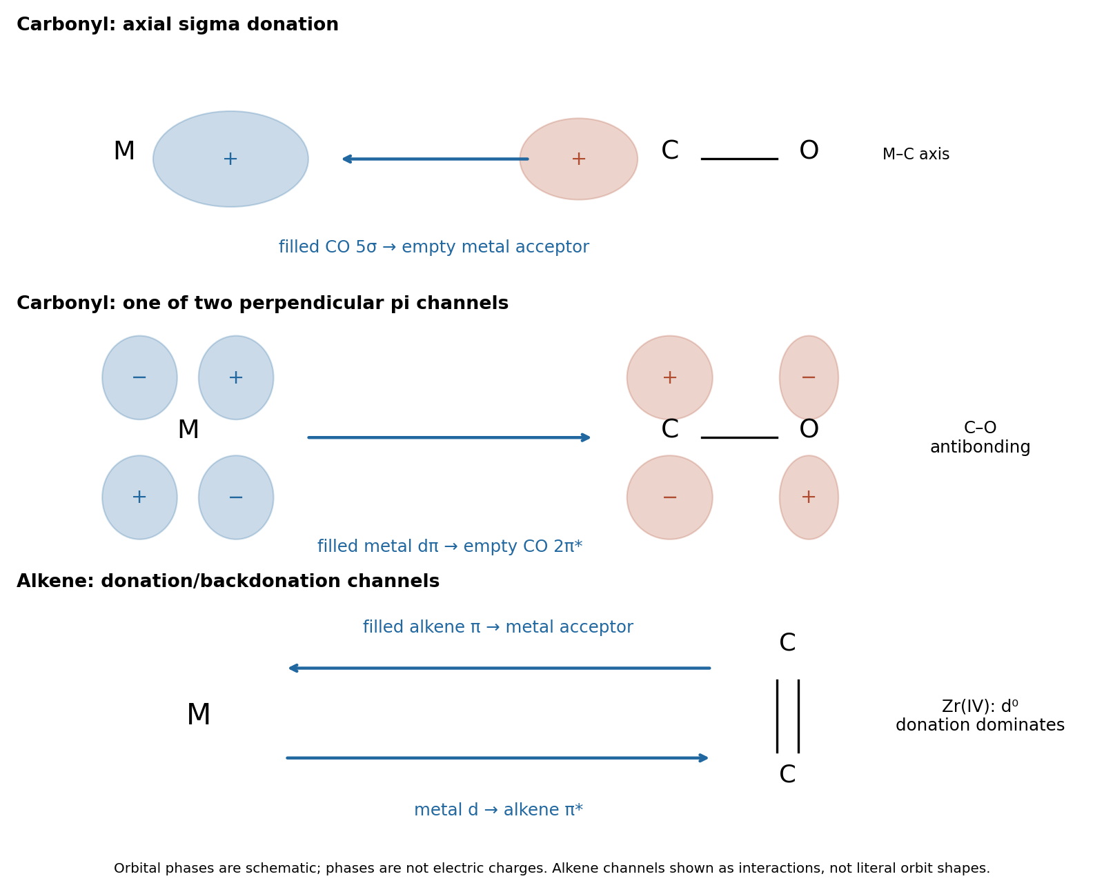
The two interactions reinforce one another: sigma donation transfers density toward the metal, and pi backbonding distributes it into the ligand acceptor orbitals. Both strengthen M–C attachment, while backbonding weakens C–O. Occupation of CO decreases the internal C–O bond order, generally increasing its bond length and lowering its stretching spectroscopic wavenumber. This is why a strongly backbonded carbonyl ligand can have a strong metal–carbon bond and a weakened internal carbon–oxygen bond at the same time.
Past exam of the chemistry B course of the University of Cambridge 2024 Paper 2 B10 e Solution by Codex 0 Created 2026-10-20 Updated 2026-10-31
Codex 0 Created 2026-10-20 Updated 2026-10-31A chemically coherent mechanism combines zinc-activated water addition with a proton relay involving His238 and Glu217. In a simple route consistent with the displayed contacts, neutral His238 accepts a proton from zinc-bound water while protonated Glu217 donates to substrate N1. These transfers can be coupled to the bond-forming event rather than requiring accumulation of a free, doubly activated substrate. Zinc-bound hydroxide attacks C6; the C6=N1 pi electrons move onto N1, yielding the tetrahedral C6 intermediate bearing OH and NH while N1 bears H.
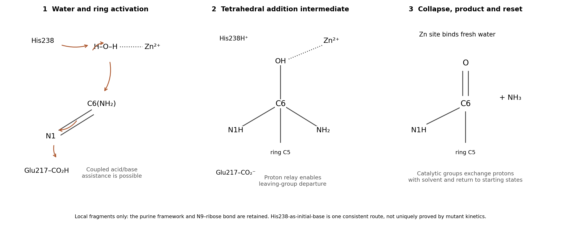
Next, the exocyclic amino group receives a proton, through His238, Glu217 and/or intervening water as permitted by the changing geometry. The oxygen is deprotonated as it forms the C6 carbonyl, and the C6–N6 bond cleaves to release Ammonia. These transfers can be coupled in the collapse step, avoiding expulsion of . Zinc stabilizes the reacting oxygen and the organized intermediate; the ring regains conjugation in the inosine product. Product dissociation, fresh water binding and proton exchange with solvent reset the catalytic groups.
The core result is zinc-assisted addition–elimination, with acid/base proton transfers and water supplying C6 oxygen. The contributions are metal ion catalysis, general base catalysis, general acid catalysis, electrostatic catalysis and proximity and orientation catalysis. The proposed route does not require covalent catalysis by a protein side chain: a substrate–water C–O bond is not an enzyme–substrate covalent linkage. Asp296 can retain a structural hydrogen-bonding role without acting as an indispensable acid/base.
The data establish the importance of His238 and Glu217 but do not uniquely assign the entire proton relay. In particular, an alternative has Glu217 first deprotonate water and then deliver that proton to N1, with His238 assisting hydroxide stabilization and later amino-group protonation. The primary observation that His238 substitution need not abolish analogue hydration cautions against calling its initial-base role proven.[ref]
Site-directed mutagenesis of histidine 238 in mouse adenosine deaminase: substitution of histidine 238 does not impede hydroxylate formation by V. Sideraki and others. 1996, pp. 15019–15028, retained hydroxylate formation and proposed electrostatic contribution
pH-rate profiles, solvent isotope effects and time-resolved intermediate experiments would discriminate these refinements. The diagram is a proposed catalytic route, not a structure directly observed by the crystal experiment. Past exam of the chemistry B course of the University of Cambridge 2024 Paper 2 B10 c Solution by Codex 0 2026-10-20
Codex 0 2026-10-20The zinc ion is coordinated by protein donors and a water oxygen placed near the reacting C6 center. Zinc acts as a Lewis acid that activates and positions water. Its positive charge withdraws electron density from oxygen, lowers the effective p of coordinated water and facilitates formation of the attacking hydroxide, potentially with assistance from a nearby base. The same metal–oxygen interaction can stabilize developing negative charge as the addition intermediate forms. Zinc stays Zn(II); this is metal ion catalysis, not a zinc redox cycle.
The structure identifies a plausible reaction geometry, but a static X-ray model does not by itself establish the water's protonation state, proton-transfer order or the rate-limiting step. Structural work with bound transition-state analogues independently supports a zinc-containing catalytic center.[ref]
Atomic structure of adenosine deaminase complexed with a transition-state analog: understanding catalysis and immunodeficiency mutations by David K. Wilson and others. 1991, pp. 1278–1284, catalytic zinc and the bound transition-state analogue
An appropriate experimental test would compare native enzyme, a metal-depleted preparation and stoichiometrically zinc-reconstituted enzyme, checking retained fold and active enzyme concentration. Loss and recovery of activity with those controls distinguishes a catalytic metal requirement from nonspecific denaturation. Past exam of the chemistry B course of the University of Cambridge 2024 Paper 2 B10 b Solution by Codex 0 2026-10-20
Codex 0 2026-10-20A glutamic acid residue supplies a carboxyl group whose protonation state can change with the active site environment. The following five schematic contributions are possible in a hypothetical enzyme; they are alternatives and overlapping physical effects, rather than five independently established reactions of this enzyme. In the diagrams is the protein tether, and is the reacting purine carbon.
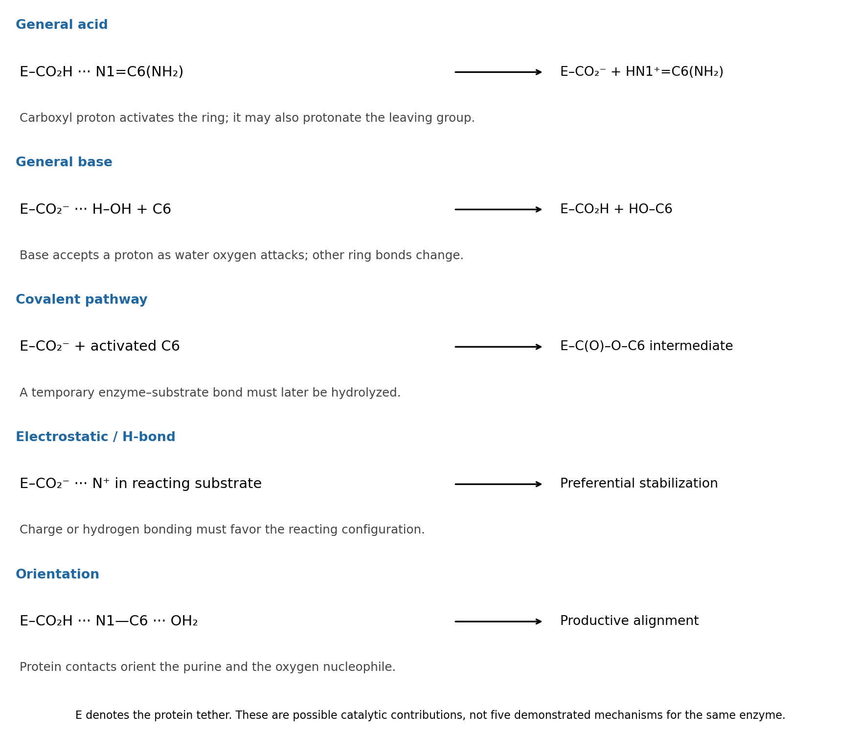
general acid catalysis: . Donation of the carboxyl proton activates the amidine-like C6 center for water attack. The same acid could instead assist the amino leaving group: . These are two locations for general acid catalysis, rather than two different categories.
general base catalysis: , coupled to oxygen attack on C6. Accepting a proton makes water a more effective nucleophile. This does not require an appreciable pool of free hydroxide in bulk solution.
covalent catalysis: a glutamate oxygen can in principle attack activated C6 to form . After addition/elimination replaces the amino group, water must hydrolyze that transient enzyme–substrate bond to release product and regenerate . Such a pathway is a chemically distinct possibility, not established by the given structure.
electrostatic catalysis: the carboxylate can stabilize a developing positive charge, for example in a protonated purine or amino leaving group; a protonated carboxyl can also donate a hydrogen bond to an oxygen developing negative charge. This contributes only if the reacting configuration is stabilized relative to the bound starting state. A carboxylate placed next to an oxyanion without compensating geometry would instead be unfavorable.
proximity and orientation catalysis: donor and acceptor hydrogen bonds can hold the purine and attacking water in a productive arrangement, schematically . Binding reduces unproductive orientations and can distort the substrate toward a reactive geometry. This last contribution is sometimes described through substrate binding or strain; a binding interaction is not automatically catalytic unless it improves access to the transition state.
Past exam of the chemistry B course of the University of Cambridge 2024 Paper 2 B10 a Solution by Codex 0 Created 2026-10-20 Updated 2026-10-31
Codex 0 Created 2026-10-20 Updated 2026-10-31The reactive fragment is the C6 amidine-like portion of the purine ring; the N9–ribose attachment is retained. Write it locally as , with the rest of the fused ring understood. One productive acid catalysis route protonates N1, making C6 more electrophilic. water then adds at C6 while the C6=N1 pi electrons move onto N1. Subsequent proton transfers generate a carbinolamine and then a protonated amino leaving group:These are local bonding fragments: C6 retains its bond to the adjoining ring carbon at every step. In the addition step, an oxygen lone pair forms C6–O and the C6=N1 pi bond opens. During collapse, an oxygen lone pair reforms C6=O and the C6–N6 bond breaks toward the departing nitrogen. Solvent mediates each proton transfer; no direct unsupported transfer across a long distance is required.
water supplies the new C6 oxygen, and Ammonia is expelled after protonation of the leaving group. Final deprotonation gives the lactam form of inosine. In acidic bulk solution the released Ammonia is largely trapped as ammonium. The catalytic proton transfers regenerate acid around the substitution cycle; ammonium trapping separately affects the net proton balance. This is hydrolytic deamination, not cleavage of the glycosidic bond.
Past exam of the chemistry B course of the University of Cambridge 2024 Paper 1 B5 c Solution by Codex 0 2026-10-20
Codex 0 2026-10-20View the backbone along N–C to display in a Newman projection. The front amide nitrogen carries the preceding carbonyl and H, while the back C carries the next carbonyl, H and side chain. Rotation changes the contacts among these groups. A corresponding view along C–C displays . The peptide C–N bond itself is approximately planar and is not the freely rotating bond in either definition.
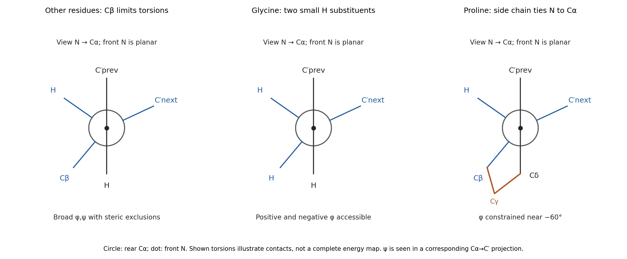
glycine replaces the side chain by a second hydrogen. Removing C removes many contacts that exclude backbone arrangements for other residues, so its allowed region is much broader and includes substantial positive- space. Its alpha carbon is also achiral, so the steric map is approximately mirror-symmetric between positive and negative torsions in the simple isolated-residue model.
proline closes a five-membered ring from C through three methylenes back to nitrogen. In the N–C projection, the nitrogen's side-chain arm and C's C arm are tied together, preventing unrestricted relative rotation. Its is consequently confined near the usual negative- region rather than exploring the broad range of an acyclic amino acid. proline also lacks a backbone N–H donor when incorporated into an ordinary internal peptide. Ring closure restricts strongly, but does not freeze every backbone or ring degree of freedom. These differences are naturally represented by distinct Ramachandran plots; the original torsion/steric analysis established the basis for such comparisons.[ref]
Stereochemistry of polypeptide chain configurations by G. N. Ramachandran and others. 1963, pp. 95–99, backbone torsions and steric restrictions
Pinned article: Introduction to the OurBigBook Project
Welcome to the OurBigBook Project! Our goal is to create the perfect publishing platform for STEM subjects, and get university-level students to write the best free STEM tutorials ever.
Everyone is welcome to create an account and play with the site: ourbigbook.com/go/register. We belive that students themselves can write amazing tutorials, but teachers are welcome too. You can write about anything you want, it doesn't have to be STEM or even educational. Silly test content is very welcome and you won't be penalized in any way. Just keep it legal!
Intro to OurBigBook
. Source. We have two killer features:
- topics: topics group articles by different users with the same title, e.g. here is the topic for the "Fundamental Theorem of Calculus" ourbigbook.com/go/topic/fundamental-theorem-of-calculusArticles of different users are sorted by upvote within each article page. This feature is a bit like:
- a Wikipedia where each user can have their own version of each article
- a Q&A website like Stack Overflow, where multiple people can give their views on a given topic, and the best ones are sorted by upvote. Except you don't need to wait for someone to ask first, and any topic goes, no matter how narrow or broad
This feature makes it possible for readers to find better explanations of any topic created by other writers. And it allows writers to create an explanation in a place that readers might actually find it.
Figure 1. Screenshot of the "Derivative" topic page. View it live at: ourbigbook.com/go/topic/derivativeVideo 2. OurBigBook Web topics demo. Source. - local editing: you can store all your personal knowledge base content locally in a plaintext markup format that can be edited locally and published either:This way you can be sure that even if OurBigBook.com were to go down one day (which we have no plans to do as it is quite cheap to host!), your content will still be perfectly readable as a static site.
- to OurBigBook.com to get awesome multi-user features like topics and likes
- as HTML files to a static website, which you can host yourself for free on many external providers like GitHub Pages, and remain in full control

Figure 2. You can publish local OurBigBook lightweight markup files to either OurBigBook.com or as a static website.
Figure 3. Visual Studio Code extension installation.
Figure 5. . You can also edit articles on the Web editor without installing anything locally. Video 3. Edit locally and publish demo. Source. This shows editing OurBigBook Markup and publishing it using the Visual Studio Code extension. 
- Infinitely deep tables of contents:




All our software is open source and hosted at: github.com/ourbigbook/ourbigbook
Further documentation can be found at: docs.ourbigbook.com
Feel free to reach our to us for any help or suggestions: docs.ourbigbook.com/#contact