Cell differentiation is the acquisition of a specialized cell state, involving changes in structure, function and gene expression. A neuron and a red blood cell illustrate different outcomes: signaling and oxygen transport. Different functions usually do not require a different inherited DNA sequence for each function. The shared-genome principle concerns nucleated precursors and most nucleated descendants: a mature mammalian red blood cell has lost its cell nucleus and mitochondria, so it is not an example of two mature biological cells both retaining the same nuclear DNA. [ref]
Biology 2e by Mary Ann Clark and others. Biology 2e, §40.2, Red Blood Cells: specialization and absence of a nucleus and mitochondria at mammalian maturity
The puzzle is how a shared set of genes supports different jobs. Gene expression changes which RNA and proteins a biological cell produces, and in what amounts. A transcription factor can regulate several genes; different combinations of regulators can interpret the same signal differently. Stable regulatory patterns can persist through the cell cycle, providing memory of developmental history. Specialization therefore usually changes use of inherited information, rather than replacing the genome wholesale. [ref]
Molecular Biology of the Cell, 4th edition by Bruce Alberts and others. Alberts et al., The Molecular Genetic Mechanisms That Create Specialized Cell Types, subsections The Expression of a Set of Genes Can Be Coordinated by a Single Protein and Stable Patterns of Gene Expression Can Be Transmitted to Daughter Cells, and Summary
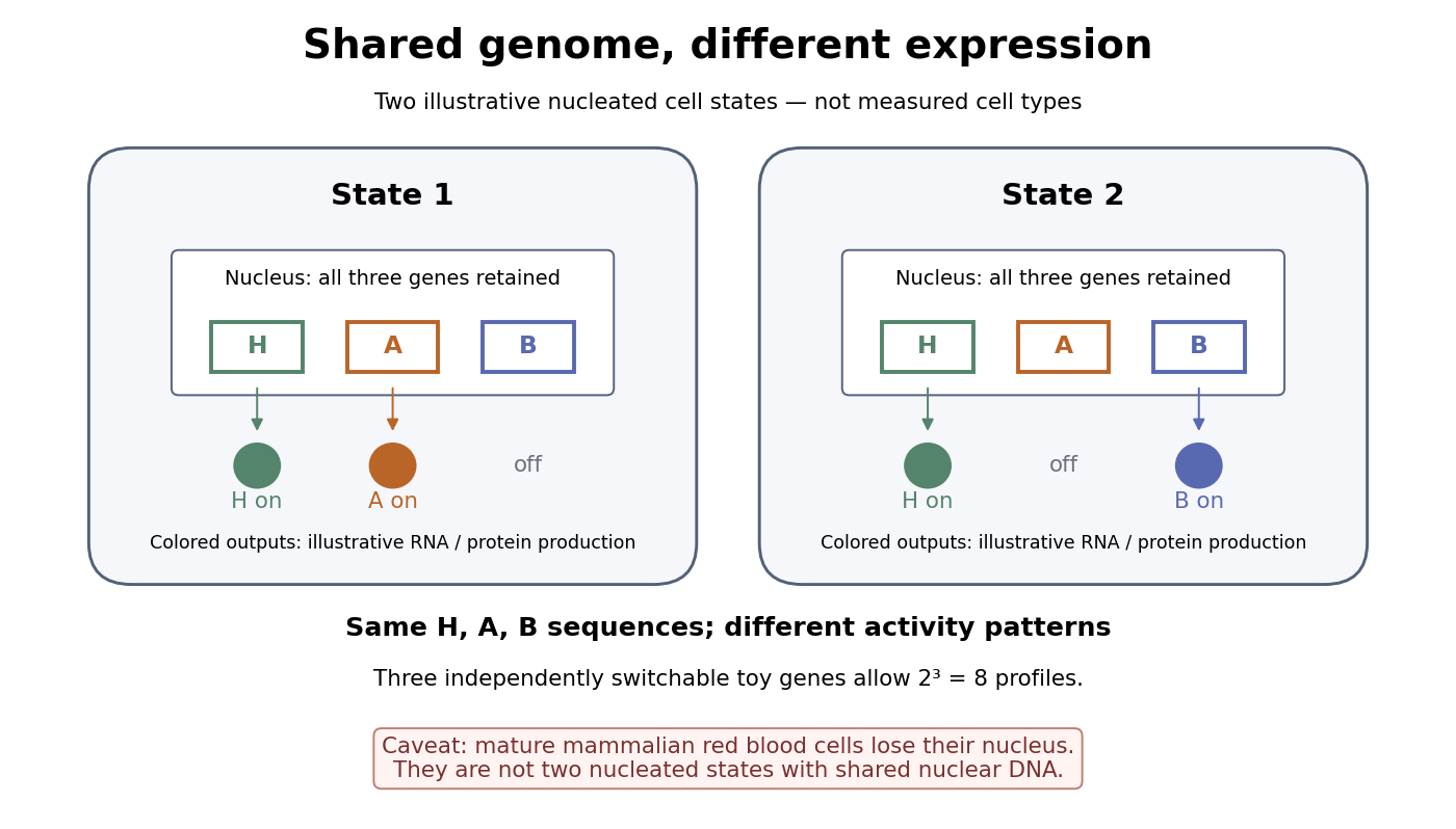
A small counting example separates sequence from activity. Suppose the same three toy genes are present in every nucleated biological cell, and independently assign each gene an on/off state. There are possible expression profiles with no DNA replacement. The figure shows only two: H and A on versus H and B on. Real gene expression is graded and coordinated, so this count illustrates possible descriptions, not a prediction of eight actual cell types. The interesting consequence is that a stable cell state can depend on how a shared record is used.
Stem cells combine self-renewal with production of differentiated descendants; progenitor cells generally have more restricted developmental options and renewal capacity. Neither term means that a biological cell must give rise to every cell type. Terminal differentiation denotes advanced specialization within a lineage, often accompanied by exit from the cell cycle. Cell fate concerns the developmental outcome of a biological cell or its descendants, whereas cell state describes its current properties. A temporary change in gene expression after signal transduction is not, by itself, proof of a new cell fate. [ref]
Anatomy and Physiology 2e by J. Gordon Betts and others. Anatomy and Physiology 2e, §3.6, Stem Cells and Differentiation: renewal, restricted differentiation potential and regulation of gene expression
A transition toward epithelial organisation and behaviour, potentially contributing to metastatic colonisation in particular cancer models.
The ability of cells to move among epithelial, mesenchymal and intermediate regulatory states. The benefit of a state can vary between dissemination, persistence and metastatic outgrowth.
A transition from an epithelial organisation toward more individually migratory cellular behaviour, involving changes in adhesion and polarity.
The developmental interval during which a cell can make a specified response to a signal. Absence of response outside the interval need not mean that the signal is absent.
Specification that depends on a cell's interactions with its surroundings. Neighbour exchange and controlled signal perturbation can test it causally.
Specification driven by information inherited within a cell, such as asymmetrically partitioned maternal material. It can coexist with external inputs later in development.
Cell fate is the developmental outcome of a biological cell or its descendants in a specified setting. What it normally becomes and what it can become after an experimental intervention are different questions. The intestinal nucleus transfer experiment tests potential after cellular reprogramming, rather than directly tracing the normal cell fate of the donor.
Persistence of a developmental choice. cell specification tests behaviour in a neutral environment; cell determination tests persistence in a viable conflicting environment.
Context-dependent capacity to change differentiated identity or developmental potential. Demonstrating a transition requires distinguishing conversion of a defined starting cell from selection of a rare pre-existing population.
Changes in intestinal epithelial potential that allow previously committed cells to restore a renewing stem-cell compartment under particular conditions.
Conversion between differentiated cellular identities. Whether a progenitor-like intermediate is traversed is an experimental question rather than part of a universal mechanism.
A return from a more committed state toward a less committed or progenitor-like state. Re-expression of one marker is weaker evidence than sustained renewal and functional lineage output.
A commitment demonstrated by retaining a fate in a conflicting environment that is viable and supplies the alternative developmental cues.
A provisional developmental commitment demonstrated operationally by adopting a fate in an appropriate neutral environment. It is weaker than cell determination.
A description of the normal developmental descendants of identified embryonic regions or cells. It reports realised fate, not the entire potential revealed by experimental transplantation.
Terminal differentiation is advanced specialization at the end of a developmental lineage. It often includes withdrawal from the cell cycle; the term does not mean that every nuclear gene needed for another cell fate has been deleted. A mature mammalian red blood cell is an especially restrictive example because it lacks a cell nucleus; the intestinal nucleus transfer experiment used nucleated donors.
Cellular reprogramming changes an established cell state so that a different developmental program becomes accessible. Experimentally induced cellular reprogramming must be distinguished from the ordinary progression of cell differentiation. An ovum's cytoplasm can reset aspects of a transferred cell nucleus; this does not mean that a specialized donor normally returns to an early developmental state in its original surroundings. [ref]
Mature cells can be reprogrammed to become pluripotent by Nobel Assembly at Karolinska Institutet. Nobel Assembly, Mature cells can be reprogrammed to become pluripotent, Scientific background: Gurdon nuclear transfer and distinction between differentiation and reprogramming
Large-scale changes in chromatin-associated regulatory states and DNA methylation during a developmental transition. Loss of methylation is not by itself a measurement of reduced transcription.
Somatic cell nuclear transfer introduces a body-cell cell nucleus into an ovum whose own nuclear contribution has been removed or inactivated. The donor nuclear genome and recipient cytoplasm have different origins; successful development does not show that the donor cell nucleus acts independently of the recipient's machinery.
Can a specialized donor still supply information for other cell types? The 1962 The Developmental Capacity of Nuclei taken from Intestinal Epithelium Cells of Feeding Tadpoles by J. B. Gurdon used mid-intestinal cells of feeding Xenopus laevis tadpoles. Donors were recognized by columnar shape, basal cell nuclei and a striated absorptive border. Dissociation used Versene solution; this made individual donors accessible. The readout was subsequent development, including normal feeding tadpoles, rather than just division or survival. [ref]
The Developmental Capacity of Nuclei taken from Intestinal Epithelium Cells of Feeding Tadpoles by J. B. Gurdon. Gurdon (1962), Material and Methods and Normal tadpoles; inspected primary text reproduced in the indexed Paperity preview
Apparatus and intervention. A microscope with ultraviolet illumination directed light at the surface region containing the unfertilized ovum's chromosomes, inactivating the recipient nuclear contribution. Irradiation also softened the surrounding jelly so a glass micropipette could enter. The pipette delivered donor nuclear material into recipient cytoplasm. In the technique's validation, fertilizing irradiated eggs produced haploid embryos, supporting destruction of the maternal nuclear contribution. This is inactivation, not a diagram of surgically removing an intact cell nucleus. [ref]
The Egg and the Nucleus: A Battle for Supremacy by J. B. Gurdon. Gurdon, The Egg and the Nucleus: A Battle for Supremacy, The Technique of Nuclear Transfer in Xenopus; indexed authored Nobel lecture text inspected
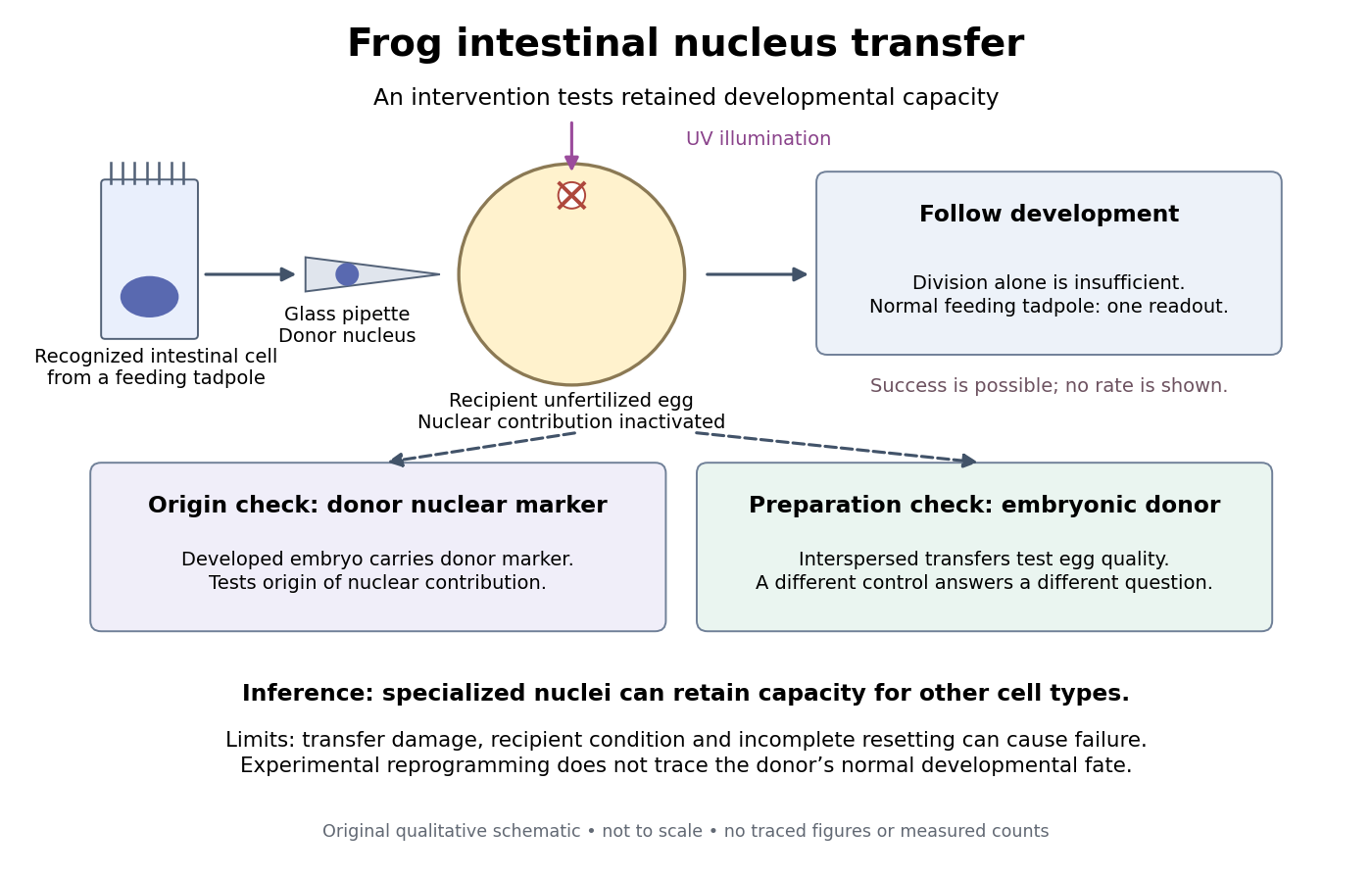
Somatic cell nuclear transfer: intervention, developmental readout and distinct controls
. Original apparatus-and-inference schematic, not a tracing of an experimental photograph. The ultraviolet beam indicates inactivation of recipient nuclear material; the injection pipette supplies a donor cell nucleus. Donor nuclear markers test origin; interspersed embryonic-donor transfers test recipient quality. Successful development is a possible outcome, not an asserted success rate. The figure omits exact dimensions, light dose and many technical steps. Details.Controls ask different questions. Donor cell nuclei carried a marker absent from recipients; post-blastula embryos carried that marker. The marker distinguished diploid donors with one nucleolus per cell nucleus from the usual two-nucleolus recipient background, rather than mistaking one nucleolus for proof of a haploid state. [ref]
The Egg and the Nucleus: A Battle for Supremacy by J. B. Gurdon. Gurdon, Nobel lecture, The Technique of Nuclear Transfer in Xenopus, p. 233: one-nucleolus diploid marker and its use to establish donor nuclear origin
Interspersed transfers of blastula or later embryonic cell nuclei checked variable recipient quality. A positive embryonic-donor control thus tests the preparation, while the marker tests donor origin. Abnormal development remained common. Neither control establishes universal transfer competence. [ref]The Developmental Capacity of Nuclei taken from Intestinal Epithelium Cells of Feeding Tadpoles by J. B. Gurdon. Gurdon (1962), Material and Methods (nuclear marker), Results (interspersed embryonic controls), and Normal tadpoles (abnormal outcomes); primary text inspected through the indexed preview
Inference and limits. A successful marked donor demonstrates that specialization had not irreversibly removed all information required to build other cell types. Failure is less decisive: transfer damage, recipient condition and incomplete cellular reprogramming can also prevent development. The marker identifies nuclear origin; it is not a complete DNA sequence comparison. The experiment tests donor nuclear potential in an altered cytoplasm, rather than normal donor cell fate, a universal capacity of every donor, or development from a mature mammalian red blood cell. The Nobel account also describes serial transfers through embryonic intermediates; these must not be silently combined with direct transfers into one success rate. [ref]
Mature cells can be reprogrammed to become pluripotent by Nobel Assembly at Karolinska Institutet. Nobel Assembly, Scientific background, Gurdon's nuclear-transfer experiments and serial transplantation


