Topics (384k) Articles (398k) Users (410) Discussions (242) Comments (383) Files (13k) New article

An aortic valve commissure is a region where adjacent aortic valve leaflets meet at their high peripheral attachment to the aortic root. In the usual three-leaflet valve, three commissures rise toward the sinotubular junction. They are high points of the crown-shaped attachment, distinct from the low landmarks defining the virtual basal ring.[ref]
Surgical anatomy of the aortic valve and root—implications for valve repair by Ruggero De Paulis and Andrea Salica. General considerations; Figure 2 and leaflet attachment description
A commissural attachment should also be distinguished from coaptation. The commissure identifies a peripheral supporting location; coaptation describes apposition of mobile leaflet surfaces during closure. Knowing three attachment positions does not establish the entire contact area or exclude an intervening gap.
An original geometric model illustrates why the spatial arrangement matters. Put three attachment points on a circle of radius with equal angular spacing. The distance between neighbors isFor mm, mm. Keeping the same radius but moving two adjacent points to separation changes their distance to mm. A fixed circular diameter alone therefore does not determine every span between attachment sites.
Real aortic valve geometry includes leaflet shape, attachment height and deformation, not just points on a flat circle. The calculation explains which information is lost by replacing a spatial arrangement with one diameter; it does not provide a surgical target. Locating each aortic valve commissure helps make anatomical descriptions and imaging views comparable without identifying the whole valve with a single ring.
The Breitenlohner-Freedman bound permits a scalar field in Anti-de Sitter spacetime to have a moderately negative squared mass without the instability that the same parameter would imply in flat spacetime. In of curvature radius , the bound is for the standard scalar dynamical stability problem with appropriate boundary conditions.
The near-boundary exponents satisfy , hence . The bound keeps these exponents real. The spacetime geometry and boundary conditions are part of the dynamical stability statement; a negative squared mass cannot be classified without them.[ref]
Stability in gauged extended supergravity by Peter Breitenlohner and Daniel Z. Freedman. Original scalar-field stability result; explicit AdS dimension and curvature-radius convention
Coronary angiography images the coronary arteries using a contrast medium and X-rays. In conventional invasive coronary angiography, a catheter delivers contrast to the coronary circulation while the imaging system records the contrast-filled vessel paths. The contrast improves visibility of the blood-filled lumen against surrounding tissue.[ref]
Heart Tests by the National Heart, Lung, and Blood Institute. Coronary angiography subsection, contrast and X-ray images
The apparatus connects fluid delivery to imaging geometry. An X-ray source and detector record the contrast signal during its passage through the vessels; changing viewing angle changes how overlapping arterial segments project into the image. The result is primarily a description of the contrast-filled lumen. It is not a direct measurement of every feature of the arterial wall or of how much oxygen reaches the myocardium.
An original geometric example shows why one apparent width can be incomplete evidence. Suppose a local lumen has an elliptical area with diameters and . Its area ishalf that of a circular lumen. One viewing direction can nevertheless show the same projected width as that circle, while an orthogonal view shows . A projected width does not uniquely determine lumen area without a shape and viewing model. These chosen dimensions are not a diagnostic threshold.
Timing and motion matter too: a moving vessel and changing contrast concentration can alter the recorded signal. A geometric narrowing, a pressure difference and downstream blood delivery are related observations but are not interchangeable. A physiological assessment needs evidence beyond one static projection.
Coronary CT angiography uses a different acquisition geometry: many X-ray views are reconstructed into a volume, commonly after contrast administration through a peripheral vein. The NHLBI account distinguishes cardiac computed tomography from the catheter-based angiographic description.[ref]
Heart Tests by the National Heart, Lung, and Blood Institute. Cardiac CT scan and Coronary angiography subsections
Naming the method makes clear whether a claim concerns a projection sequence or a reconstructed three-dimensional image.Tumour angiogenesis is the growth of new blood vessels from pre-existing vessels in response to a tumour and its surrounding tissue. It supplies a route for oxygen and nutrients to reach growing cells. Vessel formation and effective perfusion are distinct: a vascular image alone does not measure the delivery of oxygen to every part of a tumour.
A foundational experiment by Judah Folkman, Ezio Merler, Charles Abernathy and Gretchen Williams tested tumour-derived material in a rat dorsal air sac assay. A membrane wick delivered separated material locally; after 48 hours the investigators inspected the vascular response with a stereoscope and took tissue sections. A wick without the active preparation provided a negative control. The response was graded from 0 to 5+, rather than measured as an absolute endothelial cell count.[ref]
Isolation of a Tumor Factor Responsible for Angiogenesis by Judah Folkman and others. 1971, printed p.279, Biological Assay of TAF and Reading the Air Sac Assay; Figures2–4
This connected an isolated preparation to a reproducible biological effect. It did not identify a unique modern molecule or establish that inhibiting one signal would cure all cancers.An original diffusion model explains why access to vessels matters. Approximate a vessel-free tumour as a sphere of radius with uniform oxygen consumption per unit volume per unit time. Let be a constant diffusion coefficient and fix the surface oxygen concentration at . At steady state, the diffusion equation with consumption and spherical symmetry becomesFiniteness at the centre removes the singular integration term. Integrating again and imposing givesIf a stipulated threshold is required to avoid hypoxia, the model predictsFor illustration, take , and . The limiting radius is approximately , or . These are chosen model inputs, not measurements from the historical study or a universal size limit. Quadrupling consumption halves the limiting radius. A predicted negative concentration indicates failure of the assumed uniformly consuming viable sphere, not physically negative oxygen.
The calculation isolates a transport constraint; it does not model vessel branching, changing consumption or delivery by flowing blood. Signals such as vascular endothelial growth factor can influence endothelial cells, but their action and the resulting perfusion require biological measurements beyond this spherical model.
The rat dorsal air sac assay is an experimental bioassay of angiogenesis in which locally presented material is evaluated by the vascular response of tissue bordering a dorsal air space. It was used to test whether material derived from tumours could promote new blood vessels, connecting biochemical separation with a visible biological effect.
In the 1971 study by Judah Folkman, Ezio Merler, Charles Abernathy and Gretchen Williams, early experiments used membrane chambers containing live tumour cells. For testing separated material, the apparatus used silicone tubing with side openings covered by a short membrane sleeve. That sleeve acted as a diffusing wick, not as a filter. After 48 hours the vascular response was inspected with a stereomicroscope at magnification, followed by collection of sections for histology.[ref]
Isolation of a Tumor Factor Responsible for Angiogenesis by Judah Folkman and others. Printed p.279, Biological Assay of TAF and Reading the Air Sac Assay; Figure1
The investigators graded vascular proliferation from 0 to 5+. Their illustrations show a wick without the active preparation as a negative control and progressively stronger vascular responses to active material.[ref]
Isolation of a Tumor Factor Responsible for Angiogenesis by Judah Folkman and others. Figures2–4 and captions
The scientific question is whether the preparation alters the vascular response relative to an appropriate control. A visible response is not automatically a count of endothelial cells, a measured cell proliferation rate or evidence of functional perfusion.An original scoring example shows why numerical coding must follow the measurement. Suppose one group receives grades 1, 1 and 4, and another receives 2, 2 and 2. Both arithmetic means are 2, although the response patterns differ. Moreover, the ordered relabeling , , preserves the ranking but changes those means to 4 and 2. An ordered grade alone therefore does not define an interval scale on which differences have a fixed physical meaning. This example is not a reconstruction of the historical observations.
A useful contemporary comparison would specify the test material, solvent control, observation time and scoring rule, and keep the observer unaware of group assignment. Histology and explicit vessel measurements can help distinguish new vascular growth from dilation or local tissue disturbance. Such refinements answer additional measurement questions; this assay does not by itself identify a single angiogenic molecule or demonstrate a treatment effect in human cancer.
Pinned article: Introduction to the OurBigBook Project
Welcome to the OurBigBook Project! Our goal is to create the perfect publishing platform for STEM subjects, and get university-level students to write the best free STEM tutorials ever.
Everyone is welcome to create an account and play with the site: ourbigbook.com/go/register. We belive that students themselves can write amazing tutorials, but teachers are welcome too. You can write about anything you want, it doesn't have to be STEM or even educational. Silly test content is very welcome and you won't be penalized in any way. Just keep it legal!
Intro to OurBigBook
. Source. We have two killer features:
- topics: topics group articles by different users with the same title, e.g. here is the topic for the "Fundamental Theorem of Calculus" ourbigbook.com/go/topic/fundamental-theorem-of-calculusArticles of different users are sorted by upvote within each article page. This feature is a bit like:
- a Wikipedia where each user can have their own version of each article
- a Q&A website like Stack Overflow, where multiple people can give their views on a given topic, and the best ones are sorted by upvote. Except you don't need to wait for someone to ask first, and any topic goes, no matter how narrow or broad
This feature makes it possible for readers to find better explanations of any topic created by other writers. And it allows writers to create an explanation in a place that readers might actually find it.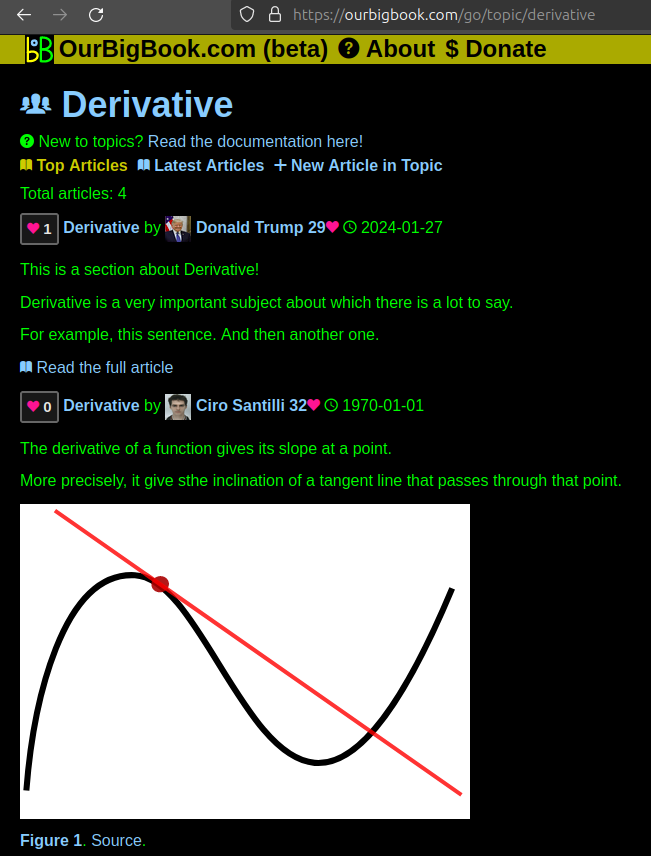
Figure 1. Screenshot of the "Derivative" topic page. View it live at: ourbigbook.com/go/topic/derivativeVideo 2. OurBigBook Web topics demo. Source. - local editing: you can store all your personal knowledge base content locally in a plaintext markup format that can be edited locally and published either:This way you can be sure that even if OurBigBook.com were to go down one day (which we have no plans to do as it is quite cheap to host!), your content will still be perfectly readable as a static site.
- to OurBigBook.com to get awesome multi-user features like topics and likes
- as HTML files to a static website, which you can host yourself for free on many external providers like GitHub Pages, and remain in full control
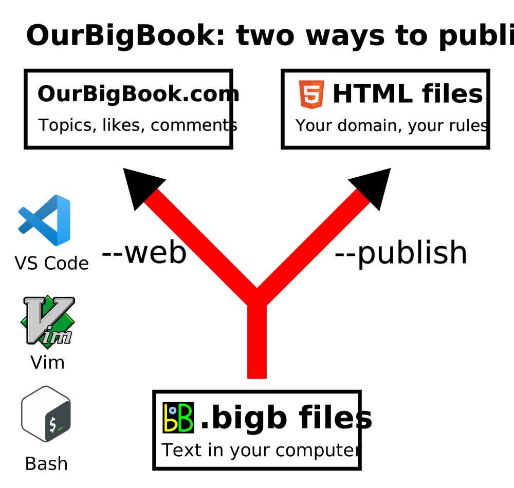
Figure 2. You can publish local OurBigBook lightweight markup files to either OurBigBook.com or as a static website.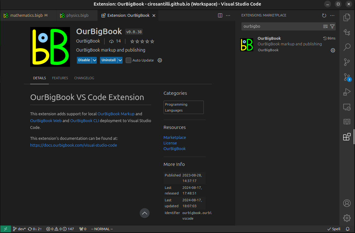
Figure 3. Visual Studio Code extension installation.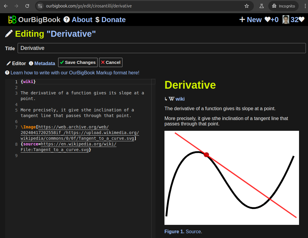
Figure 5. . You can also edit articles on the Web editor without installing anything locally. Video 3. Edit locally and publish demo. Source. This shows editing OurBigBook Markup and publishing it using the Visual Studio Code extension. 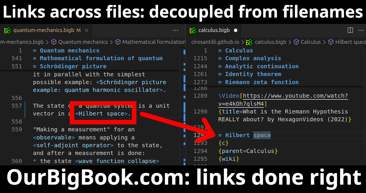
- Infinitely deep tables of contents:
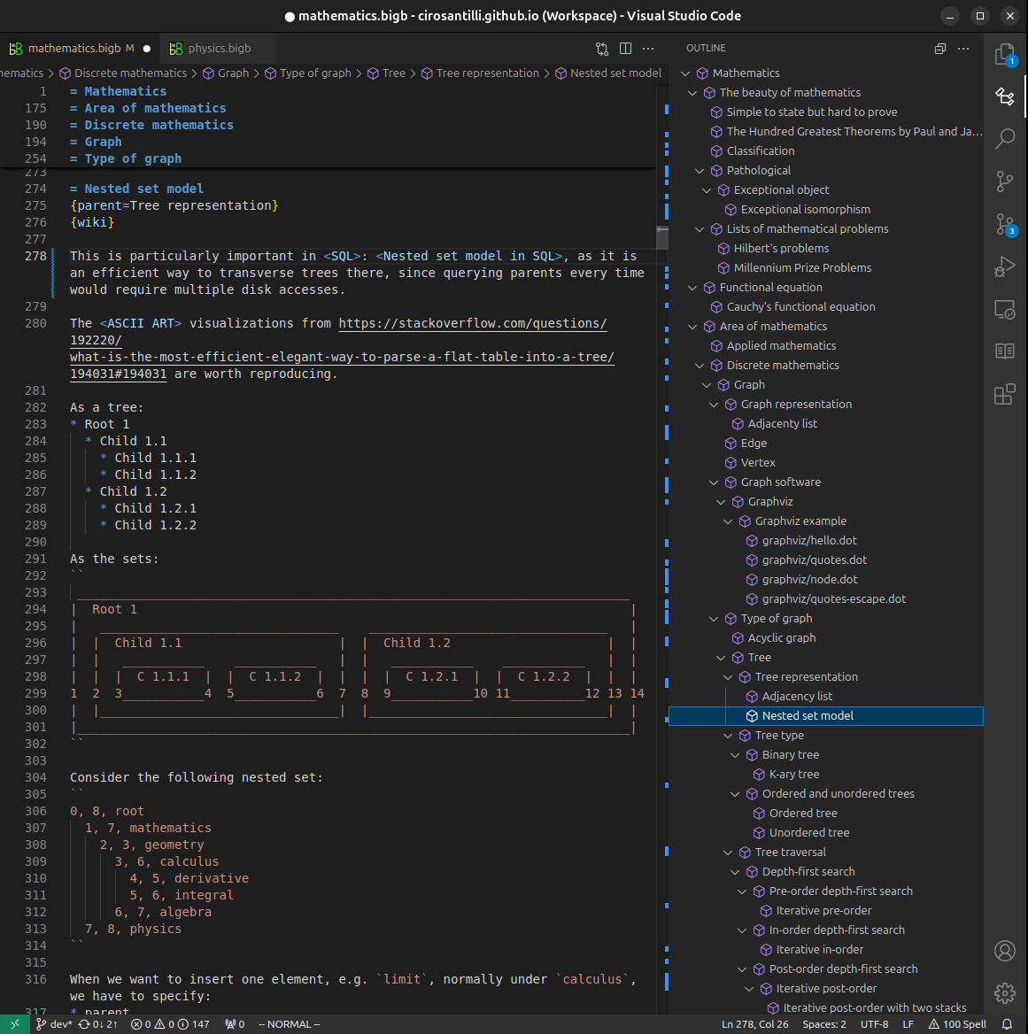
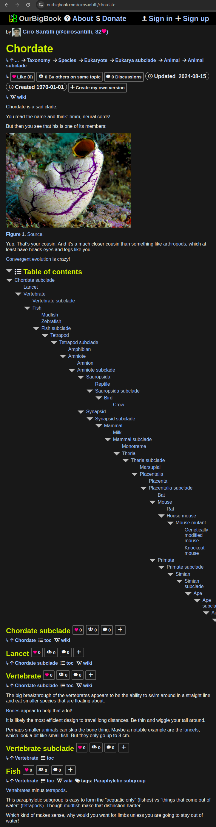
All our software is open source and hosted at: github.com/ourbigbook/ourbigbook
Further documentation can be found at: docs.ourbigbook.com
Feel free to reach our to us for any help or suggestions: docs.ourbigbook.com/#contact