Histology studies the microscopic organization of tissues. Thin sections, imaging and histological staining reveal how cells and extracellular matrix form a working structure. A two-dimensional section samples a three-dimensional arrangement, so section orientation and preparation influence interpretation.
Microdissection is the isolation of small selected structures or regions from a specimen under microscope guidance. It links the spatial organization studied in histology with assays of a chosen cell population. Unlike a microtome, which prepares thin sections, microdissection selects a particular region or constituent from material being examined.
Selection can be performed with fine mechanical tools or with instrument-assisted methods. A concrete landmark is the 1996 primary demonstration of laser capture microdissection by Michael R. Emmert-Buck and colleagues. Their apparatus placed a transparent ethylene-vinyl acetate thermoplastic film on a tissue section and used a carbon-dioxide laser pulse to activate the film over chosen cells. Adhesion allowed those cells to be collected. The paper demonstrated subsequent DNA/RNA amplification and enzyme recovery.[ref]
Laser capture microdissection by Michael R. Emmert-Buck and others. Abstract; Science 274, 998–1001 (8 November 1996), DOI 10.1126/science.274.5289.998
This describes that published apparatus; capture and cutting methods need not use the same laser or transfer mechanism.Target purity versus cell-state change in microdissection explains why spatial selection matters. In an original simple assay model, suppose target and background cells contribute calibrated signals and per cell. For target-cell fraction , the per-cell mean of an equally weighted sample is . With stipulated , , changing purity from 10% to 50% changes the mean from 10 to 50 units without any change in the target cells' own signal. An apparent increase can reflect enrichment rather than altered gene expression.
Conversely, a selected region need not be chemically pure. The footprint can include neighbouring cells, and tissue preparation or transfer can affect recovery of DNA, RNA and proteins. Normalization per cell, per total extracted RNA and per tissue area therefore asks different quantitative questions.
A useful experiment records the specimen image before and after selection, the chosen boundaries, collection yield and relevant background controls. Microdissection improves the connection between location and molecular measurement; it does not by itself prove cell identity, eliminate all contamination or establish that a recovered molecular difference is intrinsic to one cell type.
Target purity versus cell-state change in microdissection is the distinction between changing which cells enter an assay and changing the molecular state of the target cells themselves. Microdissection can enrich a selected cell population while its per-cell gene expression stays unchanged. Conversely, a measured change can combine enrichment, a genuine cell state difference and altered molecular recovery during preparation.
An original two-population model makes the ambiguity explicit. For equal per-cell weighting, let be the target-cell fraction and the target and background per-cell signals. The measured mean isWith and , changing from to raises from to without any target-cell change. If the target signal instead falls to at the second measurement, is still , above its original . A rising mixture signal therefore does not establish increased expression within the target cell population.
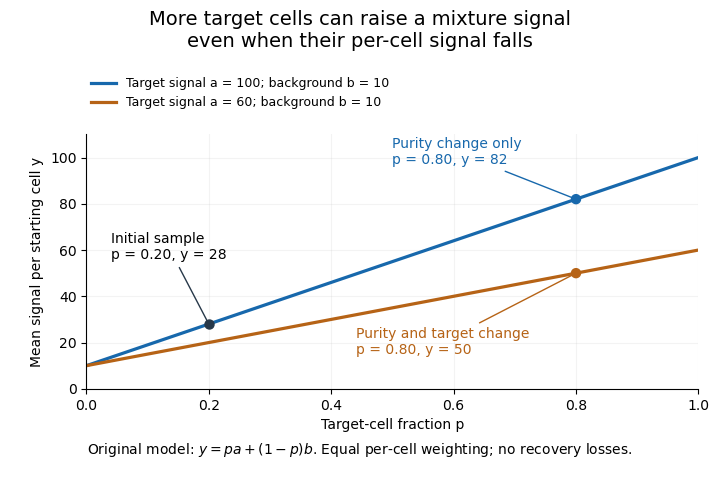
Original two-population model separating enrichment from per-cell signal change during microdissection
. Lines are stipulated mixture equations, not experimental observations. The initial point is p=0.20, a=100, b=10; the two final points share p=0.80 but have different target-cell signals. Details.If and are independently known, then . Holding those inputs fixed, a small signal error propagates as . Purity amplifies that error fivefold, whereas amplifies it by only . Estimating purity is useful, but an uncertain purity or an unrepresentative background introduces further error.
Normalization also changes the mixing weights. If target and background cells contribute total extracted RNA amounts per cell, their fraction of the extracted RNA isAt and , of the RNA comes from target cells despite target-cell purity. A signal per unit total RNA is therefore not automatically the same observable as a signal per starting cell.
Preparation can alter the analyte without creating an original in-vivo cell state difference. In a primary study, Mathieu Clément-Ziza and colleagues compared conditions preserving RNA integrity during laser capture microdissection, including an argon atmosphere and an ethanol-based preparation. Their time-dependent RNA-quality measurements address preservation, not proof that all living-cell transcription stayed unchanged.[ref]
Useful comparisons separately document selected regions, target fraction, recovery and the normalization denominator. Before/after specimen images test what was removed; molecular quality controls test what survived; matched biological comparisons test the proposed cell state change. No one of those observations substitutes for all the others.
Laser capture microdissection selects small regions or cell populations from a tissue section under microscope guidance, using a laser-mediated collection step. Its purpose is to connect visible histology with molecular measurements from a chosen location. Selection is not the same as preparing the original thin section, and a visible target boundary does not by itself guarantee pure or complete recovery.
The 1996 demonstration by Michael R. Emmert-Buck and colleagues placed a transparent ethylene-vinyl acetate thermoplastic film against a section on a glass slide. A carbon dioxide laser pulse activated the film locally above the desired cells. Focal adhesion enabled transfer of the selected tissue. The reported mechanism was capture by the activated film, rather than simply assuming that the target cells were vaporized. Other laser-assisted cutting arrangements need not use that same transfer mechanism.[ref]
Laser capture microdissection by Michael R. Emmert-Buck and others. Abstract, Science274 (1996), pp.998–1001: film, carbon-dioxide pulse and focal adhesion
The original paper demonstrated amplification of recovered DNA and RNA and recovery of enzyme activity. Those tests connect the collection apparatus with useful downstream assays; they do not prove that every molecular species survives equally or that every captured region consists of one cell type.[ref]
Laser capture microdissection by Michael R. Emmert-Buck and others. Abstract: amplification and enzyme recovery
An original selection model separates purity from yield. Suppose a section contains 100 target cells and 900 background cells. If capture retains of targets but of background, the recovered group has 80 targets and 9 background cells. Target purity is , while target recovery is . Enrichment can therefore be strong even when some desired cells are lost. In general, with initial target fraction and capture probabilities ,provided the denominator is positive. This model assumes independent per-cell accounting, not that an actual laser footprint captures each cell independently.
A measurement record can preserve specimen images before and after collection, the intended boundaries and an assay-specific blank. Images can show spatial removal; a blank can reveal film or collection-background contributions. Target purity versus cell-state change in microdissection then explains why a molecular signal increase after collection may reflect altered population composition rather than altered expression within each target cell.
Preparation time is a separate experimental variable. The RNA-preservation study by Mathieu Clément-Ziza and colleagues tested dry-environment and ethanol-based methods while measuring RNA integrity over the collection period. Keeping a recognizable histological staining pattern and recovering intact RNA are related requirements, but they are distinct measurements.[ref]
Stabilization of RNA during laser capture microdissection by performing experiments under argon atmosphere or using ethanol as a solvent in staining solutions by Mathieu Clément-Ziza and others. Abstract and Figures1–2: RNA-quality comparison and visual selection
Selection recovery versus purity in tissue profiling separates how many desired cells are retained from how strongly those cells dominate the collected material. In laser capture microdissection, high target-cell purity can coexist with low target recovery. Neither quantity alone describes the biochemical amount recovered or guarantees that the retained cell population represents every target state.
Let the starting specimen contain target and background cells, and suppose selection retains . Definewhen the denominators are positive. measures target recovery and target purity. The analogous notions in a classified set are precision and recall, but here the quantities describe a physical collection process, not merely its digital labels.
For an original specimen containing 100 targets and 900 background cells, a broad selection recovering 60 targets and 40 background cells has and . A stricter selection recovering 20 targets and one background cell instead has and . It gives a cleaner mixture but only one third as many target cells as the first selection. The suitable balance depends on the downstream assay and the sampling question.
The enrichment can be expressed as an odds identity. If and the respective recovery fractions are , thenThe ratio multiplies the starting target odds. This is a direct consequence of counting recovered targets and background, not a claim that neighbouring cells in a laser footprint are independently sampled.
Even perfect target purity can hide selection bias within the desired population. Suppose two target states initially occur equally often, but their recovery fractions are and . Among recovered targets, the first state accounts for . If its per-cell marker signal is 100 units and the second state's is 20, the selected mean is 92 units instead of the original 60, although neither state's own signal changed. Target identity, representative state sampling and gene expression are separate checks.
The original capture experiment demonstrated selected tissue transfer together with DNA/RNA amplification and enzyme recovery. Those biochemical tests establish useful recovered material, but do not supply every target/background count needed for the above recovery and purity measures.[ref]
Laser capture microdissection by Michael R. Emmert-Buck and others. Abstract, selected-cell procurement and downstream recovery demonstrations
Before/after images, target counts and matched assay-background controls provide complementary evidence. Target purity versus cell-state change in microdissection adds the distinction between changed composition and changed per-cell biology.The supporting tissue associated with a cellular tissue or lesion; its composition and relationship to the other cells require anatomical context.
A blood film is a thin layer of blood spread for microscopic examination, often with histological staining. It permits comparison of blood-cell size, shape and staining. The visible appearance also depends on spreading and preparation, so a preparation artifact should not be mistaken for a biological cell difference.
Immunohistochemistry localizes molecular targets in prepared tissue using antibodies. A visible signal depends on target availability, antibody binding and the detection method. Appropriate negative and specificity controls are needed before interpreting staining as proof of one unique molecular identity.
Histological staining uses chemical or molecular differences to make selected tissue features visible. Contrasts can reveal nuclei, extracellular material or specific targets. A positive stain reports the behavior of the preparation and method, so controls are needed before inferring unique molecular identity.
A fungal stain highlights structures used to recognize fungi in a specimen. Different methods exploit cell-wall chemistry or other features. Visibility can support identification, but stain appearance alone need not establish species identity or distinguish colonization from invasive tissue growth.
A thin preparation of biological tissue for microscopy. Its appearance depends on cutting direction, thickness, preservation and histological staining.
A tissue is an organized population of cells together with their associated extracellular material. Cooperation among the components supports a biological function. A tissue differs from an organ, which typically combines several tissue types in a larger spatial arrangement.
Mineralized tissue is biological tissue in which mineral forms a substantial structural component of an organic matrix. Bone, dentine and enamel are familiar examples, but they differ markedly in composition and architecture. In bone, mineral based on hydroxyapatite is associated with a collagen-rich extracellular matrix; biological cells form and maintain that matrix. Mineralization describes a material feature, not the absence of living activity.[ref]
A mineralized tissue is therefore also a biological composite material. Mineral amount, organic matrix, water, porosity and orientation can each affect the response. The Young's modulus of an entire specimen cannot be inferred from mineral content alone: constituent geometry changes load sharing, just as it does in the isostress rule of mixtures and the longitudinal rule of mixtures. These ideal mixture models clarify the role of geometry without being exact models of a real bone.
Even the phrase “mineral fraction” needs a specified measurement. Consider a hypothetical dry two-component specimen with of mineral of density and of organic material of density . Its mineral mass fraction is , but its mineral volume fraction isThese chosen numbers describe a bookkeeping example, not a measured tissue composition. Adding water or pores changes which total volume belongs in the denominator. A mechanical mixture calculation needs the appropriate volume fractions, not a mass percentage silently substituted for them.
An instructive primary structural study examined elephant ivory, a form of dentine, with complementary X-ray scattering, polarized-light microscopy and scanning electron microscopy. The investigators found that mineralized collagen orientations differed between the outer and inner parts of the tusk, including an outer wrapping orientation and a predominantly axial, plywood-like arrangement farther inside.[ref]
The three-dimensional arrangement of the mineralized collagen fibers in elephant ivory and its relation to mechanical and optical properties by M. Albéric and others. Primary abstract, complementary methods and region-dependent fibre orientations
The result motivates combining methods: a bright pattern in polarized light, an electron-microscope image and a scattering pattern provide different evidence about the same organized mineralized tissue. Observing orientation is not by itself a direct measurement of its full mechanical constitutive equation.Nervous tissue, also called neural tissue, is the organized tissue of the nervous system, containing neurons and glial cells. Neurons specialize in communication, including action potentials and signaling between cells. Glial cells maintain the local environment, form insulating myelin in particular locations, and perform other active functions. The brain, spinal cord and peripheral nerves contain different arrangements of these components; a drawing of one isolated neuron is not a complete picture of nervous tissue.[ref]
Anatomy and Physiology 2e by J. Gordon Betts and others. Section12.2, Nervous Tissue: neurons, glial cells and their organization
A useful example is the median eminence, where hormone-releasing axon endings interact with tanycytes and blood-vessel endothelium. In a 2004 experiment, investigators compared female-rat tissue explants after 30 minutes with or without 500 micromolar arginine, a precursor for nitric oxide production. Electron microscopy showed identified gonadotropin-releasing hormone terminals close to the vascular boundary after treatment, with fewer intervening tanycyte processes than in controls.[ref]
Vascular endothelial cells promote acute plasticity in ependymoglial cells of the neuroendocrine brain by Sandrine De Seranno and others. Figure7 and Results: treated versus untreated hypothalamic explants
The observation makes tissue organization experimentally meaningful: changes in the position of a glial cell process can alter a neuron's access to a vascular interface. This specialized arrangement should not be mistaken for the structure of every synapse or every region of nervous tissue.
Erectile tissue is vascular tissue whose filling and outflow properties permit a substantial change in volume or stiffness. Its behavior depends on blood flow and smooth-muscle regulation, linking vascular physiology to organ mechanics. It is not simply ordinary muscle that becomes rigid by contracting.
The corpus spongiosum is erectile tissue surrounding the penile urethra and continuing into the glans. Its arrangement differs from the paired corpora cavernosa. Supporting the urethral passage is mechanically compatible with erectile change without requiring identical pressure in all compartments.
Connective tissue supports, connects or separates other tissues through cells embedded in extracellular matrix. Its forms include loose supporting tissue, tendons, cartilage and bone. Variation in matrix composition and organization explains their very different mechanical behavior.
An elastic fibre is an extracellular structural element that stretches and recoils. Networks of these fibers contribute reversible deformation to tissues such as vessel walls. Their recoil role differs from the high tensile restraint supplied by many collagen-rich structures.
A sheet-like elastic component of a vessel wall, especially prominent in the tunica media of elastic arteries.
Fascia is connective tissue forming sheets, envelopes or partitions around and between anatomical structures. It organizes movement, force transmission and pathways for vessels and nerves. Naming a fascial plane identifies a spatial relationship rather than an empty gap between unrelated organs.
The membranous component of superficial perineal fascia.
The periosteum is the connective-tissue covering of the external bone surface except at appropriate articular regions. It supports vascular supply, attachment and growth or repair processes. It is distinct from the inner marrow cavity and the cartilage on joint surfaces.
A tendon is dense connective tissue transmitting force from muscle to an attachment, commonly a bone. Parallel organization of its matrix supports tensile loading. Elasticity and geometry affect force transmission, so a tendon is not an absolutely rigid connector.
The popliteus tendon connects the popliteus muscle to its femoral attachment and has close relations to the lateral knee structures. Its course helps explain the muscle’s role in controlling rotation near the extended knee. Tendon continuity and joint-cavity relations are separate anatomical questions.
The quadriceps tendon transmits force from the quadriceps femoris to the patella. The distal patellar ligament continues the chain to the tibia. This arrangement lets the patella alter the leverage of the knee-extension apparatus.
Muscle is tissue specialized for force generation through contractile machinery. Skeletal muscle, smooth muscle and cardiac muscle differ in cellular organization and control. Shortening is one possible outcome: a muscle can also generate force while its overall length remains nearly constant.[ref]
Structure of Skeletal Muscle by the National Cancer Institute. Contractile tissue and connective-tissue organization
The piriformis muscle is a deep skeletal muscle connecting the anterior sacrum to the upper femur. It passes through the greater sciatic foramen and helps rotate the extended thigh outward and abduct the flexed thigh. Its position makes it a useful landmark: the sciatic nerve usually emerges below it, although the relationship varies between people.[ref]
An anatomical variant alone does not establish a cause of pain. Adam L. Bartret and colleagues classified sciatic nerve routes on 783 hip magnetic resonance imaging examinations; variants occurred in about 19%, without a statistically significant association with a recorded diagnosis of piriformis syndrome. This observation separates a visible anatomical difference from evidence that the difference explains symptoms.[ref]
The external anal sphincter is the ring of skeletal muscle surrounding the distal anal canal. Its contraction helps maintain faecal continence; its motor neurons allow voluntary squeezing as well as reflex control. It works alongside the smooth muscle of the internal anal sphincter, so continence depends on coordinated structures rather than one isolated ring.[ref]
Anatomy and Physiology 2e by J. Gordon Betts and others. Section 23.5, The Large Intestine, Anal Canal
Branches of the pudendal nerve supply the external anal sphincter. Dissection of 28 cadavers found a local nerve plexus near the sphincters in five, illustrating why a schematic nerve route need not capture every individual's branching. Preserving muscle alone therefore does not guarantee preservation of its motor supply.[ref]
Anatomy of the pudendal nerve and its terminal branches: a cadaver study by Steven E. Schraffordt and others. Abstract, Results: inferior rectal and perineal interconnections
Cardiac muscle is the specialized striated muscle of the heart. Its contractile cells are mechanically connected and electrically coupled at intercalated discs. Mechanical connections transmit force, while gap junctions help electrical excitation spread between cells.[ref]
Excitation and contraction are related but distinct stages. Changes in membrane potential regulate calcium handling, and calcium enables the actin-myosin machinery to generate force. A sinoatrial node normally sets the pace for the coordinated sequence, rather than an independent motor neuron commanding each beat.
This separation explains why an electrical recording and a measurement of pumping answer different questions. An electrocardiogram reports summed electrical activity; contraction also depends on cellular mechanics, filling and the loads against which the heart pumps.
A spasm is an involuntary episode of muscular contraction. It describes the observed motor event rather than a unique neural or muscular cause. Its location, duration and relation to other findings are needed to interpret the mechanism.
The stylopharyngeus is a muscle running from the styloid region into the pharyngeal wall. It elevates the pharynx during swallowing and is supplied by the glossopharyngeal nerve. Its innervation distinguishes it from several neighboring pharyngeal muscles.
The serratus anterior links the ribs to the medial border of the scapula. It protracts and assists upward rotation of the scapula while holding it against the thoracic wall. Its function therefore supports arm elevation without directly crossing the glenohumeral joint.
The pectoralis minor runs from upper ribs to the coracoid process. It stabilizes and moves the scapula, rather than attaching directly to the humerus. Its deep position also makes it a landmark for nearby axillary vessels and nerves.
The popliteus is a posterior knee muscle connecting the proximal tibia with the lateral femoral region through its tendon. It contributes to controlling rotation and initiating flexion from extension. Its action depends on which skeletal segment is relatively fixed.
The hamstrings are posterior thigh muscles with important actions at the hip and knee. In the usual human grouping they include semitendinosus, semimembranosus and the long head of biceps femoris. Their multi-joint geometry links hip position to knee-flexion mechanics.
The semimembranosus is a medial hamstring muscle contributing to hip extension, knee flexion and medial rotation of the flexed leg. Its tendon also gives an expansion associated with the oblique popliteal ligament. One muscle can therefore contribute to both active motion and passive support.
The quadriceps femoris is the anterior thigh muscle group transmitting force through the quadriceps tendon, patella and patellar ligament to extend the knee. Its components do not all cross the same joints; rectus femoris also crosses the hip.
The external urethral sphincter is striated muscular tissue contributing to control of the urethral outlet. It works with bladder and other outlet mechanisms rather than determining continence alone. Its arrangement differs between male and female anatomy.
The superficial transverse perineal muscle crosses toward the perineal body from the lateral perineal region. It contributes to stabilization of this central attachment. Its small size does not make it equivalent to the deeper pelvic diaphragm.
The ischiocavernosus is a superficial perineal muscle associated with the crus of erectile structures. Contraction influences the mechanical and vascular conditions of those structures. It is related to the bulbospongiosus region but has different attachments.
The bulbospongiosus is a superficial perineal muscle associated with the bulb and nearby erectile structures, with sex-specific arrangements. Its contractions contribute to local support and expulsion of fluid in relevant anatomy. It is distinct from the deeper urethral sphincter complex.
The obturator internus arises from the internal pelvic surface and turns through the lesser sciatic region to reach the proximal femur. It contributes to hip rotation and stabilization. Its pelvic fascia also forms the pudendal canal, linking muscular and neurovascular anatomy.
The adductor magnus is a large medial thigh muscle with regions differing in attachment, action and innervation. It contributes to hip adduction, while its posterior component also supports extension. Regional anatomy explains why one named muscle need not behave as a single uniform actuator.
The obturator externus runs from the external obturator region toward the proximal femur. It contributes to lateral rotation and stabilization of the hip. Its side of origin distinguishes it from obturator internus despite the similar names.
The gracilis is a long superficial medial thigh muscle crossing both hip and knee. It contributes to hip adduction and to flexion and rotation at the knee. Its multi-joint course makes its action depend on the positions of both joints.
The adductor longus is a medial thigh muscle extending from the pubic region to the femur. It contributes to hip adduction and has position-dependent effects on flexion. Its relationship to nearby muscles defines an important part of the medial thigh architecture.
The adductor brevis is a short medial thigh muscle deep to adductor longus. It contributes to hip adduction and helps stabilize the femur relative to the pelvis. Its deep location is an anatomical distinction rather than evidence of a wholly separate movement type.
The psoas major extends from the lumbar region toward the proximal femur and joins the iliacus contribution to the iliopsoas apparatus. It is a major hip flexor. Its connection across the lumbar-pelvic region also links spinal posture with lower-limb movement.
The anconeus is a small muscle on the posterolateral elbow. It assists elbow extension and stabilization and is supplied by the radial nerve. Its action complements the much larger triceps rather than producing an unrelated elbow movement.
The supinator muscle wraps around the proximal radius and contributes to forearm supination. A deep branch of the radial nerve passes through its region. Its rotational action differs from elbow flexion even though both can accompany turning the palm upward.
The brachioradialis spans the lateral elbow to the distal radius and flexes the elbow, particularly with the forearm near a neutral rotational position. It is supplied by the radial nerve. Its flexor action therefore does not imply the innervation pattern typical of every other elbow flexor.
The buccinator muscle forms an important muscular part of the cheek. It keeps food positioned against the teeth during mastication and helps control oral pressure. Its role differs from the jaw-closing muscles even though they cooperate during eating.
The temporalis muscle extends from the temporal region to the mandibular coronoid process. It elevates the mandible, with posterior fibers contributing to retraction. Its broad origin and converging insertion explain how different fiber regions can have different pulling directions.
The lateral pterygoid muscle has attachments involving the sphenoid region and mandibular joint apparatus. It contributes to protrusion and coordinated mandibular movements. Its roles differ from those of a simple jaw-closing elevator and depend on bilateral coordination.
An adductor muscle moves a body part toward a defined reference axis or plane. Hip adductors move the thigh toward the body midline. The movement name must be interpreted relative to the specified joint and reference, rather than inferred from muscle position alone.
The levator veli palatini muscle elevates the soft palate. Its action contributes to closing the nasal route during swallowing and appropriate speech sounds. Elevation is distinct from the tension-related contribution of tensor veli palatini muscle.
A diaphragm is a partition spanning an opening or separating regions; in muscular anatomy, the name commonly refers to the respiratory diaphragm. The broader structural term also appears in pelvic diaphragm. Location and composition should therefore be specified rather than assuming every anatomical diaphragm is the same sheet.
The attachment conventionally designated as the insertion of a muscle, commonly the more mobile or distal attachment. The convention must be distinguished from observations of a particular movement.
The attachment conventionally designated as the origin of a muscle, commonly the more fixed or proximal attachment. It is distinct from the muscle insertion.
Muscle contraction is active force generation by muscular contractile machinery. A muscle can shorten, lengthen under load or maintain nearly constant length while active. Force production and visible shortening are therefore different observations, linked by the load and mechanical constraints.
A force exerted by an active or passively stretched muscle. Its contribution to rotation about an anatomical joint depends on the muscle moment arm, rather than its magnitude alone.
The perpendicular distance from a muscle force's line of action to the relevant axis of an anatomical joint. It can change with posture; multiplying it by the muscle force gives the corresponding torque.
An intercostal muscle spans adjacent ribs and contributes to thoracic mechanics. The layered arrangement includes external intercostal muscles and internal intercostal muscles. Fiber direction and regional attachments affect how each layer influences rib movement.
An internal intercostal muscle is part of the deeper intercostal layer. Its action depends on the portion considered: interosseous and interchondral regions can contribute differently to expiration and inspiration. Treating the entire layer as one uniform expiratory actuator loses that anatomical distinction.
An external intercostal muscle spans neighboring ribs in the superficial intercostal layer. It supports the intercostal spaces and can assist rib elevation during inspiration. Its fiber orientation differs from the deeper internal layer.
An abdominal muscle contributes to movement or stabilization of the trunk and to control of abdominal pressure. Different layers can cooperate during forceful expiration and postural tasks. Pressure generation and movement depend on coordinated activity rather than isolated shortening of one layer.
A scalene muscle connects cervical vertebrae with upper ribs. The group contributes to neck movement and can assist inspiration by acting on the upper thorax. Spaces between its members also form important routes for vessels and the brachial plexus.
The sternocleidomastoid muscle connects the sternum and clavicle with the mastoid region. Unilateral activity contributes to side-bending and rotation of the head, while bilateral actions depend on the head and neck position. It can also act as an accessory respiratory muscle.
The tensor veli palatini muscle contributes to tension of the soft palate and opening of the auditory tube. Its tendon changes direction around a bony pulley region. Its trigeminal motor supply distinguishes it from several neighboring muscles of the palate and pharynx.
The stylopharyngeus muscle is the stylopharyngeus, a pharyngeal elevator supplied by the glossopharyngeal nerve. It links the styloid region to the pharyngeal wall. The expanded name identifies the same anatomical muscle rather than an additional neighboring structure.
A contractile assembly in a skeletal-muscle fibre consists of serial sarcomeres. Their coordinated shortening contributes to shortening of the fibre; each sarcomere contains overlapping thin actin and thick myosin filaments.
fibrosis is persistent accumulation and remodeling of extracellular material in tissue. In mechanobiology, altered matrix architecture and resistance can change the forces experienced by resident cells.
A tissue forming a lining or covering. Its normal organization and differentiation state matter when comparing epithelial tumors with adjacent tissue.
The retinal pigment epithelium, or RPE, is a single layer of specialized epithelial cells at the back of the retina, closely apposed to the light-sensitive photoreceptors. It supports photoreceptor metabolism and participates in the visual cycle, the chemical regeneration needed to sustain light detection. Support of a receptor and detection by that receptor are distinct cellular tasks.[ref]
Molecular Mechanisms Section by the National Eye Institute. About our work: RPE monolayer, photoreceptor dependence and vitamin-A metabolism
A 1998 genetic experiment made that dependence concrete. Mice lacking RPE65 showed altered vitamin-A-related chemistry and lacked normal rod cell responses measured by electroretinography, although cone responses remained. They also lacked rhodopsin despite retaining its opsin protein component. The comparison connected defective regeneration chemistry with loss of functional visual pigment rather than simply absence of all photoreceptor protein.[ref]
Rpe65 is necessary for production of 11-cis-vitamin A in the retinal visual cycle by T. M. Redmond and others. Abstract: retinoid composition,electroretinography,rhodopsin and opsin
An illuminating experimental comparison therefore measures both sides: a chemical assay tests the available pigment-related molecules, while an electrical recording tests the retina's response to light. Either observation alone leaves more possible explanations. The result also shows why a layer that does not itself provide the main light-triggered signal can be essential to vision.
The cellular lining of blood and lymphatic vessels. Its polarized surfaces and junctions participate in vascular lumen formation.
Transient blood-forming endothelium that generates haematopoietic cells through an endothelial-to-haematopoietic transition.
An epithelium organized into multiple cell layers. Cell production, differentiation and loss must be balanced across the layers.
An epithelium characterized by flattened surface cells. Stratified examples contain renewing basal populations and differentiating suprabasal layers.
Progression away from the basal compartment toward specialized surface layers. Differentiation, delamination and subsequent cell loss need not occur simultaneously.
Removal of a cell from an epithelial sheet through coordinated behaviour of that cell and its neighbours. Extrusion and apoptosis are distinct events and need not occur in the same order.
An epithelial cell producing enamel during tooth development. Loss of the enamel-producing cellular layer after eruption limits replacement of worn crown enamel.
Bone is living mineralized connective tissue forming major parts of the vertebrate endoskeleton. Its extracellular matrix combines organic material with mineral, supporting both toughness and stiffness. Cells continually remodel it, so a bone is a regulated tissue rather than an inert stone.
The patella is a sesamoid bone within the knee-extension apparatus. It receives the quadriceps tendon proximally and connects through the patellar ligament distally. By changing the geometry of this force-transmission path, it influences the leverage of knee extension.
An epiphysis is the end region of a developing long bone, associated with a secondary ossification center. During growth, the growth-plate region separates it from the shaft-side region. The developmental term should be distinguished from the unrelated historical use for the pineal organ.
The mineral-rich residue left after combustion of bone organic material. It contains calcium phosphates and should not automatically be treated as pure tricalcium phosphate.
An osteophyte is a bony outgrowth, commonly at a joint margin in a remodeling or degenerative process. Its presence is a structural finding rather than a direct measure of pain or functional impairment. Location and relation to adjacent structures affect its significance.
The zygomatic bone forms part of the cheek prominence and lateral orbital boundary. Its connections with neighboring bones help transmit forces through the face. Its external prominence and contribution to the orbit are different aspects of one structural element.
The lacrimal bone is a small bone in the medial orbital wall associated with the lacrimal drainage region. Its position contributes to the space around the lacrimal sac. It is structurally distinct from the secretory lacrimal gland despite the shared name.
The ethmoid bone contributes to the skull base, nasal cavity and medial orbital walls. Its perforated superior region permits passage of olfactory nerve fibers. One bone therefore participates in several anatomical boundaries and in a sensory pathway.
The sphenoid bone is a central skull-base bone with contributions to the orbit and middle cranial region. Its openings and processes relate to multiple nerves, vessels and muscular attachments. That central position makes it important for understanding neighboring skull compartments.
Bone marrow is tissue within the internal spaces of bones. Hematopoietic regions produce blood cells, while adipose-rich regions have a different composition. Marrow is therefore not simply an empty bone cavity or the mineralized bone matrix itself.
A parietal bone forms much of the superior and lateral cranial vault. The paired bones meet near the midline and join other skull bones through sutures. Their broad surfaces contribute protection while the junctions have distinct developmental relationships.
The frontal bone forms the forehead and contributes to the orbital roofs and anterior cranial floor. Its internal and external surfaces relate respectively to intracranial and facial structures. A surface landmark therefore does not reveal every deeper boundary made by the same bone.
Formation of bone. Dermal, perichondral and endochondral modes should be distinguished from mineral deposition in cartilage.
A skull-roof bone formed by integration of frontal and parietal components, characteristic of frogs and some other vertebrates.
A skeletal element associated with the vertebral axis. Reduction of free ribs is part of the characteristic frog body organization.
The maxilla is a major upper-jaw bone contributing to the upper dental arch, hard palate, nasal region and orbit. Its several processes connect these regions structurally. The term refers to the bone, rather than all soft tissues of the upper jaw.
The premaxilla is the anterior upper-jaw skeletal element that carries or is associated with the front tooth-bearing region in many vertebrates. Its adult separation or fusion varies among lineages. Comparative identity cannot therefore be inferred solely from whether a visible suture persists.
Articles were limited to the first 100 out of 133 total.

