Topics (384k) Articles (398k) Users (410) Discussions (242) Comments (383) Files (13k) New article

Ole Storstein is first author of a 1959 experimental study separating airway resistance from lung compliance. The investigators recorded mouth-to-esophageal pressure differences, breathing volume and airflow to analyze the mechanical behavior of respiration.[ref]
Airway Resistance and Lung Compliance by Ole Storstein and others. Primary byline; Methods, simultaneous pressure, volume and airflow recordings
The apparatus used an intra-esophageal balloon, a differential pressure transducer and a pneumotachograph with volume integration. The paper selected corresponding points of no mouth airflow for its compliance calculation and used paired observations during inflation and deflation to estimate resistance.[ref]
Airway Resistance and Lung Compliance by Ole Storstein and others. Methods, Sanborn recording apparatus and definitions of compliance and resistance
Simultaneous signals matter because a pressure observed during flow includes a different mechanical contribution from pressure observed after that flow term vanishes.For an original one-compartment model with a chosen zero-volume baseline, writewhere is lung compliance and is airway resistance. This neglects inertial and regional effects. At stipulated , L, and L/s, the elastic contribution is 4 and the resistive contribution is 1 cm of water. Total modeled pressure is 5 cm of water.
Dividing volume by that total would give , below the chosen compliance. At the same modeled volume and zero flow, pressure becomes 4 cm of water and the ratio recovers 0.10. A time point and the definition of the pressure difference are therefore part of the observable, rather than optional details.
The primary paper explicitly notes the limitation of treating a nonuniform mechanical system as uniform.[ref]
Airway Resistance and Lung Compliance by Ole Storstein and others. Discussion, interpretation of the simplified mechanical analysis
Zero net airflow at the mouth need not establish that every region has identical local behavior. Storstein's documented contribution combines apparatus, simultaneous observations and a stated mechanical interpretation, rather than equating one pressure/volume ratio with every aspect of lung function.Robert H. Anderson studies cardiac anatomy, including the three-dimensional arrangement of the aortic root and aortic valve. His primary anatomical exposition distinguishes the semilunar leaflet attachments from the anatomical junction between ventricular and arterial walls.[ref]
The surgical anatomy of the aortic root by Robert H. Anderson. Primary byline; Abstract, leaflet attachments and ventriculo-aortic junction
A shared use of the word annulus does not guarantee that two investigators have selected the same structure.The paper describes leaflet attachments extending between basal ventricular positions and the more distal sinutubular region, forming a crown-like arrangement rather than one flat circular line.[ref]
The surgical anatomy of the aortic root by Robert H. Anderson. Abstract, three-dimensional crown and terminology
This makes anatomical terminology part of the measurement definition: diameter, circumference and attachment length require a specified object and plane.An original geometric model illustrates how projection can hide the distinction. Represent a three-lobed closed curve byIts projection onto the horizontal plane is a circle of circumference , independent of . Its actual curve length isFor and , this length exceeds . Identical projected circles can therefore represent spatial curves of different lengths. This is an explanatory mathematical crown, not a fitted human valve or a proposed replacement for anatomical landmarks.
The example shows why documenting the chosen attachment, junction or imaging plane is necessary before comparing dimensions. It does not establish that every aortic measurement must follow this curve. Anderson's contribution is to clarify the structures that those measurements and surgical descriptions intend to represent, keeping a named anatomical boundary distinct from a convenient geometric surrogate.
Frederick C. Cobey studies quantitative cardiac imaging, including three-dimensional assessment of how the mitral valve leaflets meet during closure. He is first author of a 2014 primary study using transesophageal echocardiography to measure the valve's coaptation area and relate it to functional mitral regurgitation.[ref]
Quantitative assessment of mitral valve coaptation using three-dimensional transesophageal echocardiography by Frederick C. Cobey and others. Primary byline, affiliations and Abstract
The published name and affiliations establish this contribution without expanding the middle initial.The study examined 25 patients with functional mitral regurgitation undergoing cardiac operations. It used intraoperative two- and three-dimensional echocardiography, followed by offline measurement of the end-systolic coaptation zone and valve area.[ref]
Quantitative assessment of mitral valve coaptation using three-dimensional transesophageal echocardiography by Frederick C. Cobey and others. Abstract, Methods
The three-dimensional observation addresses an extended surface, rather than assuming that a single two-dimensional slice captures its full geometry.The mitral coaptation index divides the measured coaptation zone area by the measured valve area :An original normalization example takes . Valve areas of 6 and then give indices 0.10 and 0.06, respectively. Equal coaptation area need not imply an equal proportion of the valve area. These are stipulated illustrations, not individual patient measurements from the paper.
If every reconstructed length is multiplied by the same scale factor , both areas multiply by and the index is unchanged. The ratio consequently removes that common scale dependence. It does not remove a segmentation error affecting only the coaptation zone, nor does it compensate for measuring a different cardiac phase.
The primary study found an association between the index and the severity of functional mitral regurgitation in its examined sample.[ref]
Quantitative assessment of mitral valve coaptation using three-dimensional transesophageal echocardiography by Frederick C. Cobey and others. Abstract, Results and Conclusions
Cobey's documented work connects an anatomical contact surface to a quantitative imaging observable. Its interpretation still depends on acquisition, surface identification and the population studied.Brian L. Graham is first author of the 2019 official American Thoracic Society and European Respiratory Society statement standardizing spirometry. The document specifies equipment checks, acquisition quality and the definitions used to compare forced expiratory measurements.[ref]
Standardization of Spirometry 2019 Update by Brian L. Graham and others. Title page, primary byline; Equipment and Device Quality Assurance; FVC Maneuver
Standardization connects an instrument's numerical output to a reproducible physiological observation.A basic distinction is between airflow and accumulated expired volume. For a flow-sensing instrument with correct zero and a defined start time,An original flow example takes for , with expressed in seconds. It gives L, about 2.36 L at one second and 5.19 L at four seconds. The model is not a reference human expiratory curve; it illustrates why a time-specific volume and the initial flow rate are different quantities.
If the flow estimate has a constant zero error , integration adds to the volume. A stipulated adds 0.080 L over four seconds even if repeated samples of the same biased flow are very precise. Gain errors and timing errors have different effects and need separate checks.
The primary statement describes spirometer calibration verification with a 3-L syringe at multiple flow rates.[ref]
Standardization of Spirometry 2019 Update by Brian L. Graham and others. Calibration and Quality Control, printed p.e75; Table 3
Testing several delivery rates addresses more than matching one number at one speed. It probes whether the measurement system gives compatible integrated volumes across its specified operating conditions. This is measurement calibration and quality assurance, not a diagnosis from a hypothetical flow curve.Graham's documented contribution is the shared technical framework that makes these observations comparable. Instrument response, zero setting, acquisition quality and quantity definitions remain relevant even when a computer performs the integration automatically.
Pierre Tawfik is first author of a 2022 pilot study comparing static and dynamic estimates of respiratory-system compliance and driving pressure during passive mechanical ventilation.[ref]
Static and Dynamic Measurements of Compliance and Driving Pressure: A Pilot Study by Pierre Tawfik and others. Primary byline; Study Design
The distinction concerns how pressure and volume are sampled, rather than two names for an automatically identical material constant.The paper compared methods using equilibrated plateau pressure, the first zero-flow pressure, the pressure-volume slope during constant-flow inflation and expiratory time constants.[ref]
Static and Dynamic Measurements of Compliance and Driving Pressure: A Pilot Study by Pierre Tawfik and others. Introduction and Figure 1, methods A–D
Its results concern the respiratory system, including the lungs and chest wall; they should not silently be relabeled as lung compliance alone.An original quasistatic model illustrates the boundary distinction. If the same volume change stretches the lungs and chest wall, their required pressure changes add:Here is lung compliance and chest-wall compliance. At stipulated and , the system compliance is approximately . A 0.50 L change needs 7.5 cm of water across the combined elastic system, of which 5 belongs to the lung term. These are chosen linear-model values, not measurements from Tawfik's cohort.
During flow, an additional resistive pressure contribution can occur. Relaxation after flow stops can also change the observed pressure. Consequently, specifying system boundaries and sampling time is necessary before interpreting a computed volume/pressure ratio. The primary experiment tests those methodological distinctions in the same clinical setting; the simple series-compliance calculation isolates only one reason why different quantities must be kept separate.
Warren M. Gold, with Laura L. Koth, coauthored Pulmonary Function Testing, a technical account of how respiratory measurements relate to the mechanical and gas-exchange behavior of the lungs.[ref]
Pulmonary Function Testing by Warren M. Gold and Laura L. Koth. Primary chapter byline and sections on lung volumes
The contribution is a quantitative synthesis of methods and their interpretations; the jointly authored chapter does not assign its individual methods to one coauthor.An important distinction is between a change in lung gas volume and the absolute amount left behind after expiration. The chapter explains that spirometry measures volume changes, while residual lung volume requires an indirect determination. It also shows that reduced vital capacity can accompany different underlying volume configurations.[ref]
In an original inventory example, let total capacity be and residual volume be . The available change between those endpoints isThe pairs L and L both give L. Measuring the same volume change does not identify which absolute-volume pair produced it. Adding an arbitrary constant to both endpoints leaves their difference unchanged.
A separate measurement that constrains one endpoint can remove that particular ambiguity. For example, knowing L as well as L gives L. That does not make every indirect volume method interchangeable: tracer equilibration, pressure/temperature conventions and communicating compartments still enter their measurement models.
Gold's documented technical work helps distinguish the physiological quantity from its instrument signal. More reproducible measurements of an unchanged difference cannot recover an unknown offset merely by averaging. The observed quantity, the supplementary information and the model connecting them all belong in a scientific account of a lung-function test.
Laura L. Koth is a pulmonologist whose institutional profile describes research on lung inflammation and macrophage activation.[ref]
Laura L. Koth profile at UCSF Health. About me, clinical specialty and research focus
With Warren M. Gold, she coauthored the technical chapter Pulmonary Function Testing, which explains how instrument observations are converted into respiratory quantities.[ref]Pulmonary Function Testing by Warren M. Gold and Laura L. Koth. Primary chapter byline and Gas Dilution Methods
A useful example is the chapter's closed-circuit helium dilution method. A known tracer amount mixes with communicating lung gas; its final concentration provides information about the initially unlabelled gas volume. The chapter also distinguishes such indirect measurements from the volume changes available through spirometry.[ref]
Pulmonary Function Testing by Warren M. Gold and Laura L. Koth. Gas Dilution Methods and Figure 25-10
For an original ideal balance, suppose a chamber initially contains volume with helium fraction , and the connected lung compartment contains no helium. If there is complete equilibration, no leak or tracer loss, and all volumes use the same pressure and temperature basis,Choosing L, and final fraction gives L. These are stipulated values, not a clinical test protocol or measurements from Koth's work.
The model identifies the gas volume that equilibrates under its assumptions. An additional compartment that does not communicate with the tracer would not contribute to that same dilution signal. Better precision in the final concentration does not establish that the entire anatomical volume was sampled. This is why measurement mechanism, equilibration and the intended physiological quantity must be stated together.
Koth's documented contribution combines pulmonary research with quantitative interpretation of lung-function measurements. The chapter is jointly authored; its individual methods are not attributed exclusively to one coauthor.

Marcus Carlsson investigates cardiac pumping using magnetic resonance imaging. His 2007 primary study measured longitudinal atrioventricular-plane displacement and related it to stroke volume in twelve controls, twelve elite triathletes and twelve patients with dilated cardiomyopathy.[ref]
The study combined a longitudinal displacement with a short-axis area to estimate the longitudinal contribution to ejected volume.[ref]
Atrioventricular plane displacement is the major contributor to left ventricular pumping in healthy adults, athletes, and patients with dilated cardiomyopathy by Marcus Carlsson and others. Abstract, calculation from mean epicardial area and displacement
A shorter displacement need not imply a proportionally smaller contribution when the chamber's transverse dimensions differ.An original constant-area illustration givesWith stipulated and cm, the modeled contribution is 40 mL. Doubling area to while halving displacement to 0.5 cm gives the same 40 mL. If total stroke volume is 80 mL in both examples, both longitudinal fractions are one half. These are geometric examples, not group values from the study.
This explains why comparing displacement alone and comparing its contribution to stroke volume are different questions. The experimental paper supplies actual images and a specified geometric estimator, while the toy model isolates the multiplication responsible for that distinction. Neither a displacement nor its volume fraction alone determines every aspect of ventricular function; ejection fraction also requires the corresponding chamber volume.
Marcus Carlsson, named portrait linked from his Karolinska Institutet profile
. C. Carlhäll is first author of a 2007 experiment quantifying how mitral annulus motion contributes to left ventricular filling. The study implanted markers in the ventricular wall and around the annulus in six sheep, then used biplane fluoroscopy to reconstruct three-dimensional marker motion.[ref]
Contribution of mitral annular dynamics to LV diastolic filling with alteration in preload and inotropic state by C. Carlhäll and others. Byline; Abstract, marker implantation and biplane reconstruction
This makes boundary movement an experimentally measured part of chamber filling, rather than treating the valve plane as stationary.The investigators estimated enclosed volumes using multiple tetrahedra defined by the reconstructed markers.[ref]
Contribution of mitral annular dynamics to LV diastolic filling with alteration in preload and inotropic state by C. Carlhäll and others. Abstract, tetrahedral volume estimates
Changing marker positions can change both the annular area and its excursion. Their contributions cannot generally be recovered from one distance alone.An original geometric example takes a tetrahedron with one vertex at the origin and the other vertices at , and cm. Its volume isMoving only the last vertex to cm increases this to . Translating all four vertices by the same vector leaves the volume unchanged, because the relevant vectors are differences between vertices. This illustrates the reconstruction geometry, not measured animal dimensions.
A boundary-swept volume and the amount of blood crossing that boundary are nevertheless distinct observables. Interpreting their relationship requires a consistent moving-boundary model and matching cardiac phases. The primary work compared annular dynamics under altered loading and contractile conditions; its contribution is the quantitative experimental connection between three-dimensional motion and filling. The original initial-only byline is preserved without inferring an expanded identity from a separate profile.
Arthur B. DuBois is first author of the 1956 primary report introducing a rapid plethysmographic measurement of thoracic gas volume and comparing it with a nitrogen-washout measurement of functional residual capacity in normal subjects.[ref]
A rapid plethysmographic method for measuring thoracic gas volume by Arthur B. DuBois and others. Title and primary byline, JCI 35, pp.322–326
The experiment turns a gas-compression relation into an observable pressure-volume slope.The apparatus enclosed the subject in a sealed chamber, used a mouthpiece shutter and recorded chamber and mouth-pressure signals with capacitance manometers on a cathode-ray oscillograph. Chamber calibration related its pressure signal to changes in body volume.[ref]
A rapid plethysmographic method for measuring thoracic gas volume by Arthur B. DuBois and others. Method, p.323 and Figure 1
This supplied simultaneous signals rather than an estimate from one uncalibrated pressure reading.An original Boyle's law example follows the paper's derivation. For an approximately isothermal closed gas compartment,Here is the signed change in gas volume, not a raw chamber-pressure signal. If , the approximation is . The original article derives the same exact-to-small-change distinction.[ref]
For stipulated kPa, compression L and pressure rise kPa, the exact expression gives 3.03 L and the small-change approximation gives 3.00 L. Their 0.03 L difference is an approximation effect in the chosen gas model, not observed instrument error in the study.
The experiment also depends on the relationship between mouth and alveolar pressure. The paper explicitly discusses failures when that relationship is disrupted.[ref]
A rapid plethysmographic method for measuring thoracic gas volume by Arthur B. DuBois and others. Method, p.323, unsatisfactory tracings
DuBois's contribution combines the physical law, pressure sensing, chamber calibration and tests of the assumptions needed to interpret the signal.Nirav R. Bhakta is first author of the 2023 European Respiratory Society/American Thoracic Society technical statement standardizing measurement of lung volumes. The report defines lung-volume subdivisions and distinguishes body plethysmography from gas-dilution measurements.[ref]
Standardisation of the measurement of lung volumes, 2023 update by Nirav R. Bhakta and others. Primary byline; Definitions and subdivisions of lung volume, Figure 1; Measurement of FRC
This is a contribution to making physiological measurements comparable across acquisition systems and laboratories.The relationships among functional residual capacity, inspiratory capacity, expiratory reserve volume, total lung capacity and residual lung volume are also a useful test of data reduction. For an original accounting example, writeSubtracting gives vital capacity . At stipulated FRC 3 L, IC 2 L and ERV 1 L, the derived values are TLC 5 L, RV 2 L and VC 3 L. These are illustrative inventories, not clinical reference values.
The same recorded FRC enters TLC and RV with the same coefficient. If independent FRC, IC and ERV estimates each have standard uncertainty 0.10 L, the derived TLC and RV each have variance , but their covariance is . HenceThe standard uncertainty is about 0.141 L, matching direct propagation through IC + ERV. Incorrectly treating the two derived capacities as independent gives 0.20 L instead.
Bhakta's technical work helps make the definitions and acquisition conditions behind such comparisons explicit. The algebra does not establish that a gas-dilution estimate and a plethysmographic estimate sample exactly the same accessible gas in every circumstance. A consistent volume identity and an appropriate measurement mechanism are separate requirements.
William A. Zoghbi chaired the 2017 technical report on noninvasive evaluation of native heart valve regurgitation, developed by the American Society of Echocardiography with the Society for Cardiovascular Magnetic Resonance. The primary title page names him as chair.[ref]
Recommendations for Noninvasive Evaluation of Native Valvular Regurgitation by William A. Zoghbi and others. Title page and author byline
His documented contribution includes a shared quantitative framework connecting valve anatomy, Doppler echocardiography and volumetric measurements.The report defines complementary ways to assess regurgitant volume and regurgitant fraction, rather than equating a jet's visual size with the whole amount of backward blood flow.[ref]
Recommendations for Noninvasive Evaluation of Native Valvular Regurgitation by William A. Zoghbi and others. Section II.B.6; Figure 8, volumetric assessment
The quantities depend on which surfaces, time intervals and volume conventions the observations represent.An original measurement example distinguishes shared and unequal scale errors. Suppose total ventricular stroke volume is 90 mL and matched forward volume is 60 mL, giving RF . If both estimates are multiplied by the same gain , their ratio remainsIf only the forward estimate is 10% high, however, the apparent RF becomes . A normalization removes one common factor but does not remove every acquisition or reconstruction error. These are chosen values, not an accuracy result from the report.
This illustrates why combining measurements requires knowing their common inputs and their independent limitations. Anatomical imaging, integrated flow and chamber-volume reconstruction supply different information. Zoghbi's verified report leadership belongs to that methodological integration, without attributing every coauthor's technique or a hypothetical error model to him individually.

John Gorcsan III is first author of the 1994 experimental study comparing automated echocardiography with known ventricular volume and measured pressure to estimate ventricular compliance. The publisher explicitly supplies his full name. The experiment used six perfused dog hearts, an intraventricular balloon, a micromanometer catheter and computer-linked short-axis border measurements.[ref]
Estimation of left ventricular compliance using on-line echocardiographic automated border detection and pressure data by John Gorcsan III and others. Publisher byline; abstract, isolated-heart apparatus and study objective
The methodological question is how a two-dimensional image can track a three-dimensional chamber. The balloon supplies a volume reference, while the ultrasound boundary supplies an area; agreement must be tested rather than assumed from visual resemblance.
An original geometry example makes the distinction quantitative. Under uniform scaling of an unchanged shape, area scales as and volume as , so for a shape-dependent constant . A ten-percent area increase then predicts a volume factor , rather than 1.10. This is a homothetic model, not the study's asserted ventricular shape or an independent volume measurement.
The paper reports strong correspondence between its pressure-area and pressure-volume relationships but explicitly leaves clinical application to further validation.[ref]
Estimation of left ventricular compliance using on-line echocardiographic automated border detection and pressure data by John Gorcsan III and others. Abstract, comparison results and final clinical-validation qualification
Gorcsan's verified contribution here is leadership of that measurement study, not a claim that every image-area change is already a calibrated volume change.William A. Mandarino is a named coauthor of the 1994 study estimating ventricular compliance from automated echocardiography and pressure measurements in an isolated-heart preparation. The full publisher byline establishes this identity and the shared contribution; it does not assign a separate experimental task to each coauthor.[ref]
The method pairs image-derived ventricular area with simultaneously acquired pressure, using a balloon volume as an experimental reference. A micromanometer's pressure zero and the chamber's external pressure are different inputs to interpreting that relation.
For an original measurement model, suppose the recorded pressure is , with constant offset . Then : a fixed zero offset moves a curve vertically without changing its local derivative. By contrast, a gain error givesIf , inferred ventricular compliance is about 9.09% low even though the curve retains the same qualitative shape. These stipulated errors explain why measurement calibration matters; they are not errors reported for Mandarino's apparatus.
The study's repeated paired measurements therefore concern a quantitative mapping, not just acquisition of a recognizable heart image. Its experimental results support the tested preparation, while the abstract explicitly reserves validation in clinical use.[ref]
Estimation of left ventricular compliance using on-line echocardiographic automated border detection and pressure data by John Gorcsan III and others. Abstract, simultaneous acquisition, repeated volume runs and stated limitation
Lee G. Deneault coauthored the 1994 primary experiment relating image-derived ventricular area, measured pressure and balloon-reference volume. The publisher gives the full name, and the paper's abstract describes automated boundary detection linked to simultaneous computerized pressure-area acquisition.[ref]
This work addresses an important distinction between correlation and numerical agreement. A surrogate can track changes extremely consistently while retaining a scale or offset error. For original reference volumes 10, 20 and 30 mL, let a hypothetical estimator give mL. It yields 17, 29 and 41 mL. The Pearson correlation coefficient is exactly one, yet every estimate is biased upward and the change scale is 20% high.
Likewise, a strong relation between pressure-area and pressure-volume measurements does not by itself make area and volume interchangeable. Their units and geometric conversion differ. A useful validation compares the actual relationship, its repeatability and the conditions under which the surrogate is applied.
The primary abstract reports both correlations and fitted-curve comparisons, then states that clinical application remains to be validated.[ref]
Estimation of left ventricular compliance using on-line echocardiographic automated border detection and pressure data by John Gorcsan III and others. Abstract, regression comparisons and final qualification
This establishes Deneault's participation in a concrete imaging-validation study without attributing the illustrative error model or an individually invented role to him.Shigeki Morita is a coauthor of the 1994 isolated-heart study comparing automated echocardiography with a pressure-volume reference for ventricular compliance. The exact full name appears in the publisher byline. The experiment combined intraventricular balloon volumes, micromanometer pressure and image-derived short-axis area.[ref]
The study's replication has two levels: three runs of changing volume were performed in each of six hearts. Repeated observations help describe a curve within a preparation, while different hearts supply biological replication. The reported 1,080 simultaneous measurements are not 1,080 independent hearts.[ref]
An original error model shows why that distinction matters. Let , with independent heart-level effects of variance and independent within-heart errors of variance . For hearts and observations per heart,Adding more measurements to the same heart reduces the second term but not the first. This stipulated model is not a reanalysis of the study's observations; it illustrates how the apparatus and replication structure shape the scientific question.
Morita's verified contribution is coauthorship of that measurement experiment. Its evidence supports the tested preparation and does not establish universal interchangeability of imaging area and chamber volume.
Akihiko Kawai is a verified coauthor of the 1994 study estimating ventricular compliance by automated echocardiography and paired pressure measurements. The publisher supplies the full name. An intraventricular balloon provided volume, a micromanometer supplied pressure and short-axis boundary detection supplied ventricular area in six perfused dog hearts.[ref]
Estimation of left ventricular compliance using on-line echocardiographic automated border detection and pressure data by John Gorcsan III and others. Publisher full byline; abstract, experimental preparation
The scientific target is a chamber's pressure-volume behavior. A chamber can have the same volume in two mechanical states while requiring different distending pressures; volume alone does not identify compliance.
For an original pair of linear passive models, take and , with volume in mL and pressure in mmHg. At mL, the pressures are 4 and 8 mmHg. The corresponding compliances are and mL/mmHg. Equal volume therefore coexists with a twofold difference in this derivative. These are explanatory chambers, not measurements or reference values from Kawai's study.
The experiment tested whether the image-derived pressure-area relation could track a known pressure-volume reference under its acquisition conditions. The abstract reports favorable agreement but reserves clinical validation.[ref]
Estimation of left ventricular compliance using on-line echocardiographic automated border detection and pressure data by John Gorcsan III and others. Abstract, comparison and clinical-validation limitation
That evidence documents Kawai's participation in a specific physiological measurement problem without guessing a separate personal task, date of birth or broader biography.Robert L. Kormos is a named coauthor of the 1994 primary study estimating ventricular compliance from automated echocardiography and pressure measurements. The publisher supplies the full name and describes the shared isolated-heart measurement experiment.[ref]
Estimation of left ventricular compliance using on-line echocardiographic automated border detection and pressure data by John Gorcsan III and others. Publisher full byline; abstract, study objective
The apparatus makes the reference quantity unusually concrete: an intraventricular balloon supplies known volume changes, rather than inferring every volume from the same image being tested. A micromanometer records pressure, and a computer interface pairs it with short-axis ventricular area.
An original calibration example separates absolute volume from its increments. If a reference has a fixed offset , then and . The offset does not disappear from the reported absolute volumes. If instead , the inferred ventricular compliance is multiplied by . With true compliance 4 mL/mmHg and , it becomes 4.2 mL/mmHg. Agreement about increments is therefore different from agreement about the curve's absolute location.
The primary experiment varied balloon volume repeatedly and compared image-area and volume-based pressure relations. Its favorable experimental correspondence and explicit need for clinical validation document a methodological contribution, without establishing an identical result for every chamber or loading condition.[ref]
Ahmet Baydur is first author of the 1982 primary study testing an esophageal balloon method for estimating pleural pressure. His official faculty profile identifies respiratory mechanics and control of ventilation among his research interests.[ref]
A Simple Method for Assessing the Validity of the Esophageal Balloon Technique by Ahmet Baydur and others. Primary byline; abstract, experimental objective
[ref]The esophageal balloon occlusion test experiment occluded the airway at end expiration in ten subjects and compared changes of esophageal and mouth pressure during ensuing inspiratory efforts. It tested sitting, supine and lateral positions. The reported pressure-change ratio was near unity in sitting and lateral positions; some supine deviations could be reduced by repositioning the balloon. This made balloon placement and posture testable parts of the measurement rather than assuming that any esophageal pressure trace was an adequate surrogate.[ref]
A Simple Method for Assessing the Validity of the Esophageal Balloon Technique by Ahmet Baydur and others. Abstract, occlusion protocol, posture comparison and repositioning observations
For an original reduction, suppose an occluded effort changes mouth pressure by cm of water and esophageal pressure by cm of water. The change ratio is . If repositioning gives cm of water at the same mouth change, the ratio becomes 0.98. These invented numbers illustrate the comparison; they are not observations from the paper or a prescribed acceptance threshold.
A change ratio also cannot determine a constant offset. If , then for fixed . Agreement of changes therefore addresses pressure transmission under the tested conditions, rather than proving equality of every absolute pressure reading.
Ahmet Baydur, identified portrait linked from his USC faculty profile
. Basil Martin Wright was a physician and instrument developer associated with the portable Wright peak flowmeter. The primary museum catalogue explicitly connects B. M. Wright with the full name Basil Martin Wright, and the 1959 paper with C. B. McKerrow presents maximum forced expiratory flow as a measurable index with a new portable instrument.[ref]
Wright Peak Flowmeter at the Wood Library-Museum of Anesthesiology. Named inventor description; catalogue Author field, accession 1998-02-25-1
[ref]Maximum Forced Expiratory Flow Rate as a Measure of Ventilatory Capacity by Basil Martin Wright and C. B. McKerrow. Primary byline and instrument-report title
The practical motivation was portability: the museum describes the earlier available lung-volume apparatus as bulky, while the Wright instrument enabled measurements outside the laboratory. Its dial reports peak expiratory flow in litres per minute. Portability changes where an observation can be made, while dynamic response determines what part of a rapid flow event is preserved.
An original sensor model illustrates that second issue. Suppose a hypothetical readout obeys , initially at zero. A rectangular input of height lasting produces a maximumFor , the reading reaches only about 63.2% of the actual peak before the input stops. This is a deliberately simplified low-pass sensor, not a claim that the Wright meter has that transfer function. It shows why static measurement calibration alone does not establish accuracy for a short expiratory peak.
Wright's documented contribution makes a useful distinction between physiological quantities and their instrumentation: a conveniently retained peak is valuable because it can be measured repeatedly, but it is still a peak rather than a full record of volume, timing and flow. The collection's tentative dating of its particular specimen is not treated as an exact invention date.
Pinned article: Introduction to the OurBigBook Project
Welcome to the OurBigBook Project! Our goal is to create the perfect publishing platform for STEM subjects, and get university-level students to write the best free STEM tutorials ever.
Everyone is welcome to create an account and play with the site: ourbigbook.com/go/register. We belive that students themselves can write amazing tutorials, but teachers are welcome too. You can write about anything you want, it doesn't have to be STEM or even educational. Silly test content is very welcome and you won't be penalized in any way. Just keep it legal!
Intro to OurBigBook
. Source. We have two killer features:
- topics: topics group articles by different users with the same title, e.g. here is the topic for the "Fundamental Theorem of Calculus" ourbigbook.com/go/topic/fundamental-theorem-of-calculusArticles of different users are sorted by upvote within each article page. This feature is a bit like:
- a Wikipedia where each user can have their own version of each article
- a Q&A website like Stack Overflow, where multiple people can give their views on a given topic, and the best ones are sorted by upvote. Except you don't need to wait for someone to ask first, and any topic goes, no matter how narrow or broad
This feature makes it possible for readers to find better explanations of any topic created by other writers. And it allows writers to create an explanation in a place that readers might actually find it.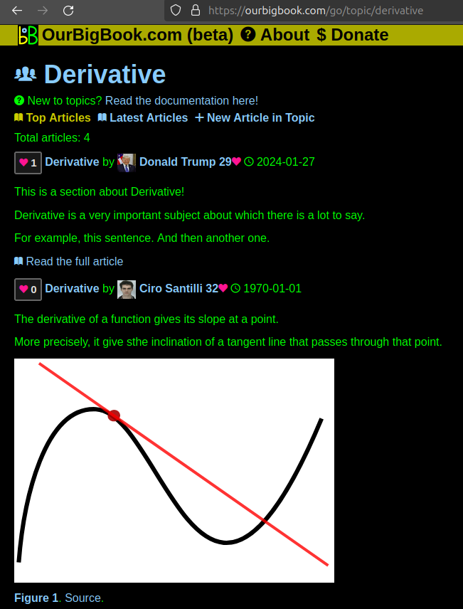
Figure 1. Screenshot of the "Derivative" topic page. View it live at: ourbigbook.com/go/topic/derivativeVideo 2. OurBigBook Web topics demo. Source. - local editing: you can store all your personal knowledge base content locally in a plaintext markup format that can be edited locally and published either:This way you can be sure that even if OurBigBook.com were to go down one day (which we have no plans to do as it is quite cheap to host!), your content will still be perfectly readable as a static site.
- to OurBigBook.com to get awesome multi-user features like topics and likes
- as HTML files to a static website, which you can host yourself for free on many external providers like GitHub Pages, and remain in full control

Figure 2. You can publish local OurBigBook lightweight markup files to either OurBigBook.com or as a static website.
Figure 3. Visual Studio Code extension installation.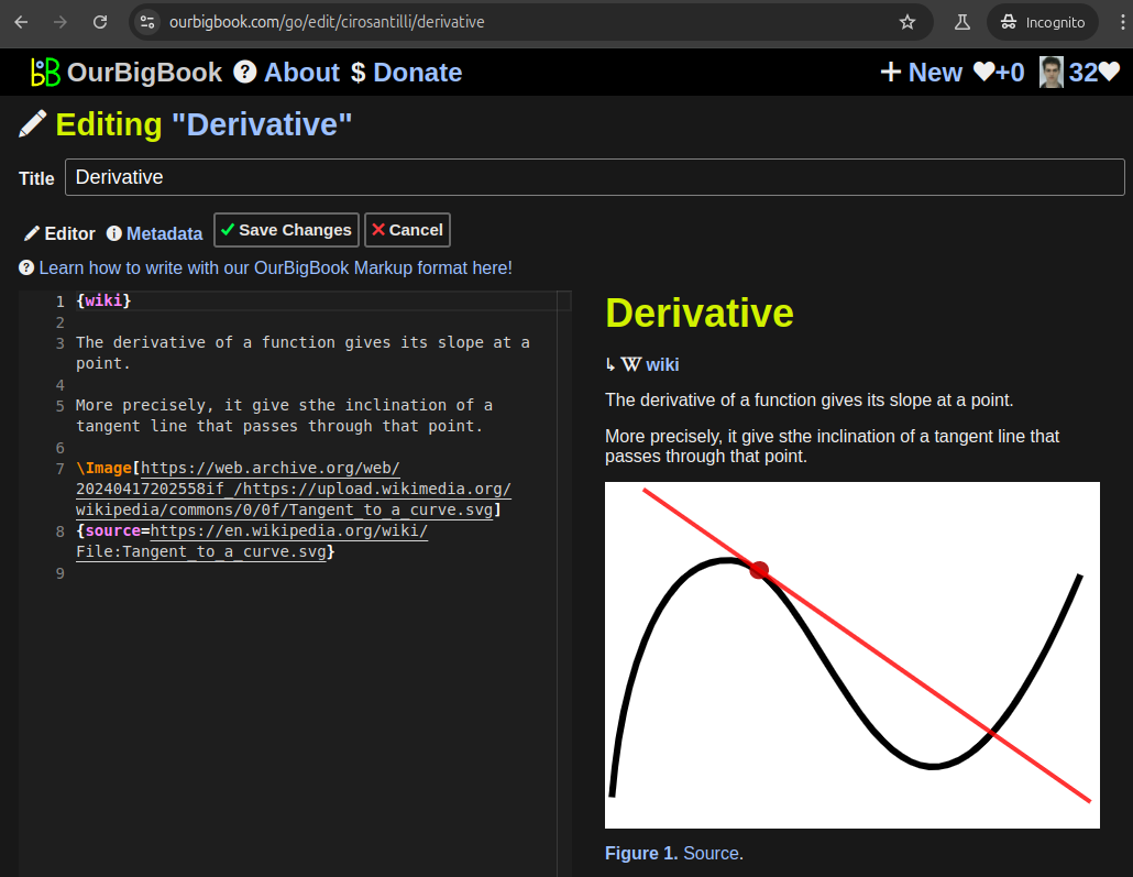
Figure 5. . You can also edit articles on the Web editor without installing anything locally. Video 3. Edit locally and publish demo. Source. This shows editing OurBigBook Markup and publishing it using the Visual Studio Code extension. 
- Infinitely deep tables of contents:
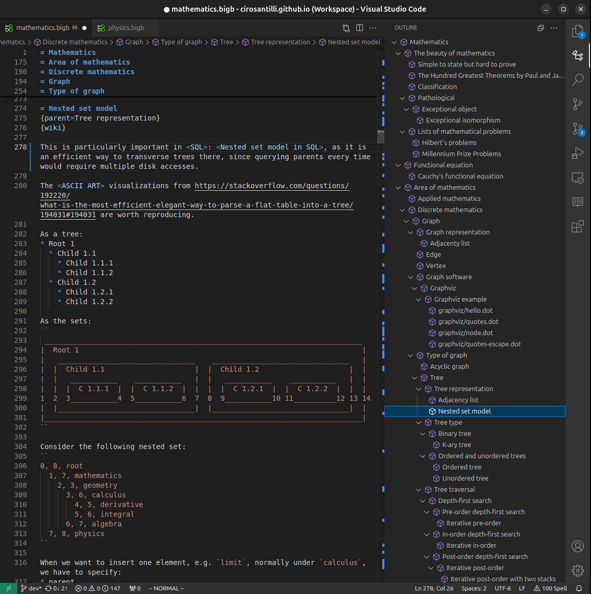

All our software is open source and hosted at: github.com/ourbigbook/ourbigbook
Further documentation can be found at: docs.ourbigbook.com
Feel free to reach our to us for any help or suggestions: docs.ourbigbook.com/#contact