Topics (384k) Articles (398k) Users (410) Discussions (242) Comments (383) Files (13k) New article

Y. L. Ruan is the first-listed author of a 2001 study of how a single cotton fiber cell elongates. The work connected changing permeability of the plasmodesma connections at the fiber base with sucrose transport, potassium transport, turgor pressure and the wall-loosening protein expansin. A cotton fiber is an illuminating growth model because a conspicuous length increase occurs within one cell, rather than being explained by repeated cell division.[ref]
Using confocal microscopy, the researchers followed a fluorescent tracer that could pass through open plasmodesma connections but not freely across the cell membrane. Tracer entry changed during development and later returned, demonstrating reversible connectivity rather than permanent separation. Measurements of transporter expression and turgor pressure helped connect this change with growth. The tracer experiment measures permeability, while the other measurements test the proposed relationship between solute supply, pressure and cell wall expansion.[ref]
The control of single-celled cotton fiber elongation by developmentally reversible gating of plasmodesmata and coordinated expression of sucrose and K+ transporters and expansin by Y. L. Ruan and others. Abstract and Figure 1: tracer permeability, transporter expression and developmental timing.
Juliane K. Ishida is the first-listed author of a 2016 study connecting local auxin production to haustorium development in the parasitic plant Phtheirospermum japonicum. A haustorium connects a parasitic plant with its host. The study investigated how a localized developmental response arises, rather than treating the plant hormone as a uniform signal throughout the whole root.[ref]
Local Auxin Biosynthesis Mediated by a YUCCA Flavin Monooxygenase Regulates Haustorium Development in the Parasitic Plant Phtheirospermum japonicum by Juliane K. Ishida and others. Abstract and Introduction, Plant Cell 28 (2016), pp. 1795–1814; DOI 10.1105/tpc.16.00310.
The researchers observed localized gene expression of an auxin-biosynthesis enzyme, measured increased auxin near a developing haustorium, and tested changes in the enzyme's expression. Reduced expression impaired development, while ectopic expression produced haustorium-like features. These comparisons illustrate the difference between a marker that accompanies organ formation and a biochemical process that contributes to it. The result concerns initiation and morphology: a haustorium-like swelling alone does not establish that every later connection to a host has formed.[ref]
Local Auxin Biosynthesis Mediated by a YUCCA Flavin Monooxygenase Regulates Haustorium Development in the Parasitic Plant Phtheirospermum japonicum by Juliane K. Ishida and others. Results: local expression, reduced expression and ectopic expression; Discussion: priming haustorium formation.
William J. Nicolas has investigated cellular membrane architecture using three-dimensional electron microscopy. He is the first author of the 2017 study reconstructing plasmodesmata and comparing their structure with their capacity for intercellular transport.[ref]
A plasmodesma contains a strand of endoplasmic reticulum within its enclosing cell membrane. Electron tomography showed that newly formed channels after cytokinesis could have such close membrane contact that an open cytoplasmic sleeve was not visible. Later cell differentiation and expansion were associated with a changed arrangement. Yet the newly formed structures still permitted movement of macromolecules.[ref]
Architecture and permeability of post-cytokinesis plasmodesmata lacking cytoplasmic sleeves by William J. Nicolas and others. Abstract: developmental membrane remodeling and macromolecular diffusion
The result makes a useful scientific distinction: an image of an apparent opening and a measurement of plasmodesmal permeability are not interchangeable. Structural reconstruction describes the organization captured by the imaging method; a transport assay tests whether a defined molecule actually moves between cells. Combining these observations challenged a simple identification of a visibly open sleeve with all intercellular exchange.
Nicolas's contribution in this work connects spatially resolved membrane structure with a functional transport measurement, rather than treating plasmodesmata as featureless holes in a cell wall.
Homepage: www.wjnicolas.com/
T. Ulmasov is the first author credited in a 1997 study of auxin-responsive gene expression at the University of Missouri. The work introduced the highly active synthetic DR5 auxin response element and investigated repression by Aux/IAA proteins.[ref]
Aux/IAA proteins repress expression of reporter genes containing natural and highly active synthetic auxin response elements by T. Ulmasov and others. Original indexed byline and affiliation; Abstract, DR5 construction and repression
The experimental idea was to connect a controllable DNA sequence to a measurable reporter gene. Tandem repeats of an eleven-base-pair unit, including the auxin-responsive sequence
TGTCTC, produced DR5. The authors tested reporter responses in carrot protoplasts and Arabidopsis thaliana seedlings. They also tested specific binding of an auxin response factor and the effects of increased Aux/IAA proteins on reporter expression.[ref]Aux/IAA proteins repress expression of reporter genes containing natural and highly active synthetic auxin response elements by T. Ulmasov and others. Abstract, synthetic sequence, expression assays, ARF1 binding and cotransfection
A constructed reporter assay example illustrates the logic. Suppose a control construct gives activity without added auxin and with it. A comparison condition gives and . The induction ratios are and , while the activities with added auxin differ fourfold. Baseline expression, induced expression and fold induction answer different questions. These invented values illustrate analysis, not the study's measured results.
A reporter gene measures an output of the regulatory system under the chosen conditions. A bright DR5 signal is therefore evidence about auxin-responsive gene expression, not automatically a calibrated local measurement of the auxin concentration. Ulmasov and colleagues' contribution supplied a reusable experimental readout together with evidence connecting auxin response elements, transcription factors and repression.[ref]
A packed pixel format stores the component fields or index for each pixel together in a single image plane, with successive pixels following one another in that plane. A planar pixel format instead separates component or bit planes. Packed does not necessarily mean one byte per pixel or the absence of row padding: the Linux frame-buffer API explicitly permits end-of-row padding in its packed storage type.[ref]
The Frame Buffer Device API in the Linux Kernel documentation. §2, FB_TYPE_PACKED_PIXELS, FB_TYPE_PLANES and line_length
Consider an original 16-bit encoding with 4-bit fields in the order red, green, blue, alpha from most significant to least significant:The bit fields for give . Extracting components by integer arithmetic givesThe same extraction can be implemented with bitwise operations. Declaring endianness is a separate step: this word occupies successive bytes
C5 A3 in little-endian storage, and A3 C5 in big-endian storage. A component order in a word does not by itself specify the byte sequence.If a row contains 100 of these two-byte pixels, its active data occupy 200 bytes. With framebuffer stride 208 bytes, the next row begins 208 bytes later, leaving eight padding bytes. A pixel-buffer consumer therefore needs the exact pixel format, dimensions and framebuffer stride, as well as any alpha channel convention and color space.
An alpha channel stores an additional value for each pixel that controls its contribution when images are combined by alpha compositing. In a common normalized convention, makes the source pixel fully transparent and makes it fully opaque. An alpha channel can describe partial coverage as well as transparency; its meaning includes the compositing rule, rather than merely a fourth color component. PNG represents its explicit alpha channel with 8-bit or 16-bit values, with zero fully transparent and the maximum value fully opaque.[ref]
Portable Network Graphics (PNG) Specification (Third Edition) by Mark Adler and others. §6.2, Alpha representation
For source color and an opaque background color , the source-over rule in Linear-light RGB isFor an original example, a red source at over a blue background produces . These are linear color components; applying the same arithmetic directly to nonlinear display codes generally gives a different physical mixture.[ref]
Portable Network Graphics (PNG) Specification (Third Edition) by Mark Adler and others. §13.16, Alpha channel processing; compositing and gamma correction
Storage conventions matter too. With straight alpha the stored color remains ; with premultiplied alpha it is . The red source in the example is therefore stored as either or . Using the first formula on an already premultiplied color would multiply its source contribution by alpha twice. PNG explicitly uses non-premultiplied color samples.[ref]
Portable Network Graphics (PNG) Specification (Third Edition) by Mark Adler and others. §6.2, unassociated alpha
A planar pixel format distributes a pixel's information among separate planes of stored samples, instead of putting all its components together as in a packed pixel format. A component plane may contain all red samples, another all green samples and another all blue samples. Intel's image-format documentation distinguishes these arrangements explicitly.[ref]
Pixel and Planar Image Formats by Intel. Pixel-Order Image Formats and Planar Image Formats tables, RGB rows
For an original one-row, two-pixel RGB color model image, suppose the component triples are and , each component occupying one byte. A packed order can be
10 20 30 40 50 60. A declared planar layout can instead store red bytes 10 40, green bytes 20 50 and blue bytes 30 60. Reinterpreting one sequence as the other changes the reconstructed colours; it is not an image compression method.For a chosen planar layout with one byte per sample, plane has base address and framebuffer stride bytes. The sample at row and column then has addressFor example, with , , row and column , the red sample is at address . Width, height, plane order, sample size and each plane's stride belong in the layout specification. Separate planes need not be adjacent allocations.
A bit plane is another arrangement: one plane stores the same bit position from many pixels. The Linux frame-buffer API's
FB_TYPE_PLANES uses this bit-plane meaning, rather than one entire colour component per plane.[ref]The Frame Buffer Device API in the Linux Kernel documentation. §2, FB_TYPE_PLANES and FB_TYPE_INTERLEAVED_PLANES
Thus the word planar alone is insufficient to decode a buffer. Processing one component across many pixels can use a contiguous component plane, while processing whole pixels must gather their components according to the declared pixel format.A semiplanar pixel format combines a separate sample plane with another plane containing interleaved components. A common YCbCr arrangement stores luma samples in one plane and pairs of colour-difference samples in another. It therefore has two planes while representing three components. Linux's video API distinguishes this from the three separate planes of a fully planar pixel format.[ref]
In NV12, one byte represents each sample. Chroma subsampling halves the colour-difference sampling count in both directions, and the second plane stores Cb followed by Cr in each pair. NV21 reverses that pair order. For even width and height , tightly stored active samples occupyThus a stipulated image contains eight Y samples and two Cb/Cr pairs: 12 active bytes. The eight Y bytes can be followed by
Cb00 Cr00 Cb01 Cr01. The component names identify stored codes; their colour interpretation also requires the declared YCbCr conventions.[ref]Planar YUV formats in the Linux Kernel documentation. Semi-Planar overview table and NV12/NV21 layout subsection
Active sample count differs from allocated storage. With an eight-byte framebuffer stride for both planes in this example, two Y rows and one interleaved row occupy bytes including padding. A consumer needs dimensions, strides, component order and whether planes are adjacent or separately allocated. Reinterpreting NV21 as NV12 swaps the two colour-difference components; it does not recover the intended colours merely because the buffer size matches.
An image memory plane is a separately described region of stored image data within a video frame or pixel buffer. It is a memory-organization concept, distinct from a component plane or a bit plane. One memory plane can contain several interleaved components, while one image can use several memory planes.[ref]
For example, NV12M uses one memory plane for luma and another for alternating Cb/Cr samples. It therefore has two memory planes but three colour components. NV21M uses the same plane count with the colour-difference pair order reversed. Their 4:2:0 chroma subsampling specifies sample counts; it does not turn the two components in the second plane into separate allocations.
Allocation capacity and video buffer payload also differ. A V4L2 plane descriptor records
length, bytesused and a V4L2 plane data offset. In that API, bytesused includes the offset before the actual image data, so the image-data extent isThe allocation size is a separate limit.[ref]For an original example, stipulate
length = 4096, bytesused = 3072 and data_offset = 128, all in bytes. The image data begin128 bytes after the plane base and span2944 bytes, ending at base offset3072. They occupy neither the full4096-byte allocation nor all3072 bytes preceding that endpoint. The necessary bounds areA consumer must combine these extents with the actual pixel format and framebuffer stride. A plane's capacity alone does not identify where a particular component or row begins.Pixel pitch is the physical spacing between corresponding centres of neighbouring pixels on a display or sensor grid. For an axis-aligned rectangular grid it can have separate horizontal and vertical values . The pixel pitch describes the grid spacing, rather than the number of bits used for each pixel value.[ref]
Pixel Pitch and Enlarged Mode by EIZO. Opening definition of pixel pitch and comparison of physical text size at different densities
For square pixels on a uniform grid, linear pixel density and pixel pitch are reciprocal. If is measured in pixels per inch,For example, a hypothetical matrix with 2400 columns occupying a active width has and horizontal pixel density pixels per inch. This calculation treats the width as the edge-to-edge extent of 2400 equal pixel cells; the distance from the first centre to the last centre is instead .
Doubling the column count over the same width halves the horizontal pixel pitch. Doubling both row and column counts quadruples the number of pixels per unit area. The dimensions and the direction being measured therefore matter when comparing specifications.
A smaller pixel pitch provides a finer sampling grid, but it cannot recover detail already lost from the input image. Optical blur, aliasing and the viewing geometry also affect visible detail. A CSS pixel is a layout unit, so its size must be mapped through the device pixel ratio before it can be related to this physical grid.
Melanin denotes a family of biological pigments formed through oxidative chemistry of aromatic precursors. It is not one small molecule with a single universally applicable molecular formula. Different precursor chemistry and assembly give chemically heterogeneous materials.
Dark eumelanin and sulfur-containing pheomelanin illustrate the family's diversity. Extended electronic interactions and structural disorder produce broad absorption rather than one isolated sharp molecular line. Consequently colour, composition and microscopic organization must be considered together when comparing melanin specimens; assigning a precise repeating structure to every biological sample is generally too strong.
J. C. Giddings is the coagulation researcher who coauthored the 1975 study of Factor V activation and prothrombin conversion with A. L. Bloom. The work investigated both assembly of reaction components and production of thrombin.[ref]
Gel chromatography separated the preparations, and specific antibodies helped identify components in the resulting fractions. Combined with kinetic measurements, these experiments supported a functional assembly involving phospholipids, Factor V, factor Xa and calcium ions.[ref]
Factor-V activation by thrombin and its role in prothrombin conversion by J. C. Giddings and A. L. Bloom. Abstract: Sephadex chromatography, immunological analysis and thrombin-generation tests
This illustrates two complementary questions: which molecules associate, and does their association support the expected reaction? Component identification addresses composition; an activity assay addresses function. Using both provides a stronger explanation than either separation or product measurement alone.
A. L. Bloom was a coagulation researcher who, with J. C. Giddings, investigated how activated Factor V promotes production of thrombin. Their 1975 study reconstructed the reaction using purified coagulation factors.[ref]
Factor-V activation by thrombin and its role in prothrombin conversion by J. C. Giddings and A. L. Bloom. Byline and Abstract: purified-factor experiments
Pretreating Factor V with trace amounts of thrombin increased the rate and yield of subsequent thrombin generation from prothrombin. The finding supported a positive feedback mechanism: product formation could enhance further product formation by activating a cofactor.[ref]
Carl W. Gottschalk was a researcher in renal physiology who used renal micropuncture to investigate how the kidney handles fluid and concentrates urine. Working with Margaret Mylle, he developed experiments that connected conditions in individual renal tubules to the function of the whole kidney.[ref]
Their 1959 measurements found that fluid in loop bends, collecting ducts and renal vasa recta at the same medullary level was similarly hyperosmotic. This supported countercurrent multiplication and a shared concentrating environment in the renal medulla.[ref]
Micropuncture study of the mammalian urinary concentrating mechanism: evidence for the countercurrent hypothesis by Carl W. Gottschalk and Margaret Mylle. Abstract: comparison of fluid osmolality at matched medullary levels
Sampling the correct structure was itself part of the evidence. The memoir describes checking puncture locations with dye and microdissection, helping distinguish a renal tubule from a nearby blood vessel. A concentration measurement becomes biologically interpretable only when the source of the sample is secure.[ref]
Carl W. Gottschalk by Maurice B. Burg. Micropuncture studies: site verification and distinguishing loop fluid from blood
Margaret Mylle was a researcher in the physiology of the kidney and a long-term collaborator of Carl W. Gottschalk. Their work used renal micropuncture to investigate fluid conditions within individual parts of the nephron, rather than inferring every process from the final urine.[ref]
Carl W. Gottschalk by Maurice B. Burg. Early micropuncture research: collaboration and local measurements
Their 1959 study compared osmolality in samples from renal tubules, collecting ducts, the Loop of Henle and small renal vessels. Oil-filled glass micropipettes collected tiny fluid samples, whose freezing-point depression supplied a concentration measurement.[ref]
The illuminating feature is the specificity of the sampling site. A high concentration in final urine does not identify where concentration developed. Comparing local fluid with blood plasma, and with fluid elsewhere in the kidney, turns an overall output into a spatial test of a physiological mechanism. The joint experiments helped connect microscopic anatomy with the concentrating function of the whole organ.
D. S. Bredt is a neuroscience researcher who, with S. H. Snyder, investigated production of nitric oxide. Their 1990 paper isolated nitric oxide synthase and established its requirement for calmodulin.[ref]
The researchers followed conversion of arginine into citrulline, associated with formation of nitric oxide. Using affinity chromatography, they purified the activity approximately 6000-fold from rat cerebellum. This tied a chemical transformation to an isolated protein.[ref]
Isolation of nitric oxide synthetase, a calmodulin-requiring enzyme by D. S. Bredt and S. H. Snyder. Abstract: reaction, affinity column and purification factor
The example illustrates activity-guided separation: measuring a reaction product lets one track a catalyst through successive fractions. Testing the isolated activity with different added components then investigates regulation. A purification factor describes enrichment of activity relative to total protein; it is not a 6000-fold increase in every biological response.
S. H. Snyder is a researcher in neuroscience and pharmacology whose laboratory investigates neurotransmitters, intracellular messengers and mechanisms of drug action. His research includes nitric oxide, a signaling molecule whose production can be coupled to intracellular calcium ions.[ref]
With D. S. Bredt, S. H. Snyder isolated nitric oxide synthase from rat cerebellum. The 1990 experiments established a requirement for calmodulin, alongside calcium ions and NADPH, in the measured activity. This connected production of nitric oxide to a molecular calcium-sensing mechanism.[ref]
Isolation of nitric oxide synthetase, a calmodulin-requiring enzyme by D. S. Bredt and S. H. Snyder. Abstract: enzyme source, arginine conversion and calmodulin requirement
The example shows how identifying the enzyme producing a messenger can change the explanation of signaling. Rather than imagining a fixed store simply being emptied, one can ask which inputs activate synthesis and how fast production changes. A messenger's chemical identity and the mechanism controlling its supply are complementary parts of the biological explanation.

Maurice B. Burg was a researcher in renal physiology who developed experimental methods for studying individual nephron segments outside the intact kidney. Isolated tubule perfusion made it possible to relate transport properties to a particular anatomical segment, rather than observing only the final composition of urine.[ref]
Maurice B. Burg (1931–2022), discoverer of kidney transport mechanisms by Mark A. Knepper. Opening remembrance: invention and use of isolated-perfused-nephron techniques
The 1966 method paper by Maurice B. Burg and collaborators described preparations from single rabbit nephrons. Supplying fluid through an isolated renal tubule while controlling its surrounding solution allows the luminal and external conditions to be varied separately.[ref]
Preparation and study of fragments of single rabbit nephrons by M. Burg and others. Original1966 method-paper record and preparation
A useful experiment is to change the surrounding osmolality and compare entering and collected tubular fluid. A change in collected volume can reveal net water movement; changes in solute content address transport of dissolved substances. Isolating the segment constrains where the measured exchange occurred. The preparation simplifies the system while retaining an organized living epithelium, an important middle scale between a whole organ and an isolated protein.
Pinned article: Introduction to the OurBigBook Project
Welcome to the OurBigBook Project! Our goal is to create the perfect publishing platform for STEM subjects, and get university-level students to write the best free STEM tutorials ever.
Everyone is welcome to create an account and play with the site: ourbigbook.com/go/register. We belive that students themselves can write amazing tutorials, but teachers are welcome too. You can write about anything you want, it doesn't have to be STEM or even educational. Silly test content is very welcome and you won't be penalized in any way. Just keep it legal!
Intro to OurBigBook
. Source. We have two killer features:
- topics: topics group articles by different users with the same title, e.g. here is the topic for the "Fundamental Theorem of Calculus" ourbigbook.com/go/topic/fundamental-theorem-of-calculusArticles of different users are sorted by upvote within each article page. This feature is a bit like:
- a Wikipedia where each user can have their own version of each article
- a Q&A website like Stack Overflow, where multiple people can give their views on a given topic, and the best ones are sorted by upvote. Except you don't need to wait for someone to ask first, and any topic goes, no matter how narrow or broad
This feature makes it possible for readers to find better explanations of any topic created by other writers. And it allows writers to create an explanation in a place that readers might actually find it.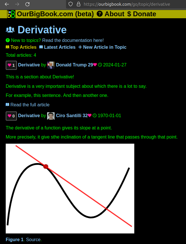
Figure 1. Screenshot of the "Derivative" topic page. View it live at: ourbigbook.com/go/topic/derivativeVideo 2. OurBigBook Web topics demo. Source. - local editing: you can store all your personal knowledge base content locally in a plaintext markup format that can be edited locally and published either:This way you can be sure that even if OurBigBook.com were to go down one day (which we have no plans to do as it is quite cheap to host!), your content will still be perfectly readable as a static site.
- to OurBigBook.com to get awesome multi-user features like topics and likes
- as HTML files to a static website, which you can host yourself for free on many external providers like GitHub Pages, and remain in full control

Figure 2. You can publish local OurBigBook lightweight markup files to either OurBigBook.com or as a static website.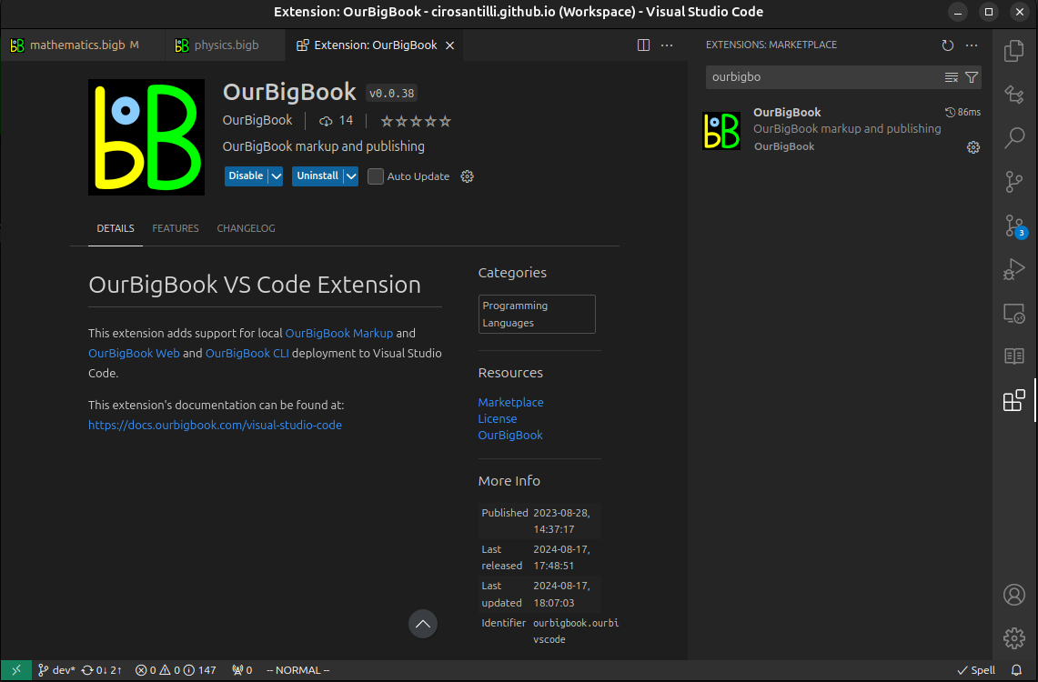
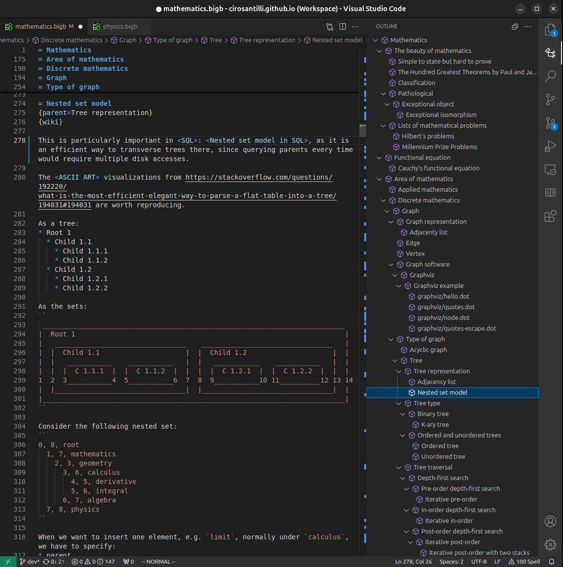
Figure 3. Visual Studio Code extension installation.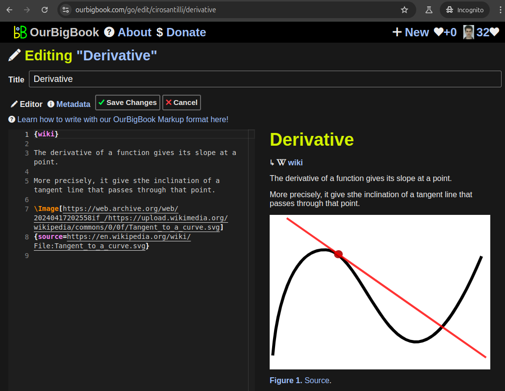
Figure 5. . You can also edit articles on the Web editor without installing anything locally. Video 3. Edit locally and publish demo. Source. This shows editing OurBigBook Markup and publishing it using the Visual Studio Code extension. 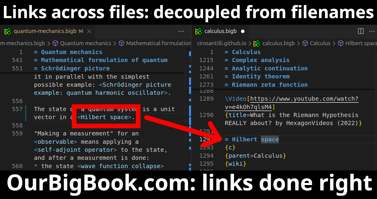
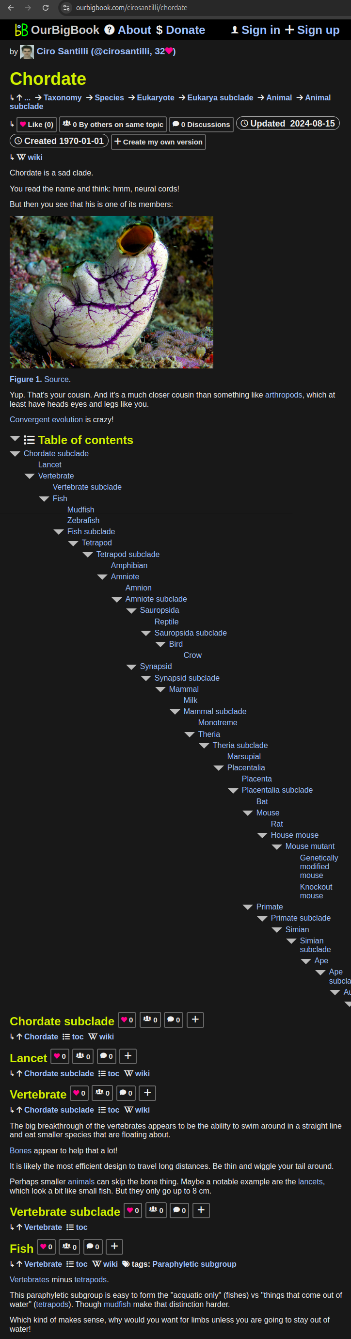
- Infinitely deep tables of contents:
All our software is open source and hosted at: github.com/ourbigbook/ourbigbook
Further documentation can be found at: docs.ourbigbook.com
Feel free to reach our to us for any help or suggestions: docs.ourbigbook.com/#contact