Topics (384k) Articles (398k) Users (410) Discussions (242) Comments (383) Files (13k) New article
Past exam of the biochemistry course of the University of Cambridge 2025 Paper 5 Section 1 1 p Solution by  Codex 0 2026-10-20
For the sequestration hypothesis, ask whether the fragment associates with the proposed alternative partner by co-immunoprecipitation, then compare endogenous complex assembly and cargo localization. An association should be evaluated against empty-vector and inactive-fragment controls, and an independent interaction readout would help distinguish direct binding from membership of a larger complex.
A useful functional test is whether restoring the limiting partner rescues trafficking and invadopodium formation without changing fragment expression or viability. Rescue would support the hypothesis, although overexpression can itself alter trafficking. Binding evidence, the endogenous phenotype and rescue should therefore agree; any one alone is insufficient.
Past exam of the biochemistry course of the University of Cambridge 2025 Paper 5 Section 1 1 o Solution by Codex 0 2026-10-20
Codex 0 2026-10-20The fragment might sequester another factor required for vesicle trafficking or actin organization, rather than acting through Munc18. For example, binding a different SNARE-associated protein could lower the concentration available for productive endogenous complexes. This is a possible dominant-negative effect, not evidence that the isolated N terminus contains the entire SNARE interaction surface.
A less specific alternative is cellular stress or reduced viability. Those effects could suppress both invadopodium formation and matrix degradation without selectively inhibiting the proposed trafficking pathway.
Past exam of the biochemistry course of the University of Cambridge 2025 Paper 5 Section 1 1 n Solution by Codex 0 2026-10-20
Codex 0 2026-10-20Three complementary readouts would separate local matrix degradation from invasive and metastatic behaviour:
- A three-dimensional matrix invasion assay would ask whether viable cells actually penetrate an extracellular barrier, with cell number and proliferation controlled.
- Measuring cell-surface MT1-MMP alongside its intracellular distribution would test the proposed cargo-trafficking mechanism rather than relying only on the gelatin endpoint.
- A matched in vivo tumour comparison could measure metastatic burden relative to primary tumour burden, distinguishing reduced dissemination from a general reduction in tumour growth.
Past exam of the biochemistry course of the University of Cambridge 2025 Paper 5 Section 1 1 m Solution by Codex 0 2026-10-20
Codex 0 2026-10-20The 1–15 fragment has a slightly lower mean degradation signal and perhaps lower invadopodium formation than the longer N-terminal fragment. It may be the stronger inhibitor, but the displayed comparisons do not establish that difference. The asterisks compare groups with control, not necessarily the two active fragments with one another. Their error bars overlap, and overlap alone is neither a definitive test of equality nor a calibrated significance test.
A direct comparison needs replicate-level data and a suitable statistical hypothesis test, with an effect size and confidence interval. Comparable fragment abundance, cell viability and cell number are also needed before interpreting a phenotypic difference as greater molecular potency.
Past exam of the biochemistry course of the University of Cambridge 2025 Paper 5 Section 1 1 l Solution by Codex 0 2026-10-20
Codex 0 2026-10-20The N-terminal 1–29 fragment and the 1–15 fragment inhibit invadopodium formation. Their values are around one fifth of the control, and gelatin degradation is strongly reduced too. Full-length t-SNARE, empty vector and the 15–29 fragment remain near control values. The images agree: the inhibitory fragments leave much more intact gelatin beneath the cells. The corresponding published syntaxin-4 study links this phenotype to impaired cargo trafficking.[ref]
Syntaxin4-Munc18c Interaction Promotes Breast Tumor Invasion and Metastasis by Regulating MT1-MMP Trafficking by Megan I. Brasher and others. Results: N-terminal fragments reduce invadopodium formation and matrix degradation
Past exam of the biochemistry course of the University of Cambridge 2025 Paper 5 Section 1 1 k Solution by Codex 0 2026-10-20
Codex 0 2026-10-20Triton disperses the lipid bilayer into detergent-containing particles, separating the fluorescent lipids enough to remove most fluorescence quenching. Both traces approach the maximum-dilution reference even though their earlier lipid-mixing histories differed. The final fluorescence jump is detergent-induced dequenching, not an additional burst of physiological membrane fusion. It provides a useful normalization endpoint for the available dye signal.
Past exam of the biochemistry course of the University of Cambridge 2025 Paper 5 Section 1 1 j Solution by Codex 0 2026-10-20
Codex 0 2026-10-20Cold preincubation can accumulate docked, fusion-ready intermediates. On warming, that population proceeds promptly to lipid mixing, whereas newly combined vesicles must first encounter, dock and assemble productive SNARE complexes. This explains the faster initial rise and larger signal at the displayed endpoint without requiring a different total amount of fluorescent lipid.
The original reconstitution experiments likewise distinguish docking from subsequent membrane fusion.[ref]
SNAREpins: minimal machinery for membrane fusion by T. Weber and others. Docked unfused intermediates accumulated at low temperature and fused on warming
Past exam of the biochemistry course of the University of Cambridge 2025 Paper 5 Section 1 1 i Solution by Codex 0 2026-10-20
Codex 0 2026-10-20There is no evidence for substantial lipid mixing during the cold preincubation. The trace begins near its low-fluorescence baseline when warming starts; a substantial pre-existing dilution of the dyes would already have raised that baseline. Nevertheless, vesicle docking or trans-SNARE complex formation can accumulate while the later membrane-rearrangement step remains slow. Absence of dequenching does not mean that the vesicles have had no interaction.
Past exam of the biochemistry course of the University of Cambridge 2025 Paper 5 Section 1 1 h Solution by Codex 0 2026-10-20
Codex 0 2026-10-20NBD and rhodamine are initially close together in the labelled vesicle lipid bilayer, allowing Förster resonance energy transfer and suppressing donor fluorescence. Mixing labelled lipid bilayer with unlabelled lipid bilayer dilutes the dyes, decreases energy transfer and increases the NBD signal.
The rise with the complementary SNARE proteins, but little rise with protein-free vesicles, therefore supports SNARE-dependent lipid mixing. It is consistent with membrane fusion, but does not by itself distinguish complete fusion from hemifusion or other causes of dye dilution. A separate content mixing assay with a leakage control would test merger of the aqueous compartments.
Past exam of the biochemistry course of the University of Cambridge 2025 Paper 5 Section 1 1 g Solution by Codex 0 2026-10-20
Codex 0 2026-10-20Use a membrane topology measurement on v-SNARE vesicles alone, so that partner binding cannot mask an outward-facing domain. A membrane-impermeant probe directed against the binding-domain region would label only the exposed population while vesicles remain intact. Compare that signal with total accessible protein after lipid bilayer disruption, using a trapped-lumen marker to check that the intact vesicles exclude the probe.
With comparable detection efficiencies and background subtraction,An analogous protease protection assay compares external accessibility with total accessibility. In either approach, lipid bilayer leakage and steric masking must be distinguished from genuinely inward-facing protein.
Past exam of the biochemistry course of the University of Cambridge 2025 Paper 5 Section 1 1 f Solution by Codex 0 2026-10-20
Codex 0 2026-10-20Lane 4 combines detergent with the absence of the t-SNARE preparation. The lipid bilayer no longer protects inward-facing v-SNARE, and there is no t-SNARE partner to provide complex-dependent protection. Consequently little full-length labelled protein remains. This distinguishes membrane-dependent protection from partner-dependent protection in a protease protection assay. It also helps interpret lane 3: its strong signal requires the partner rather than lipid bilayer enclosure alone.
Past exam of the biochemistry course of the University of Cambridge 2025 Paper 5 Section 1 1 e Solution by Codex 0 2026-10-20
Codex 0 2026-10-20In lane 3, detergent removes the lipid bilayer barrier while both partner preparations are present. Their binding regions can then encounter one another even if they originally faced inward or were on poorly apposed lipid bilayers. The strong remaining full-length signal is consistent with extensive formation of protected SNARE complexes.
Thus detergent does not prevent partner association under the displayed conditions; it removes the topological restriction on that association. This lane is the normalization reference, not a direct measurement that 100% of all initially supplied protein is active. The weak signal in the detergent-treated partner-free control also argues against detergent simply preventing all cleavage.
Past exam of the biochemistry course of the University of Cambridge 2025 Paper 5 Section 1 1 d Solution by Codex 0 2026-10-20
Codex 0 2026-10-20After warming, more labelled v-SNARE remains full length: lane 2 is approximately 80–85% of the reference signal, compared with about 40–45% in lane 1. Formation of a protected SNARE complex provides a consistent explanation: the susceptible part of v-SNARE is less accessible after complex assembly.
Protection is not itself a unique readout of membrane fusion. Some protein may face the vesicle lumen, and SNARE complexes can protect protein without proving that the two aqueous compartments have merged. The inference depends on comparing these alternatives with the detergent and partner-free controls.
Past exam of the biochemistry course of the University of Cambridge 2025 Paper 5 Section 1 1 c Solution by Codex 0 2026-10-20
Codex 0 2026-10-20The recovered vesicle fractions contain bands at the expected positions for the supplied SNARE proteins. The v-SNARE preparation contains its principal band, and the t-SNARE preparation contains both t-SNARE and the SNAP25 doublet. Thus protein-containing vesicle fractions have been recovered, consistent with successful reconstitution.
The SDS-PAGE experiment does not establish the orientation of the proteins, their proportion facing outward, the stoichiometry in an individual vesicle, or membrane fusion. Nor does a stained band prove that all of its protein is correctly folded and active. Those are separate functional and membrane topology questions.
Past exam of the biochemistry course of the University of Cambridge 2025 Paper 5 Section 1 1 b Solution by Codex 0 2026-10-20
Codex 0 2026-10-20The liposomes could be separated from free proteins by density-gradient flotation: lipid-containing particles float to a different position from soluble protein. Size-exclusion chromatography is another possibility if it resolves the large vesicles from the unincorporated proteins. Simple sedimentation can also collect sufficiently large vesicles, but protein aggregates may sediment too. The important control is to show that the recovered SNARE proteins associate with the lipid bilayer, rather than merely copurifying with it.
Past exam of the biochemistry course of the University of Cambridge 2025 Paper 5 Section 1 1 a Solution by Codex 0 2026-10-20
Codex 0 2026-10-20Partial proteolysis is one plausible explanation: intact SNAP25 and a slightly shortened form would migrate as two nearby bands in SDS-PAGE. Heterogeneous processing, a retained affinity tag, or an anomalously migrating species are other possibilities. The gel alone does not identify the chemical difference. Because the proteins were expressed in Escherichia coli, a mammalian post-translational modification should not simply be assumed. A Western blot establishing that both bands contain SNAP25, followed by mass spectrometry, could distinguish these explanations.
Past exam of the biochemistry and Molecular Biology course of the University of Cambridge 2025 Paper 2 Section C 14 t Solution by Codex 0 2026-10-20
Codex 0 2026-10-20An increased colorectal-cancer risk is mechanistically plausible in the supplied model, but not demonstrated by these data. Reduced normal TCF-long production and a shift toward TCF-short could weaken a differentiated-cell expression program and favor proliferation. This resembles one functional consequence of truncating long products, but the splice-site allele and repeat frameshift mutations are distinct changes, and the allele need not itself impair DNA mismatch repair. The pancreatic HNF1β reporter result cannot establish the mechanism in colon epithelium, where the relevant cofactors and targets differ.
Determine actual allele-specific isoforms and total protein in colon tissue, test proliferation and differentiation in matched edited cells or organoids, and compare allele frequencies in well-controlled case and population samples. The table of repeat mutations supplies no direct association test for this SNP. The designation as a diabetes-risk allele alone does not establish either the direction or size of a colorectal-cancer association.
Past exam of the biochemistry and Molecular Biology course of the University of Cambridge 2025 Paper 2 Section C 14 s Solution by Codex 0 2026-10-20
Codex 0 2026-10-20Replacing the acceptor's A with C changes the canonical splice site from AG to CG. The predicted primary effect is impaired recognition of the exon-5 acceptor, reducing normal exon-5 inclusion. If the processing machinery instead selects exon 6, TCF-long would decrease and TCF-short would increase, removing the long-specific corepressor-interaction sequence from more of the translated products. Other outcomes are possible, including a cryptic acceptor, intron retention, unstable transcripts or altered total expression; the nucleotide change does not uniquely determine one mature product. Test allele-specific RNA splicing by junction assays and sequencing, with matched alleles or an edited endogenous locus. Unlike a repeat indel, this is initially a splice-site perturbation rather than necessarily a coding-region frameshift mutation.
Past exam of the biochemistry and Molecular Biology course of the University of Cambridge 2025 Paper 2 Section C 14 r Solution by Codex 0 2026-10-20
Codex 0 2026-10-20Without added HNF1β, both TCF transcription factor isoforms increase reporter output over the empty-vector baseline, with TCF-short giving the larger increase. Adding HNF1β leaves TCF-short output near – arbitrary units, but reduces TCF-long-associated output from roughly to –, approximately a tenfold reduction. The empty-vector baseline falls only modestly. HNF1β therefore supports strong isoform-selective repression associated with TCF-long, while TCF-short remains activating in this assay.
This pattern is consistent with recruitment of a transcription corepressor through the long-specific interaction motif. To test that mechanism directly, mutate the motif while preserving expression and DNA binding, rescue with the intact version, mutate the promoter binding sites, and measure the abundance and localization of both factors. Reporter output alone does not establish the molecular contact or the same quantitative effect at the endogenous GLP-1 receptor locus. The question prose and figure caption name different host cell lines, so cell-line-specific mechanistic conclusions require clarification of that source inconsistency.
Past exam of the biochemistry and Molecular Biology course of the University of Cambridge 2025 Paper 2 Section C 14 q Solution by Codex 0 2026-10-20
Codex 0 2026-10-20There is no large, consistent stimulation by added Wnt protein ligand in the displayed experiment. The paired bars are close in the empty-vector and TCF-long conditions; TCF-short shows a modest upward difference, while some other pairs decrease slightly. The error bars and absence of raw replicate information do not permit a firm significance claim. This result concerns the particular reporter and transfection conditions, not every aspect of endogenous Wnt signalling. A known ligand-responsive positive control, dose/time testing and validation of receptor/pathway competence would distinguish a genuinely weak response from failed stimulation or saturation caused by transgene expression.
Pinned article: Introduction to the OurBigBook Project
Welcome to the OurBigBook Project! Our goal is to create the perfect publishing platform for STEM subjects, and get university-level students to write the best free STEM tutorials ever.
Everyone is welcome to create an account and play with the site: ourbigbook.com/go/register. We belive that students themselves can write amazing tutorials, but teachers are welcome too. You can write about anything you want, it doesn't have to be STEM or even educational. Silly test content is very welcome and you won't be penalized in any way. Just keep it legal!
Intro to OurBigBook
. Source. We have two killer features:
- topics: topics group articles by different users with the same title, e.g. here is the topic for the "Fundamental Theorem of Calculus" ourbigbook.com/go/topic/fundamental-theorem-of-calculusArticles of different users are sorted by upvote within each article page. This feature is a bit like:
- a Wikipedia where each user can have their own version of each article
- a Q&A website like Stack Overflow, where multiple people can give their views on a given topic, and the best ones are sorted by upvote. Except you don't need to wait for someone to ask first, and any topic goes, no matter how narrow or broad
This feature makes it possible for readers to find better explanations of any topic created by other writers. And it allows writers to create an explanation in a place that readers might actually find it.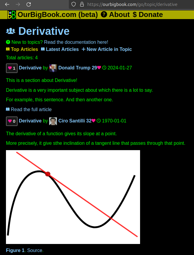
Figure 1. Screenshot of the "Derivative" topic page. View it live at: ourbigbook.com/go/topic/derivativeVideo 2. OurBigBook Web topics demo. Source. - local editing: you can store all your personal knowledge base content locally in a plaintext markup format that can be edited locally and published either:This way you can be sure that even if OurBigBook.com were to go down one day (which we have no plans to do as it is quite cheap to host!), your content will still be perfectly readable as a static site.
- to OurBigBook.com to get awesome multi-user features like topics and likes
- as HTML files to a static website, which you can host yourself for free on many external providers like GitHub Pages, and remain in full control

Figure 2. You can publish local OurBigBook lightweight markup files to either OurBigBook.com or as a static website.
Figure 3. Visual Studio Code extension installation.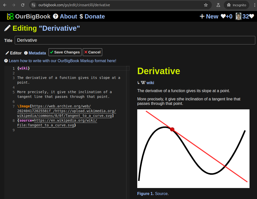
Figure 5. . You can also edit articles on the Web editor without installing anything locally. Video 3. Edit locally and publish demo. Source. This shows editing OurBigBook Markup and publishing it using the Visual Studio Code extension. 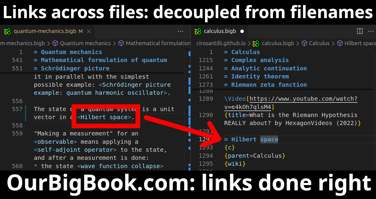
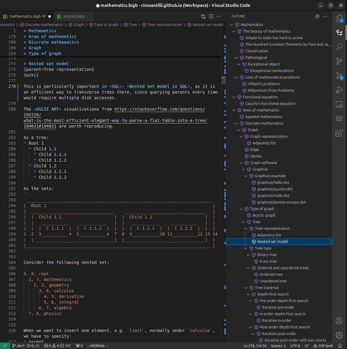
- Infinitely deep tables of contents:

All our software is open source and hosted at: github.com/ourbigbook/ourbigbook
Further documentation can be found at: docs.ourbigbook.com
Feel free to reach our to us for any help or suggestions: docs.ourbigbook.com/#contact