
Let be the Fourier transform of the initial temperature. Solving the scalar linear ordinary differential equation givesRecover using the Fourier inversion theorem, with inverse factor for this two-dimensional convention. Equivalently, take the convolution of the initial data with the two-dimensional heat kernel. On a periodic square, the same exponential applies to each discrete Fourier mode, with and .
Apply the Fourier transform of a derivative twice in each spatial coordinate, and commute the time derivative with the spatial Fourier transform. For the Laplacian,ThusThe minus sign is essential: positive thermal diffusivity damps short spatial scales rather than amplifying them. The zero Fourier mode is unchanged in the absence of a source.
Assume sufficient decay and differentiability to justify integration by parts and exchange of integrals; Schwartz functions are one convenient class. With the stated Fourier transform convention, integrate first in :The boundary contribution vanishes under the decay assumption. The Fourier transform of a derivative consequently turns constant-coefficient spatial differential operators into multipliers. Repeating it gives , so the heat equation becomes an independent temporal ordinary differential equation at each wavevector.
The photic zone is the illuminated layer supporting photosynthesis; its euphotic depth is conventionally associated with about 1% of surface photosynthetically active radiation, integrated over approximately –. This is a useful convention rather than a universal organism-specific compensation depth. Spectral filtering can be represented locally bywhere is the wavelength-dependent diffuse attenuation coefficient. It includes effects of absorption and scattering and is not simply equal to the absorption coefficient .
In clear, low-chlorophyll water, pure water removes red light strongly, while weak blue pigment and CDOM absorption permits blue light, typically around –, to penetrate furthest. The deepest usable light is consequently dim and blue-rich, and the euphotic zone can extend relatively deep. At high pigment concentrations, phytoplankton pigments preferentially remove blue and red light; associated detritus or CDOM can remove still more blue. The remaining transmission window commonly shifts toward blue-green or green wavelengths, roughly –, and the total light falls off faster, so the euphotic depth is shallower. Exact window and depth depend on the full particle/dissolved mixture, not chlorophyll alone. The diagram normalizes the two deep spectra separately to show their different shapes, not equal absolute light at a common depth.
This confines photosynthetic growth to a thinner illuminated interval in high-chlorophyll water and allows a deeper potential habitat in clear low-chlorophyll water, subject also to nutrient supply, mixing and the organism's compensation requirement. Cells near the base benefit from low-light acclimation and pigment systems that capture the remaining wavelengths. In blue-rich deep ocean water, blue-absorbing pigments and some low-light-adapted picoplankton can be favored; where the surviving light is greener, groups with effective green-absorbing accessory pigments, such as certain phycobilins, gain an advantage. A pigment appears coloured by light it fails to absorb, so green-absorbing accessory pigments need not themselves look green. There is no universal deep species determined by chlorophyll concentration alone. A deep chlorophyll maximum can partly reflect increased pigment per cell rather than a proportional maximum in carbon biomass.[ref]
Phytopia's light and color by NASA. Photic-zone discussion: the 1% convention, deep penetration in clear blue waters, and shallower illuminated habitat in productive turbid waters; pigment-based community selection here follows the surviving wavelengths and is conditional.
Chlorophyll a occurs in nearly all oxygen-producing phytoplankton groups and is central to their photosynthesis. Its concentration can be measured reproducibly by pigment extraction or calibrated optical methods, and its absorption affects ocean colour, making it observable over large areas. It is therefore a practical common index of photosynthetic standing stock across diverse communities, unlike a pigment limited to one group.
It is a proxy, not a fixed conversion to phytoplankton biomass or primary production. The carbon-to-chlorophyll ratio varies with species, light acclimation, nutrient supply and physiological condition; low-light cells often contain more pigment per unit carbon. Dead pigment and methodological differences can also affect interpretation, and nonphotosynthetic plankton are not represented. A pigment increase need not mean the same proportional increase in living carbon biomass.[ref]
Chlorophyll maps by NASA Earth Observatory. Chlorophyll alters sunlight absorption and reflection, enabling mapping of near-surface phytoplankton distributions; pigment is used as an index rather than a direct carbon measurement.
Many open-ocean chlorophyll retrieval algorithms infer chlorophyll a from the blue-to-green remote-sensing reflectance ratio. Increasing pigment generally depresses blue reflectance relative to green. In coastal waters, however, coloured dissolved organic matter and non-algal particles can independently absorb blue light, as in the sketches, and therefore mimic an increase in pigment. Their concentrations need not covary with phytoplankton biomass: river runoff and sediment resuspension can change them separately.
Moreover, the satellite measures emerging radiance, not absorption directly. Schematically,where is the backscattering coefficient; this is an explanatory approximation, not a complete retrieval formula. Suspended particles change both absorption and backscattering, and different mixtures can produce similar band ratios. Shallow-bottom reflection, land-adjacency effects and more difficult atmospheric correction introduce further coastal errors. Consequently a simple blue/green ratio can overestimate chlorophyll a when excess CDOM removes blue light, although sediment scattering and other effects mean the bias is not universally positive. Multiband or hyperspectral methods that separately constrain pigment, dissolved absorption and particles, supported by local observations, are preferable in optically complex water.[ref]
Ocean optics protocols by NASA. Revision 4, Volume I: unaccounted blue absorption by CDOM creates regional chlorophyll-retrieval uncertainty; absorption and scattering are distinct optical properties.
The principal non-water visible-light absorbers can be grouped into phytoplankton pigments, coloured dissolved organic matter (CDOM), and non-algal particles (NAP), including organic detritus and absorbing mineral material. A useful additive decomposition of the absorption coefficient isThe supplied sketch separates the three non-water contributions; its amplitudes are arbitrary and are not concentrations or measured spectra.
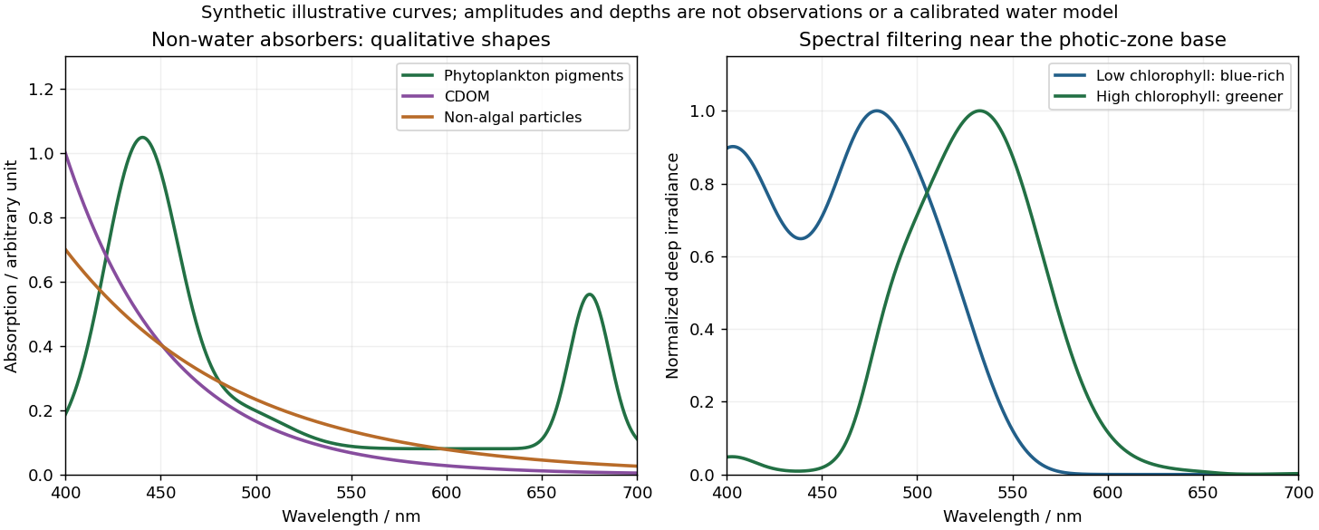
Qualitative marine absorption spectra and deep-light spectral filtering
. Original synthetic sketches: phytoplankton have blue and red pigment absorption bands, while CDOM and non-algal particles preferentially absorb short wavelengths. The right panel illustrates blue-rich deep light in low-chlorophyll clear water and a greener spectrum in more absorbing high-chlorophyll water. Spectra are normalized separately; curves and representative depths are illustrative, not observations or a calibrated radiative-transfer calculation. Details.
Phytoplankton pigments give prominent absorption maxima around and , associated with chlorophyll a, and a relative minimum in the green. Accessory pigments, including carotenoids and phycobilins, broaden or add absorption bands, so an assemblage is not just a pure chlorophyll-a solution. Intracellular packaging and cell size also modify the shape and absorption per unit pigment.
Coloured dissolved organic matter consists of dissolved light-absorbing organic compounds, often including humic material from terrestrial runoff and marine degradation. Its absorption is strongest at the blue/ultraviolet end and declines approximately exponentially toward red, without the narrow chlorophyll red peak. A common local parameterization is with positive spectral slope ; here is an optical slope, not gas saturation.
Non-algal particles include nonliving organic detritus, pigment degradation products and absorbing suspended mineral particles. Their aggregate absorption often also decreases approximately exponentially from blue to red, with variable slope and mineral-dependent departures. Suspended sediments can strongly scatter as well as absorb; bright sediment-laden water is not evidence of a red absorption peak. Chlorophyll-containing living cells belong in , so these categories should not be double counted.[ref]
Ocean optics protocols by NASA. Revision 4, Volume I, sections defining chlorophyll, CDOM and non-pigmented-particle absorption, and the importance of blue absorption to ocean-colour retrieval.
Healthy green vegetation has strong visible absorption in the blue, around –, and red, around –, mainly from chlorophylls and other photosynthetic pigments. Weaker absorption around green wavelengths produces a relative reflectance maximum near and the familiar green appearance. The red edge is the steep reflectance rise around –. In the near-infrared, weak pigment absorption and multiple scattering at internal leaf tissue–air interfaces produce high reflectance, commonly a broad plateau. This plateau is primarily a structural scattering effect, rather than chlorophyll reflecting the light.[ref]
Measuring vegetation by NASA Earth Observatory. “Normalized Difference Vegetation Index”: contrast between chlorophyll absorption of visible light and strong near-infrared scattering by leaf structure.
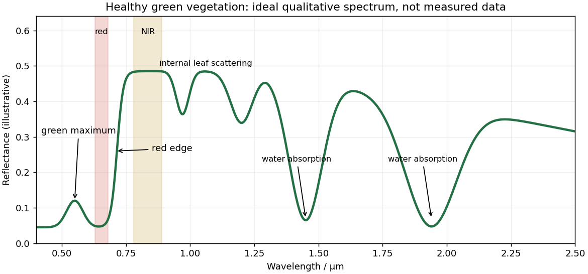
Qualitative reflectance spectrum of healthy green vegetation
. Original synthetic leaf spectrum with a green maximum, red absorption, red edge, near-infrared plateau and shortwave-infrared water absorption troughs. The curve is illustrative rather than measured; shaded bands exemplify red and near-infrared measurements for a vegetation index. Details.
Leaf water produces absorption troughs in the shortwave infrared, especially near and , with weaker features near and . Atmospheric water vapor also strongly absorbs near and , so satellite bands usually avoid the centres of those deep troughs. Drying, senescence, pigment loss and changes in canopy structure alter the spectrum; measured canopy reflectance also includes shadows and the soil background.
To highlight vegetation, assign the near-infrared band to the red display channel, red to green and green to blue: vigorously vegetated areas appear bright red in this conventional false-colour composite. Alternatively calculate the normalized difference vegetation index from atmospherically corrected, spatially registered red and near-infrared reflectances:Large positive values indicate a strong vegetation-like red/NIR contrast. For example, and give about . Mask clouds, their shadows and invalid low-signal pixels. Bare soil usually has a smaller contrast, while water often has negative values; fixed universal thresholds are inappropriate, and dense vegetation can saturate the index. A band ratio improves contrast but is not a direct count of plants or a unique measure of physiological health.[ref]
Measuring vegetation by NASA Earth Observatory. “Normalized Difference Vegetation Index”: normalized red/near-infrared contrast and interpretation of green vegetation.
For a surface-breaking reverse earthquake, expect a fault scarp or disrupted belt with the hanging wall displaced upward and commonly over the footwall. Small thrust faults, splays and backthrusts may repeat or override shallow deposits. Shortened soil, roads or other markers may buckle, while soft sediment can form pressure ridges and compression folds. Near the scarp, gravitational collapse and local bending can also produce tensile fissures and small normal faults; a reverse event need not create compression everywhere at the surface.
For a blind reverse rupture, expect coherent uplift and warping, newly tilted surfaces, or a fold scarp without a throughgoing primary fault offset. Small flexural-slip surfaces along bedding, secondary thrust faults or backthrusts, and opening cracks or small normal faults over the bent outer arc of the anticline can accompany the folding. Deformation may be distributed or localized at fold hinges. Thus a blind main fault does not imply an entirely unbroken ground surface, and secondary cracking must be distinguished from the principal rupture by its geometry and displacement pattern. Shaking-triggered landslides or soil liquefaction can accompany either case and should not automatically be mapped as primary tectonic rupture.[ref]
Blind-thrust mapping report by United States Geological Survey. Active fold scarps can arise through localized coseismic uplift and tilting without the principal thrust reaching the ground surface.
Interferometric synthetic-aperture radar compares the phase of coherent radar returns acquired before and after the earthquake from closely repeated viewing geometry. After image registration and removal of orbital and topographic phase, the residual interferometric phase constrains relative ground displacement along the radar line of sight. Defining positive toward the satellite and choosing the interferogram's phase order accordingly,One complete colour cycle therefore represents of relative line-of-sight displacement. Reversing the image order reverses the displayed phase sign. This observes a projection of both horizontal and vertical motion, not uplift alone.[ref]
Interferometry by NASA. NISAR mission explanation: remove orbit/topography contributions and interpret each complete fringe as half a radar wavelength of line-of-sight range change.
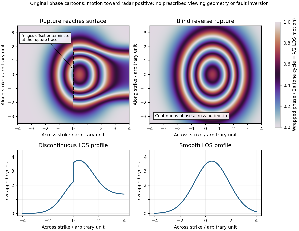
Qualitative interferograms for surface-breaking and blind reverse rupture
. Original wrapped-phase cartoons and corresponding unwrapped line profiles. They illustrate a displacement discontinuity versus a continuous deformation lobe, rather than numerical elastic fault models. The chosen phase sign is positive toward the radar. Every complete colour cycle represents half a radar wavelength of line-of-sight displacement; real fringe shape depends on viewing and rupture geometry. Details.
If the main reverse rupture reaches the surface, coherent areas on opposite sides can show a displacement step: fringes become offset, truncated or abruptly juxtaposed along a trace. A narrow belt can lose interferometric coherence where cracking, intense gradients or changed scatterers prevent reliable phase comparison. If the rupture remains blind, a simple elastic model instead predicts a continuous surface displacement field, often a broad asymmetric lobe with continuous curved or closed fringes and a smooth displacement gradient across the projected tip.
These are diagnostic tendencies, not a unique test. An ordinary wrapped-phase boundary is just a colour reset and is not itself a rupture. A step of an integer number of cycles can be hidden in wrapped phase; loss of interferometric coherence can hide either kind of near-field deformation. Vegetation, atmosphere and viewing direction also affect the pattern, and a displacement perpendicular to the radar line of sight is poorly sensed. Inspect coherence and unwrapped displacement, compare ascending and descending views where possible, and test a fault model against field observations. A sharp fold scarp above a blind rupture or a secondary shallow fault can also create localized gradients, so smooth fringes do not prove a buried tip and broken fringes alone do not prove a surface-breaking main rupture.
A blind reverse fault can transfer displacement into folding above its buried tip. Look for an asymmetric growing anticline, often with a steep forelimb and gentler backlimb, aligned with the inferred fault strike. A fault-propagation fold is one possible geometry; the fold alone does not uniquely establish the fault's depth or shape. Exposed older geological strata may be more steeply tilted than younger deposits, while growth strata thin and onlap toward an uplifting crest or fan in dip as deformation accumulates. Progressive unconformities and warped young deposits help distinguish active folding from an inherited fold.
In the landscape, seek an elongate uplifted ridge, warped or tilted river terraces and alluvial fans, localized fold scarps, incision across the rising structure, and drainage deflection or abandoned channels. Compare landforms of known relative ages: increasing deformation with age records repeated motion. Opposite fold limbs may show contrasting uplift and tilting, while active axial surfaces can concentrate deformation into narrow belts even without the main fault reaching the surface. Combine geological mapping and dating with seismic reflection or evidence from geodesy where available; an erosional ridge alone is not proof of a buried active reverse fault.[ref]
Fault slip must taper toward zero at the tips of an isolated finite fault, because its two sides are still joined beyond the tips. Accumulating more central displacement at a fixed short length steepens the displacement gradient and loads the surrounding rock, especially near the tips. In a simple elastic scaling, a fault of length and characteristic displacement requires a characteristic driving shear stress of orderwhere is the shear modulus and the omitted coefficient depends on fault geometry. The host's finite strength prevents this gradient increasing indefinitely. Tip damage, new cracking and linkage with neighboring segments allow the fault system to lengthen and accommodate more displacement without unbounded tip loading. Self-similar growth at roughly comparable driving stress consequently gives the approximate fault displacement-length scaling ; displacement also commonly increases toward the interior of a fault segment.[ref]
Fault displacement-length scaling by United States Geological Survey. 2008 study, abstract: approximately linear scaling for shearing discontinuities under constant driving stress, with near-tip stress limited by rock strength.
The word “must” describes this mechanical limitation over sustained growth. It is not a rule that every earthquake or every increment of accumulated fault slip lengthens the mapped trace: strong barriers, interactions and early establishment of fault length can permit substantial later displacement at nearly constant length.
Use compression-positive principal stresses . The two-dimensional Mohr's circle shown is the – circle: its horizontal axis is compressive normal stress , its vertical axis signed shear stress , its centre is , and its radius is . The intercepts and represent principal stress planes, on which shear stress vanishes. The top and bottom points have , the maximum shear stress.
In the rock sketch, is the counterclockwise angle from the direction to the plane's unit normal vector . Choose the clockwise unit tangent and define , with compression positive. Thus a positive produces a positive rotation on the plotted circle. The actual traction on the positive-normal face is . Reversing the tangent convention reverses the plotted shear sign, not the physical prediction.
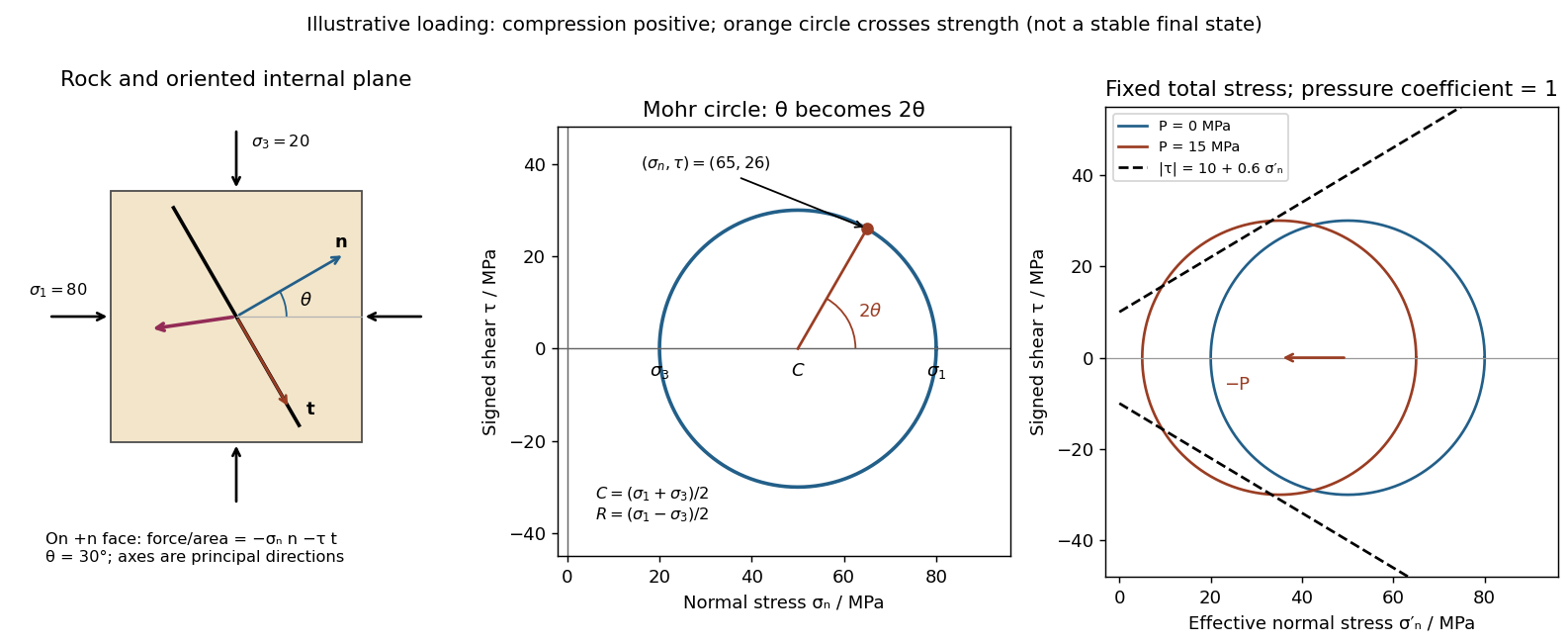
Stressed rock, Mohr circle, and the effect of pore pressure
. Original illustrative construction with principal compressions 80 and 20 MPa and a plane normal 30 degrees from the maximum compression. The circle point is approximately (65,26) MPa at twice that angle. A separate panel translates the circle left by 15 MPa pore pressure at fixed total stress, bringing it to a frictional failure envelope. Details.
In three dimensions there are circles for each pair of principal stresses. The largest is the – circle; a general three-dimensional plane requires all three principal stresses and its normal, rather than a single angle on this circle.
There are unlisted articles, also show them or only show them.