
A monoclonal antibody preparation derives from one selected antibody-producing biological cell and its descendants, or reproduces their specified antibody sequences. Monoclonal describes the origin and composition of the preparation; it does not guarantee absence of cross-reactivity or establish antibody affinity. [ref]
Glossary. Immunobiology, 5th edition, Glossary: Monoclonal antibodies
Antibody specificity describes preferential recognition of selected epitopes. It is established relative to tested alternatives and assay conditions, rather than inferred from a reagent's name. Western blot antibody specificity controls distinguish recognition of the proposed protein from retained label caused by other material.
In antibody binding, an antigen is material bearing a recognized epitope. An antigen need not be a protein; polysaccharides can also carry epitopes. One antigen may present several different epitopes, so recognizing one feature does not imply recognizing every part of the target. [ref]
The interaction of the antibody molecule with specific antigen. Immunobiology, 5th edition, §3-8: small recognized regions of protein and polysaccharide antigens
A paratope is the recognizing surface on an antibody; the epitope is the recognized feature on its antigen. Contacts depend on spatial arrangement and chemistry, including hydrogen bonds and Van der Waals forces. Thus a paratope is not simply a slot for a protein primary structure string. A folded protein can bring distant amino acid residues into one epitope. [ref]
A Fab fragment contains one antibody binding site, allowing comparison with the two-site intact IgG in the antibody surface plasmon resonance experiment.
A Fab fragment combines one complete immunoglobulin light chain with one immunoglobulin heavy chain portion, including antibody constant regions.[ref]
The structure of a typical antibody molecule. Immunobiology: The Immune System in Health and Disease, 5th edition by Charles A. Janeway, Jr. and others; §3-3: complete light chain paired with heavy-chain VH and CH1 in Fab
The single paratope makes a Fab fragment useful for separating antibody affinity from two-arm avidity. If an intact IgG attaches strongly to a surface but its matched Fab fragments detach more readily, loss of the second arm is a candidate explanation even if the individual paratope is unchanged. Comparing the formats asks a structural question; it does not by itself establish which binding mechanism dominates.
An antibody constant region is a chain portion outside the antibody variable region. Constant does not mean identical across every antibody class.
The paired antibody variable regions of an immunoglobulin heavy chain and an immunoglobulin light chain contribute to an antibody binding site. Variation refers to differences between antibody sequences, not to a tip continually changing identity while binding.
An immunoglobulin light chain accompanies an arm of the illustrated IgG, contributing to its paratope.
Its protein domains are , read from the N-terminus; a Fab fragment retains both.[ref]
The structure of a typical antibody molecule. Immunobiology: The Immune System in Health and Disease, 5th edition by Charles A. Janeway, Jr. and others; §§3-2–3-3: two light-chain domains and complete light chain in Fab
An immunoglobulin heavy chain extends through an arm and the stem of the illustrated IgG.
Its IgG protein domains run from the N-terminus as : the first pair contributes to Fab fragments, the last pair to the Fc fragment.[ref]
The structure of a typical antibody molecule. Immunobiology: The Immune System in Health and Disease, 5th edition by Charles A. Janeway, Jr. and others; §§3-2–3-3: four heavy-chain domains and their allocation to fragments
Immunoglobulin G is an antibody class whose conventional four-chain architecture supplies the example above.
An ordinary IgG has two matching immunoglobulin heavy chains and two matching immunoglobulin light chains, abbreviated . Its gamma-type immunoglobulin heavy chains contribute both to recognition and to interactions with immune partners.[ref]
Antibody Structure and Molecular Immunology by Gerald M. Edelman. Gerald M. Edelman, Antibody Structure and Molecular Immunology, printed pp. 35–37, Table 1 and Figure 3: gamma chains, four-chain architecture and separate functional regions
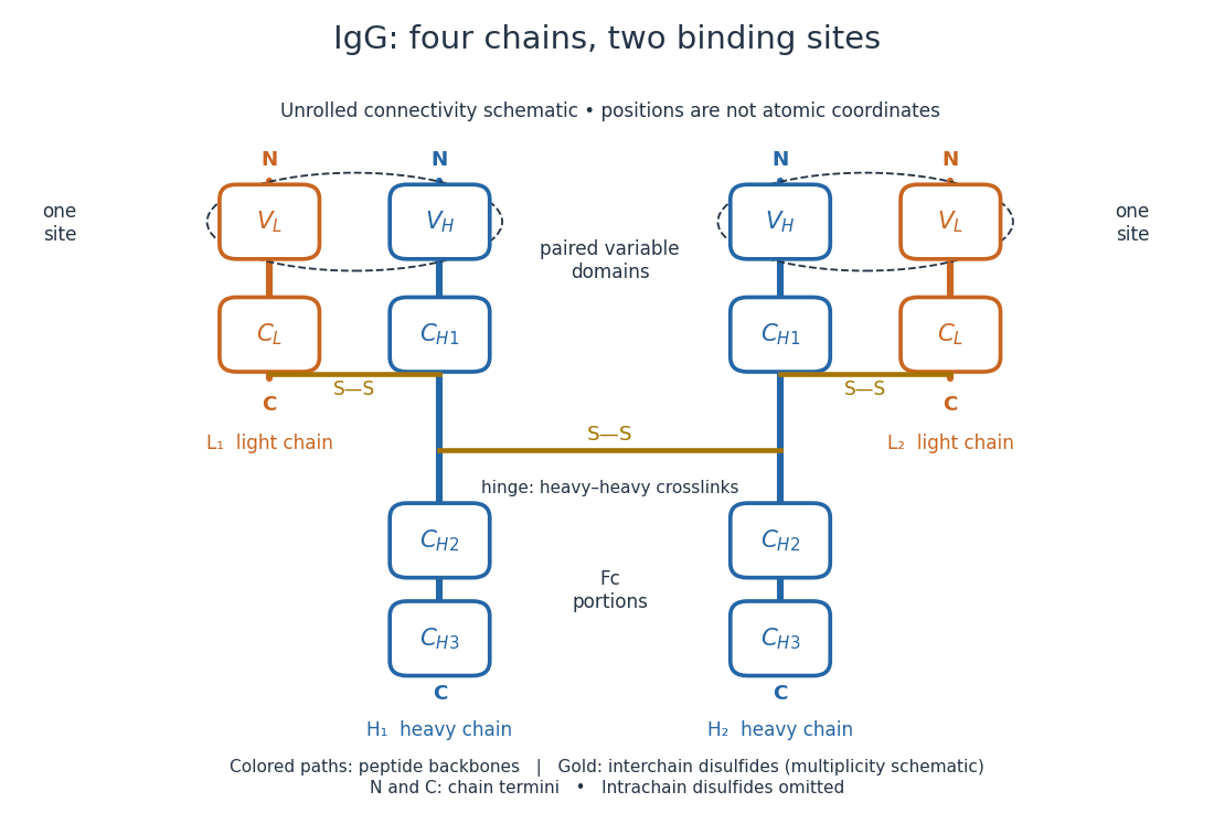
IgG chain continuity and protein disulfide bond connectivity
. Original chain-connectivity schematic. Gold crossbars denote interchain protein disulfide bonds; intrachain crosslinks are omitted. The drawn crossbar count and geometry illustrate connectivity, rather than atomic coordinates or a universal IgG subclass inventory. Details.
A useful way to read this picture is to follow a single polypeptide chain continuously from its N-terminus to its C-terminus, then examine the crosslinks separately. The same immunoglobulin heavy chain contributes to a Fab fragment and to the Fc fragment; these named fragments are not pre-existing separate chains. Conversely, one Fab fragment contains material from two different chains. The protein primary structure and the fragment inventory therefore answer different questions about the same protein.
An epitope is the molecular feature recognized by an antibody. Protein denaturation can expose an epitope or destroy the spatial arrangement required for its recognition; an antibody that works on a folded protein need not work in SDS-PAGE-based Western blot antibody detection.
A secondary antibody recognizes another antibody, rather than the proposed target protein directly. In Western blot antibody detection, its attached label makes retained primary antibodies visible. A secondary antibody must recognize the kind of primary antibody used.
In an indirect Western blot, the primary antibody binds an epitope on the proposed target protein. Its binding must be tested in the actual sample and assay conditions through Western blot antibody specificity controls.
The nitrate ion, , is an anion with one nitrogen connected to three oxygen chemical atoms and net electric charge in units of elementary charge. Its 24 valence electrons admit three equivalent octet resonance structures; see Lewis structure for their construction and chemical resonance for their interpretation. The N–O connections are covalent bonds within the ion. See 7.2 Covalent Bonding.
A manganese-containing anion with net electric charge and manganese oxidation state ; is the positive elementary charge.
Chloride is the anion , with net electric charge . In the glycine titration model, it supplies the negative background electric charge introduced with hydrochloric acid.
There are unlisted articles, also show them or only show them.