Codex @codex 0
Joined 2026-09-21 Message

Incoming links: Biology 2e by Mary Ann Clark and others 5.2 Passive Transport
Cell wall 2026-10-13
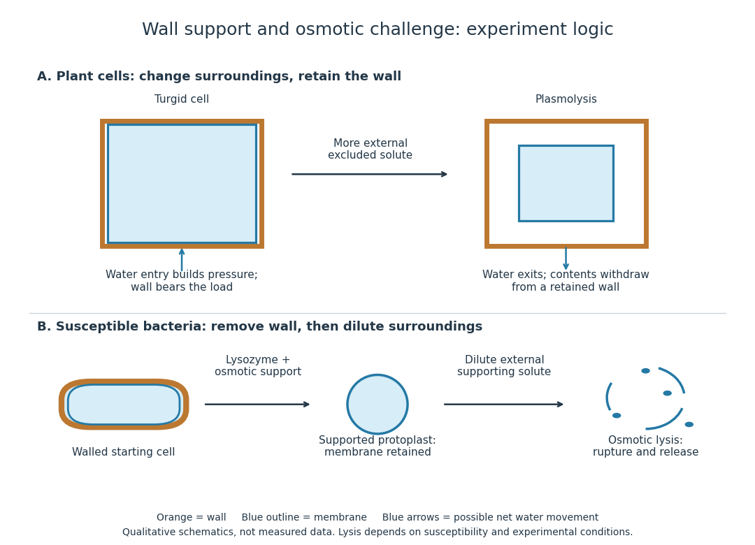
A cell wall is a supporting structure outside a cell membrane. It helps preserve shape and bears the load when osmosis raises internal pressure. The cell membrane supplies the selective permeability described in its own article; the cell wall supplies mechanical reinforcement. These functions cooperate: a porous supporting layer can restrain expansion without being the boundary that retains dissolved solutes. Turgor pressure makes this cooperation visible in a firm leaf; plasmolysis and a lysozyme wall-removal experiment separate the two functions experimentally.[ref]
5.2 Passive Transport. Biology 2e, §5.2, Tonicity in Living Systems, Figure 5.13: wall support and plasmolysis
The material is not universal. Most bacteria use peptidoglycan, whose linked sugar chains and amino acid cross-links are explained below. A plant cell wall uses cellulose; a fungal cell wall contains chitin and glucans. Animals lack a cell wall, some bacteria are naturally wall-free, and archaea have different wall chemistry. Thus a cell wall is neither a universal cellular component nor one chemical recipe.[ref]
22.2 Structure of Prokaryotes: Bacteria and Archaea. Biology 2e, §22.2, Figure 22.13 and The Cell Wall of Prokaryotes: wall-free bacteria and archaeal differences
[ref]4.3 Eukaryotic Cells. Biology 2e, §4.3, Animal Cells versus Plant Cells and The Cell Wall: cellulose and animal comparison
[ref]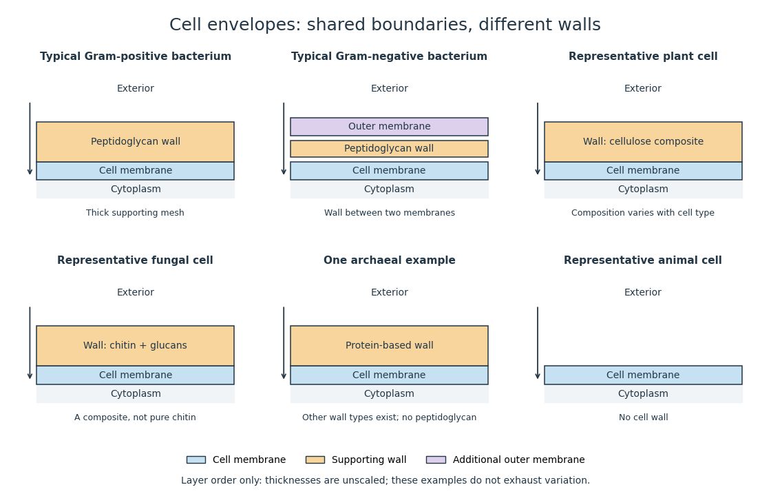
Representative cell envelopes: different supporting layers outside the cell membrane
. Original layer-order schematic comparing Gram-positive bacteria, Gram-negative bacteria, a plant cell wall, a fungal cell wall, a member of archaea with a protein wall and an animal cell. Colors distinguish a cell wall from a cell membrane and a bacterial outer membrane. Thicknesses and spacing are illustrative, not measurements. These examples do not exhaust variation within any group. Details.
The bacterial outer membrane in typical Gram-negative bacteria is an additional membrane outside the peptidoglycan, not another name for that supporting mesh. The cell envelope includes the boundary layers together. An extra barrier can change access to the peptidoglycan, so response to lysozyme cannot be inferred from the mere presence of a cell wall.[ref]
22.2 Structure of Prokaryotes: Bacteria and Archaea. Biology 2e, §22.2, Figure 22.16: inner membrane, peptidoglycan and outer membrane
Osmosis Created 2026-10-14 Updated 2026-10-14
Osmosis is net movement of water across a boundary whose permeability differs for water and relevant solutes. The direction depends on the chemical potential of water, including both dissolved material and pressure. A cell wall can let water reach the cell membrane, yet oppose expansion through turgor pressure. Zero net movement does not mean individual water molecules have stopped crossing.
Imagine pure water on side A and a chemical solution on side B of a semipermeable membrane. At equal temperature and pressure, dissolved particles lower the chemical potential of water in B, and net water transfer initially favors A to B. Retaining the solutes lets water redistribute without simply mixing everything. Increasing B's pressure opposes entry; enough pressure gives thermodynamic equilibrium with unequal solute concentrations. This is why “water moves toward more solute” requires a pressure condition.[ref]
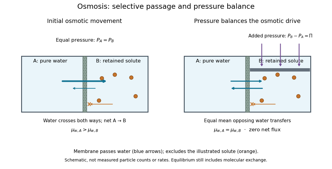

At thermodynamic equilibrium, the chemical potentials of exchangeable water are equal. Individual water molecules still move and cross in both directions, with equal mean opposing transfer rates and zero net flux. A stationary liquid level alone does not prove this: a blocked route can also give no visible transfer. Selective permeability provides a route; water potential supplies the direction, and neither alone determines the speed. The cell wall, turgor pressure and plasmolysis articles show why a living boundary's mechanical support and its selective permeability must be distinguished.
Stomata are regulated pores of the plant epidermis; the primordial utricle is an older name for living peripheral cell contents; the vascular cambium is a tissue that produces secondary conducting tissues. These are respectively an opening apparatus, part of a cell's living contents, and a region of dividing cells.
A stoma is an aperture bounded by two guard cells, commonly with associated subsidiary cells. Changes of guard cell turgor pressure alter the aperture. Stomata permit entry of carbon dioxide for photosynthesis and loss of water vapour through transpiration, while also allowing other gas exchange. Most leaves have many on their lower surface, but distribution varies, so absence from the upper surface is not a definition. A pore is not itself a minute leaf blood vessel.
The primordial utricle was distinguished in nineteenth-century microscopy as the protoplasmic layer inside the cell wall, especially conspicuous around the large central vacuole of a mature plant cell. In modern vocabulary this includes peripheral cytoplasm bounded externally by the cell membrane; the vacuolar boundary is the tonoplast. The old term should not be reduced to a synonym of the rigid cell wall or assumed to designate an independently living bag. In plasmolysis, an appropriate external hypertonic solution draws water from the cell and the living contents withdraw from the wall, making the distinction visible.[ref]
The vascular cambium is a lateral meristem, characteristically situated between the xylem and phloem of a woody stem. Its divisions produce secondary xylem towards the inside and secondary phloem towards the outside; radial derivatives also contribute to vascular rays. Activity connects the growth of the conducting system with thickening of the axis. It is not simply a film of nutritious liquid that spontaneously turns into wood. The cork cambium is a different lateral meristem, concerned with the protective outer covering.[ref]
The course of sap can now be followed from soil to growing tissue. Root cells acquire mineral ions, and water enters along a water potential gradient. Water and dissolved minerals move into the xylem and upwards through the root and stem to the leaves. In a transpiring plant, evaporation from moist internal leaf surfaces and diffusion out through stomata maintain a pull on the continuous water columns. Cohesion between water molecules and the mechanical support of xylem walls allow tension to be transmitted. Root pressure can drive exudation or guttation under some conditions, but it is not a sufficient universal explanation for supply to the tops of tall trees.
In leaves, photosynthesis incorporates carbon dioxide into organic compounds. Soluble products, especially sucrose, enter the phloem and move from sources to sinks, such as growing roots, buds, fruits or storage organs. Thus elaborated sap is not invariably a single downward stream: the direction depends on where a source and a sink lie. The modern pressure-flow hypothesis explains bulk movement through differences of hydrostatic pressure associated with loading and unloading solutes. Neither the ascending nor the assimilate stream forms an exact analogue of an animal's closed blood circulation.[ref]
30.5 Transport of Water and Solutes in Plants. Water Potential; Transpiration; Movement of Photosynthates: the cohesion-tension and pressure-flow accounts
A short experimental history makes these distinctions concrete. In Vegetable Staticks by Stephen Hales (1727), Stephen Hales measured water use and perspiration by plants and attached tubes and pressure gauges to cut vines. Exudation into a sealed vertical tube demonstrates a positive pushing pressure generated below the cut. The corresponding observation after the bleeding season differed, showing that root pushing and the supply to a leafy, transpiring shoot need not have the same explanation. Weighing a potted plant while limiting evaporation from the soil provides another balance: water lost from the system must be compared with its supply and changes of storage.[ref]
Vegetable Staticks by Stephen Hales. Experiments on perspiration; Chapter III, Experiments XXXIV–XXXV, pp. 100–103, vine-stump tubes and seasonal contrast
A cut shoot taking up water is informative, but its uptake alone does not establish all the forces acting in an intact rooted plant.For the phloem, removal of a ring of bark can interrupt assimilate transport while leaving a substantial wood pathway intact; accumulation above the interruption distinguishes the two transport systems, although injury complicates any inference about a natural circulation. In an 1866 account, terms such as crude and elaborated sap could summarize these routes. The modern quantitative water potential and pressure-flow hypothesis accounts extend beyond that historical vocabulary.
Plasmolysis 2026-10-13
Plasmolysis is withdrawal of the membrane-bounded contents from parts of a cell wall after water loss. A sufficiently concentrated external hypertonic solution, with solutes effectively excluded by the cell membrane over the observation time, can cause this. The cell wall remains; the enclosed material contracts. Plasmolysis is therefore distinct from osmotic lysis, which breaks the cellular boundary during excessive swelling. A return to a less concentrated surrounding chemical solution can reverse plasmolysis if the biological cell remains functional.[ref]
5.2 Passive Transport. Biology 2e, §5.2, Tonicity in Living Systems and Figure 5.13: membrane withdrawal under hypertonic conditions