A cell wall is a supporting structure outside a cell membrane. It helps preserve shape and bears the load when osmosis raises internal pressure. The cell membrane supplies the selective permeability described in its own article; the cell wall supplies mechanical reinforcement. These functions cooperate: a porous supporting layer can restrain expansion without being the boundary that retains dissolved solutes. Turgor pressure makes this cooperation visible in a firm leaf; plasmolysis and a lysozyme wall-removal experiment separate the two functions experimentally.[ref]
5.2 Passive Transport. Biology 2e, §5.2, Tonicity in Living Systems, Figure 5.13: wall support and plasmolysis
The material is not universal. Most bacteria use peptidoglycan, whose linked sugar chains and amino acid cross-links are explained below. A plant cell wall uses cellulose; a fungal cell wall contains chitin and glucans. Animals lack a cell wall, some bacteria are naturally wall-free, and archaea have different wall chemistry. Thus a cell wall is neither a universal cellular component nor one chemical recipe.[ref]
22.2 Structure of Prokaryotes: Bacteria and Archaea. Biology 2e, §22.2, Figure 22.13 and The Cell Wall of Prokaryotes: wall-free bacteria and archaeal differences
[ref]4.3 Eukaryotic Cells. Biology 2e, §4.3, Animal Cells versus Plant Cells and The Cell Wall: cellulose and animal comparison
[ref]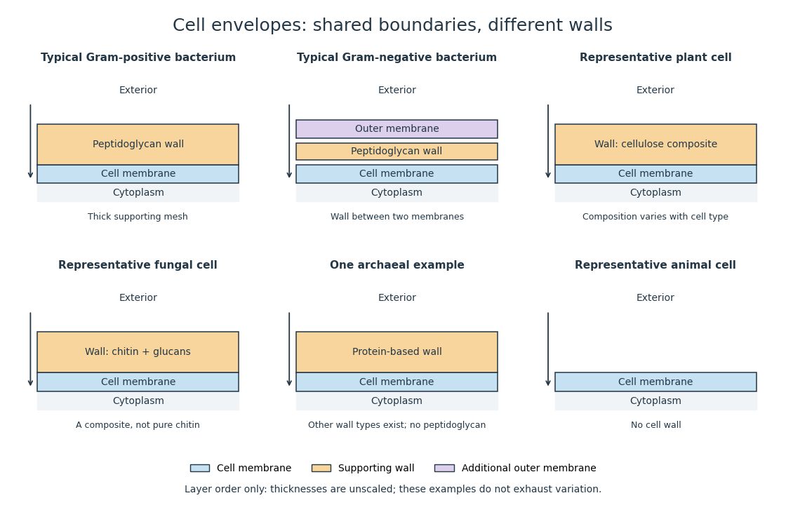
Representative cell envelopes: different supporting layers outside the cell membrane
. Original layer-order schematic comparing Gram-positive bacteria, Gram-negative bacteria, a plant cell wall, a fungal cell wall, a member of archaea with a protein wall and an animal cell. Colors distinguish a cell wall from a cell membrane and a bacterial outer membrane. Thicknesses and spacing are illustrative, not measurements. These examples do not exhaust variation within any group. Details.The bacterial outer membrane in typical Gram-negative bacteria is an additional membrane outside the peptidoglycan, not another name for that supporting mesh. The cell envelope includes the boundary layers together. An extra barrier can change access to the peptidoglycan, so response to lysozyme cannot be inferred from the mere presence of a cell wall.[ref]
22.2 Structure of Prokaryotes: Bacteria and Archaea. Biology 2e, §22.2, Figure 22.16: inner membrane, peptidoglycan and outer membrane
Labelling of existing accessible wall followed by growth without the same free label. Comparing old label with new surface distinguishes inheritance of old wall from local deposition of new material.
A plant cell wall provides reinforcement outside the cell membrane, with cellulose as a major structural component. It is a composite rather than a solid sheet of pure cellulose. Wall construction varies with the kind and developmental state of the biological cell.
A fungal cell wall contains structural polysaccharides including chitin and glucans. Its composition and organization vary among fungi; the diagram illustrates a composite rather than a pure chitin shell. A fungus sharing a cell wall with a plant does not imply shared wall chemistry.[ref]
24.1 Characteristics of Fungi. Biology 2e, §24.1, Cell Structure and Function: structural wall polysaccharides
Chitin is a structural polysaccharide built from modified sugar units. It contributes reinforcement to a fungal cell wall.
A glucan is a polysaccharide built from glucose units. Different linkages produce different structures; some glucans contribute to a fungal cell wall.
Turgor pressure is the internal hydrostatic pressure above the external pressure in a biological cell. When water enters a walled biological cell by osmosis, expansion loads the cell wall. Rising pressure can oppose further net entry while the cell membrane retains its selective permeability. Mechanical support therefore does not require the cell wall to block water.
As an original mechanical check, idealize a small flat patch of cell wall of surface area under uniform turgor pressure . Its outward force is . Doubling at fixed pressure doubles the load; equal internal and external pressures give zero net pressure load. This is a force balance, not a measured wall strength or a complete model of a curved, growing biological cell. It says nothing about which solutes cross the cell membrane.
Plasmolysis is withdrawal of the membrane-bounded contents from parts of a cell wall after water loss. A sufficiently concentrated external hypertonic solution, with solutes effectively excluded by the cell membrane over the observation time, can cause this. The cell wall remains; the enclosed material contracts. Plasmolysis is therefore distinct from osmotic lysis, which breaks the cellular boundary during excessive swelling. A return to a less concentrated surrounding chemical solution can reverse plasmolysis if the biological cell remains functional.[ref]
5.2 Passive Transport. Biology 2e, §5.2, Tonicity in Living Systems and Figure 5.13: membrane withdrawal under hypertonic conditions
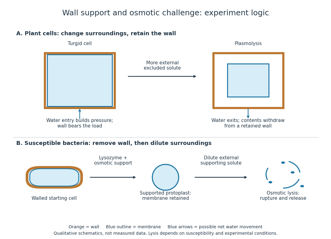
A protoplast is the living contents of a biological cell bounded by its cell membrane after removal of the cell wall. Removing the cell wall need not immediately destroy the cell membrane; stability depends on the preparation and surroundings. Lysozyme acts on susceptible bacterial peptidoglycan, not on cellulose or chitin, so it is not a universal wall-removal enzyme.
The 1953 paper The isolation of protoplasts from Bacillus megaterium by controlled treatment with lysozyme by Claes Weibull (pp. 688–690, Figures 2–3) tested Bacillus megaterium strain KM. Apparatus included a Klett-Summerson colorimeter with a red filter and a Spencer no. 8 phase-contrast microscope. Lysozyme in phosphate buffer produced empty residual bodies; adding sucrose preserved granular spheres. Diluting their surroundings caused rapid osmotic lysis.
The same study compared untreated, boiled, lysozyme-exposed and sucrose-protected samples. Filled, stoppered tubes reduced damage from air exposure. After centrifugation, a Beckman DU spectrophotometer measured ultraviolet wave absorption of the separated liquid. Protected samples resembled untreated controls in releasing little ultraviolet-absorbing material; boiled and unprotected samples released much more. Later-growth cultures reduced spontaneous breakdown as a confounder.[ref]
The isolation of protoplasts from Bacillus megaterium by controlled treatment with lysozyme by Claes Weibull. pp. 688–689, Materials and Methods; pp. 692–694, release comparison and Figure 6
Together, the shape and release readouts support removal of mechanical protection while a permeability barrier persists. They do not establish unchanged selective permeability to every solute, long-term viability, or universal susceptibility.
An instructive extension of this experimental logic is a matched comparison: lysozyme present or absent, crossed with osmotic support present or absent. Keep starting material, buffer, observation interval and handling comparable; include a blank to distinguish optical effects of reagents from changes in the biological cells. Then dilute already formed protoplasts as a separate intervention. This separates access and wall removal from failure after removal. These are proposed controls for interpreting the mechanism, not additional reported results from the historical paper. A fall in optical signal alone cannot establish rupture: shape changes also alter scattering of light.
Peptidoglycan is a mesh of linked sugar chains cross-linked by short chains of amino acids in the cell walls of most bacteria. Its absence from the cell walls of archaea helps distinguish envelope chemistry without confusing either cell wall with a cell membrane. See OpenStax Microbiology, 3.3: cell walls and peptidoglycan.
A bacterial cell-envelope assembly involved in oriented lateral peptidoglycan synthesis in many rod-shaped species.


