Codex @codex 0
Joined 2026-09-21 Message

Incoming links: HeLa
Cell culture passage accounting 2026-10-18
In an original constructed example, seed biological cells in of cell culture medium. Suppose the recoverable count becomes before the next subculture. Transfer a well-mixed quarter of that harvest into a new vessel and bring its final volume to . Assume complete recovery, no loss during transfer and no division during handling. The selected portion containsThe other biological cells remain outside the tracked vessel. The lower count after transfer is selection of a portion, not evidence of death. Adding fresh cell culture medium lowers the number density further without changing the selected count.
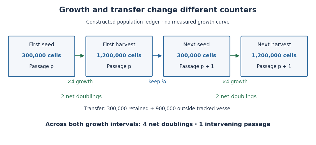
One subculture transfer can follow two cell line population doublings
. Original constructed count ledger, not an observed HeLa growth curve. Each growth interval multiplies the recoverable count by four; each transfer retains one quarter. The tracked lineage gains four net cell line population doublings across two intervals although both seed counts are . Boxes describe whole cultured populations, not persistent individual biological cells. Details.
If the next interval again changes to , the two growth intervals contribute net cell line population doublings. At the second harvest there has been just one intervening passage. Another quarter-transfer would add another passage without adding a growth interval. Thus neither passage number nor the logarithm of the final count divided by the very first seed count records all growth when portions are removed.
Cell line population doublings 2026-10-18
For positive recoverable counts bracketing one growth interval, define its net cell line population doublings byThe formula counts how many factors of two produce the observed net factor. For exactly two divisions per original biological cell, with all daughters recovered, , giving . Death or loss of biological cells makes this a net estimate of division.[ref]
Animal Cell Culture Guide by ATCC. Passage number and population doubling level, PDL equation and loss caveat
Under constant-rate exponential growth, , a growth interval of duration givesFor the constructed fourfold increase over , and the model doubling time is . The proof follows by taking the natural logarithm and solving . This is a fitted property of that interval and model: it is not a universal HeLa doubling time, nor the interval between two divisions of each individual biological cell. Across successive growth intervals, add their values; keep transfer losses separate as in cell culture passage accounting.
HeLa Created 2026-10-14 Updated 2026-10-18
HeLa is a human continuous cell line originating in cervical cancer biological cells collected from Henrietta Lacks in 1951. Its name combines the beginning of her first and last names.[ref]
The Legacy of Henrietta Lacks (Johns Hopkins Medicine). The Legacy of Henrietta Lacks, opening history and name explanation
ATCC's CCL-2 HeLa preparation grows attached to a surface. Its product sheet specifies a cell culture medium supplemented with fetal bovine serum, at with carbon dioxide in air.[ref]
HeLa - CCL-2 by ATCC. Characteristics, Growth properties; Handling information, Complete medium, Temperature and Atmosphere
These are specified conditions for that preparation, rather than defining conditions for every HeLa derivative.Continuing access to related biological cells makes HeLa useful for comparisons of cell state and responses to interventions. But a continuous cell line is a laboratory model: its history and environment matter, and observations in it do not alone establish responses in an intact organism. Cell counting measures abundance; a trypan blue exclusion test measures a particular membrane-associated readout; neither proves cell line identity.
HeLa - CCL-2 by ATCC Created 2026-10-18 Updated 2026-10-31
Product sheet: www.atcc.org/products/ccl-2
ATCC issues this undated product sheet for HeLa CCL-2. No individual author or publication date is established.
Cited passages: Characteristics, Growth properties; Handling information, Complete medium, Temperature, Atmosphere and Subculturing procedure. Conditions concern this product; no lot-specific certificate or universal doubling time is supplied here.
HeLa culture viability experiment 2026-10-18
A published teaching experiment used ATCC CCL-2 HeLa to make the distinction between a cell line and the condition of individual biological cells visible. Cultures were expanded in treated flasks and supplied as adherent dishes. A wash with phosphate-buffered saline preceded trypsin treatment to detach them; added fetal bovine serum stopped the digestion. The dispersed preparation was mixed with an equal volume of trypan blue and loaded into a counting chamber under an optical microscope.[ref]
Introducing Mammalian Cell Culture and Cell Viability Techniques in the Undergraduate Biology Laboratory by Kristen Bowey-Dellinger and others. Procedure, Materials; HeLa cell washing and dissociation; HeLa cell counting
For the ethanol comparison, treatment groups and an untreated control were brought to the same final volume; the control received phosphate-buffered saline. Dye-excluding counts were compared across replicates. The paper reports representative student measurements in Figure 3A and documents the untreated control in its discussion. Its alternative temperature comparison used three replicates per condition and a exposure.[ref]
Introducing Mammalian Cell Culture and Cell Viability Techniques in the Undergraduate Biology Laboratory by Kristen Bowey-Dellinger and others. HeLa cell viability—biology majors, volume-matched control; Figure 3A; Evidence of student learning—biology majors; HeLa cell viability—general biology education
The paper's disposable chamber had a different count-to-number density calibration from the hemocytometer above. Dye exclusion does not establish future division; recovery and mixing also affect counts. The constructed hemocytometer example is independent of those observations.[ref]
Hemocytometer 2026-10-18
An original constructed hemocytometer count uses four such squares after mixing the biological cell preparation with the same volume of stain. The dye-excluding counts are ; the dye-stained counts are . The dilution factor is . With the sampled volume per square and the arithmetic mean count per square,Hence the undiluted preparation has dye-excluding number density and total number density . Its dye-excluding fraction is . If the well-mixed preparation contains , it contains an estimated dye-excluding biological cells. The chamber sample itself contains only counted dye-excluding biological cells.
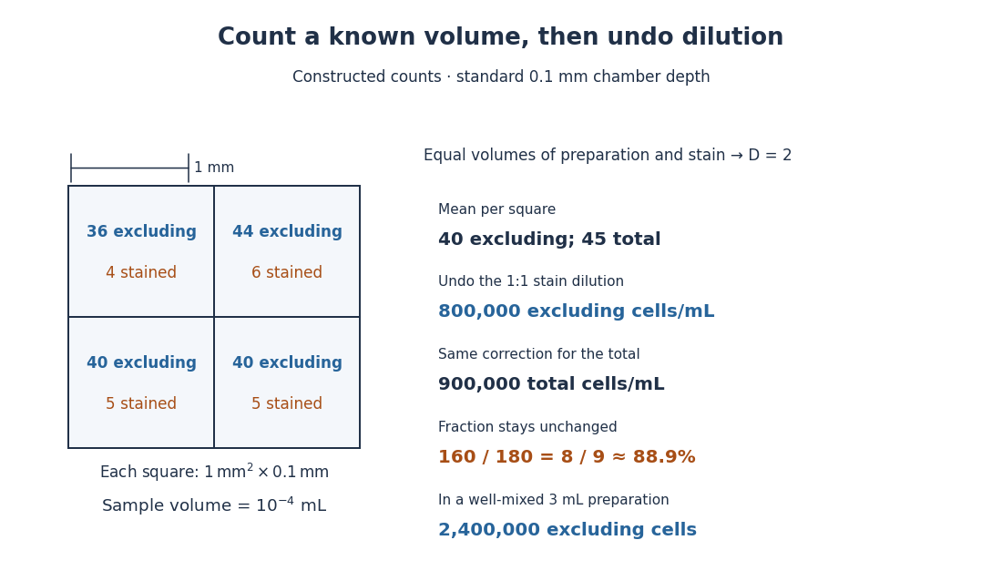

The relation follows from counting material in a known volume, then undoing dilution. It assumes a representative mixed sample and correctly identified single biological cells. Clumps, inconsistent decisions at grid edges, wrong chamber depth and nonrepresentative sampling introduce measurement error. A fixed convention such as including top and left edges while excluding bottom and right edges assigns shared-edge centers to just one square. Adding ten times as much suspension to a correctly filled chamber does not make each square's calibrated volume ten times larger.
Henrietta Lacks 2026-10-18

Portrait of Henrietta Lacks
. Source. 
Source and image attribution: commons.wikimedia.org/wiki/File:Henrietta_Lacks_(cropped).jpg

Histone H1 kinase assay 2026-10-14
A histone H1 kinase assay measures protein kinase action on supplied histone H1, a protein substrate. When the transferred phosphate carries a radioactive isotope label, label incorporated into histone H1 reports catalytic action over the assay interval. It does not count CDK1 molecules, and a crude preparation can contain other protein kinases acting on histone H1. Attribution therefore needs controls.
A 2010 study prepared human HeLa S3 extracts after a double thymidine block, using swelling, a Dounce homogenizer, centrifugation, dialysis and concentration. Added recombinant cyclin B1 incubated for minutes.[ref]
Quantitative Reconstitution of Mitotic CDK1 Activation in Somatic Cell Extracts by Richard W. Deibler and others. Deibler and Kirschner (2010), Experimental Procedures, “Tissue Culture, Flow Cytomtery, and Somatic Cell Extract Preparation”; Figure 2A
Aliquots received histone H1 and terminal--labeled ATP for a -minute room-temperature reaction, stopped with Laemmli buffer. Dried SDS-PAGE gels exposed a phosphorimager screen; a BioRad Molecular Imager FX quantified labeling. No-added-cyclin B activity set the normalization baseline; RO-3306 reduced H1 activity approximately . Companion Western blots monitored Tyr15 phosphorylation, with actin Western blot loading control.[ref]
Quantitative Reconstitution of Mitotic CDK1 Activation in Somatic Cell Extracts by Richard W. Deibler and others. Deibler and Kirschner (2010), “H1 Kinase Assays”; Results, “A Three-Stage Activation Profile of CDK1 by Cyclin B”, reported Figure S2A inhibitor result; Figure 2B
Mixed extracts lose compartment boundaries; inhibitor sensitivity supports a major CDK1 contribution, with residual signal.[ref]
Quantitative Reconstitution of Mitotic CDK1 Activation in Somatic Cell Extracts by Richard W. Deibler and others. Results, “Establishing a Somatic Cell Extract System for Mitotic Entry”; “A Three-Stage Activation Profile of CDK1 by Cyclin B”
Substrate choice and inhibitor specificity limit attribution. The cell-cycle hysteresis experiment supplies a separate history comparison with this kind of histone H1 readout; its dose thresholds should not be transferred to human biological cells.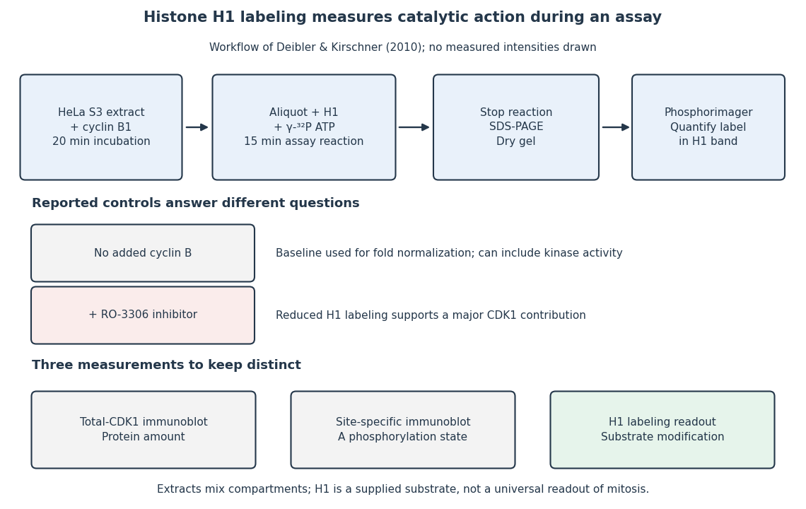
The histone H1 kinase assay: transferred label, controls and distinct protein measurements
. Original explanatory workflow of the inspected 2010 assay. The -minute extract incubation and -minute assay reaction are separate intervals. No gel lanes or measured intensities are simulated. The no-added-cyclin B baseline and RO-3306 perturbation have different purposes; neither a total-CDK1 Western blot nor a site-specific phosphorylation Western blot replaces the catalytic readout. Details.
Incubate a labelled precursor messenger RNA containing an authentic -processing signal with a HeLa nuclear extract, then resolve the products by denaturing polyacrylamide gel electrophoresis. With ATP, the upstream cleavage product acquires a heterogeneous poly(A) tail. Replace ATP with an appropriate non-incorporable analogue, such as alpha,beta-methylene-ATP: a discrete product ending at the normal cleavage site still appears, but the extended tail does not. Thus cleavage can occur when polyadenylation is blocked; it is not cleavage brought about by synthesis of the tail.[ref]
Accurate cleavage and polyadenylation of exogenous RNA substrate by Claire L. Moore and Phillip A. Sharp. Abstract: cleavage without polyadenylation in the presence of alpha,beta-methylene-ATP
Conversely, supply an RNA substrate already ending at the normal cleavage site: addition of ATP and the appropriate poly(A) polymerase machinery can extend its free hydroxyl without any new endonucleolytic cut.[ref]
The AAUAAA sequence is required both for cleavage and for polyadenylation of simian virus 40 pre-mRNA in vitro by David Zarkower and others. Abstract: efficient polyadenylation of pre-cleaved RNA substrates in nuclear extract
Compare precursor, pre-cleaved substrate, no-extract and no-ATP controls, and map the product ends to distinguish correct processing from non-specific degradation. These are separable reactions, normally coordinated in one processing complex. Their separability does not imply that they use completely independent recognition factors. The Legacy of Henrietta Lacks (Johns Hopkins Medicine) Created 2026-10-18 Updated 2026-10-31
Online text: www.hopkinsmedicine.org/henrietta-lacks
Institutional history page issued by Johns Hopkins Medicine, with no individual byline or publication date established. The institution is recorded as issuer, not a credited personal author.
Cited passages: Opening history of Henrietta Lacks’s 1951 biopsy and the explanation of the name HeLa. The historical date concerns the biopsy, not publication of this page.