Topics (384k) Articles (398k) Users (410) Discussions (242) Comments (383) Files (13k) New article

A co-receptor is a receptor that acts with another receptor in a specified recognition, signaling or entry process. The word describes a functional partnership: which partner is needed depends on the biological system and the measured response. It does not mean that every receptor involved must have the same normal ligand or physiological role.
A revealing example is entry by certain HIV-1 isolates. H. Deng and colleagues reported in 1996 that CD4 alone did not explain entry requirements. The chemokine receptor then called CC-CKR-5, now CCR5, supported entry mediated by the envelopes of tested primary macrophage-tropic HIV-1 strains. The comparison with T-cell-line-adapted viruses showed why a co-receptor must be identified in a particular virus–cell context.[ref]
Identification of a major co-receptor for primary isolates of HIV-1 by H. Deng and others. Abstract, Nature 381 (1996), pp. 661–666; DOI 10.1038/381661a0.
The conceptual experiment separates recognition from sufficiency: possessing one required cell-surface receptor does not ensure that a cell has all the components needed for entry. Nor does identifying entry cofactors by itself establish every later step of a productive infection.
Tumor necrosis factor receptor 2, abbreviated TNFR2, is a cell-surface receptor for tumor necrosis factor. In humans the encoding gene is TNFRSF1B; the protein is also known as the p75 receptor or CD120b. It is distinct from the p55 receptor, TNFR1. A cytokine and its receptor are different molecules: one supplies a signal and the other provides a binding and response apparatus.[ref]
The transmembrane protein has an extracellular ligand-binding portion, a membrane-spanning segment and an intracellular portion involved in signal transduction. Binding at the outside and a downstream cellular response are separate experimental observations. A receptor-binding measurement alone does not establish whether a particular cell proliferates, survives or dies.
A motivating molecular experiment used direct expression screening: a human lung-fibroblast cDNA library was screened with radiolabeled tumor necrosis factor alpha. C. A. Smith and colleagues isolated a clone encoding an integral membrane protein of 461 amino acids; the expressed receptor also bound TNF-beta. Its predicted cysteine-rich external region connected it to a broader receptor family.[ref]
A receptor for tumor necrosis factor defines an unusual family of cellular and viral proteins by C. A. Smith and others. Abstract, expression library, radiolabeled ligand and predicted extracellular region
This combines a functional binding screen with sequence evidence, rather than identifying a receptor from a name alone.Etanercept provides a useful engineered comparison. It joins the extracellular ligand-binding portion of the human p75 receptor to an Fc region of immunoglobulin G subclass 1, making a soluble dimeric fusion protein.[ref]
Enbrel prescribing information in DailyMed. Sections 11 and 12.1, p75 extracellular region/Fc composition and ligand interception
This retains a ligand-catching surface while omitting the native membrane attachment and intracellular signaling machinery. Its ligand capture therefore does not mean that it reproduces a membrane TNFR2 signal.An experimental comparison can measure binding and cellular response separately, using otherwise matched cells with and without the receptor and controlling for other TNF-binding receptors. A change in labeled-ligand binding tests one part of the mechanism; a change in a specified downstream readout tests another. The receptor's presence alone cannot substitute for either measurement.
Area-coverage antialiasing assigns an edge pixel a value based on the fraction of its footprint covered by a geometric shape. In a simple rasterization model with constant foreground value , constant background value , and a uniformly weighted pixel region , let be the shape. ThenEquivalently, is the average of the shape's indicator function over . This is a spatial average: a partially covered opaque shape can produce an intermediate pixel value without its material being transparent. Filtering the image over a finite region reduces the abrupt changes produced by deciding coverage at one sample point.[ref]
For a worked example, take the unit-square pixel and a white shape covering over a black background. The diagonal divides the square into equal areas, so and the pixel gets half the white value. A single center sample lies exactly on the edge and must choose one side by a convention; a tiny edge movement can flip that binary decision. The covered area instead changes continuously. The arithmetic applies to values proportional to light, before a nonlinear display encoding.
Exact geometric coverage and multiple sample tests are two ways of estimating this average; a finite set of samples can miss a sufficiently thin shape. Uniform footprint averaging also has limits: its abrupt filter boundary does not remove every high-frequency component, so it reduces aliasing without guaranteeing a perfectly band-limited image.[ref]
Physically Based Rendering: From Theory to Implementation (4th edition) by Matt Pharr and others. section 8.8.1, box filter and residual aliasing
A digital image represents visual information by discrete numerical data. In raster graphics, an image is an array of pixels, each carrying one or more channel values. A grayscale channel can encode brightness; a color image commonly stores red, green and blue channels. The encoding must specify how those numbers relate to color, rather than treating a channel value as a physical light intensity automatically.
An uncompressed image with three 8-bit channels uses bytes before headers and row padding. Increasing the pixel dimensions and increasing the number of representable channel values address different limitations: spatial detail and tonal precision. Data compression can reduce stored size without changing these nominal dimensions.
A radiologist is a physician specializing in diagnosis and treatment using medical imaging. The role includes choosing or advising on suitable imaging methods, interpreting measurements in anatomical and clinical context, and communicating their significance. Some radiologists also perform procedures under imaging guidance through interventional radiology.[ref]
What does a radiologist do? by RadiologyInfo.org. Professional-society account of physician interpretation, collaboration and minimally invasive procedures.
The distinction from image acquisition matters. A radiologic technologist often operates the acquisition equipment, while a radiologist interprets the resulting images and relates them to the question being investigated. The work is collaborative; neither the instrument's output nor a single visual feature automatically supplies a diagnosis.[ref]
What does a radiologist do? by RadiologyInfo.org. Your Radiologist and the description of the healthcare team.
A scientific example is phase-encoded imaging of cerebrospinal fluid. D. R. Enzmann and collaborators used a sequence synchronized to the cardiac cycle to distinguish motion in different fluid spaces. Interpretation required more than recognizing their outlines: opposite flow directions and different timing provided information about fluid dynamics.[ref]
Interventional radiology uses medical imaging to guide instruments into the body for diagnosis or treatment. A biopsy can obtain material for diagnosis; a catheter can provide access for a therapeutic procedure. Guidance may use fluoroscopy, ultrasound, computed tomography or magnetic resonance imaging, depending on the instrument, target and procedure.[ref]
Professions in Interventional Radiology by RadiologyInfo.org. Interventional Radiologist: imaging modalities, needles/catheters and diagnostic/therapeutic procedures.
The central measurement problem is localization. In an image-guided biopsy, seeing a suspicious region and seeing a needle separately is insufficient: the operator needs evidence that the sampling part of the needle reaches the intended region. A single X-ray projection collapses depth, so two objects can overlap on the image while remaining separated in the body. Choosing imaging views and following instrument motion addresses that geometric uncertainty. This explains why image guidance is more than taking a picture before an intervention.
An early therapeutic motivation was to use the access already provided by arterial catheters to treat obstructed vessels. Charles T. Dotter and Melvin P. Judkins's 1965 report described a percutaneous transluminal approach to arterial obstruction. Its developmental discussion connected contrast examinations and observed catheter pathways with the possibility of opening a passage through an obstructed region.[ref]
Percutaneous Transluminal Treatment of Arteriosclerotic Obstruction by Charles T. Dotter and Melvin P. Judkins. Abstract and Developmental Background, Figures1–2.
A radiologic technologist is a healthcare professional who carries out medical imaging procedures and helps obtain images suitable for interpretation. Responsibilities include positioning the patient, operating imaging equipment, checking image quality and recording acquisition details. Depending on specialization, the equipment can use X-rays, computed tomography, magnetic resonance imaging or ultrasound. The professional role connects the physical measurement to a usable clinical image.[ref]
What does a Radiologic Technologist do? by RadiologyInfo.org. Operating Equipment and Patient Care, reviewed1June2026.
Consider a radiography examination intended to show an anatomical boundary. A technically sharp image can still be inadequate if positioning superimposes the structure on another one or excludes it from the recorded field. The radiologic technologist therefore needs both knowledge of anatomy and control of the acquisition geometry. In an X-ray examination, restricting the beam to the required region also helps limit unnecessary exposure and scattered radiation.[ref]
What does a Radiologic Technologist do? by RadiologyInfo.org. Patient Care and Radiation Safety: positioning, quality checks and beam adjustment.
Acquiring an image and deciding what its findings mean are related responsibilities within a team. The radiologist interprets the study in its clinical context; a radiologic technologist helps ensure that the acquired evidence can answer the intended question. For example, motion blur can conceal a small feature without establishing that the feature is absent. Recognizing an acquisition limitation prevents a technical defect from being mistaken for biological evidence. Specific professional titles and permitted duties depend on training and jurisdiction.
Fluoroscopy produces a sequence of X-ray projection images displayed during an examination or procedure. An X-ray beam passes through the body and a detector records the transmitted pattern, allowing motion of anatomy, a catheter or an introduced contrast agent to be followed. This temporal information makes fluoroscopy useful in interventional radiology, where the position of an instrument must be checked as it moves.[ref]
Fluoroscopy by the US Food and Drug Administration. Description and Benefits/Risks: dynamic display, instrument/contrast tracking and catheter/stent applications.
A simple sampling example shows both the benefit and the limit. If a hypothetical system displays 15 distinct frames per second, successive frames are separated by seconds. An instrument moving at a constant travels about 2 mm between frames. The image sequence can therefore reveal movement that a single radiograph cannot, while still leaving motion between samples unobserved. These numbers describe an invented measurement, not a recommended operating setting.
Each projection also collapses depth: a needle and a target may overlap on the screen while remaining separated inside the body. Choosing another view or another imaging method can address that ambiguity. More frames alone do not restore the missing depth coordinate.
Because fluoroscopy uses ionizing X-rays, exposure duration and technique affect delivered radiation. Holding every other setting fixed while doubling the time doubles the accumulated exposure in a constant-rate model; actual procedures need their own dose measurements. The originating FDA description emphasizes that prolonged, complex image-guided procedures can deliver substantial doses.[ref]
Fluoroscopy by the US Food and Drug Administration. Benefits/Risks and Information for the imaging team.
Pinned article: Introduction to the OurBigBook Project
Welcome to the OurBigBook Project! Our goal is to create the perfect publishing platform for STEM subjects, and get university-level students to write the best free STEM tutorials ever.
Everyone is welcome to create an account and play with the site: ourbigbook.com/go/register. We belive that students themselves can write amazing tutorials, but teachers are welcome too. You can write about anything you want, it doesn't have to be STEM or even educational. Silly test content is very welcome and you won't be penalized in any way. Just keep it legal!
Intro to OurBigBook
. Source. We have two killer features:
- topics: topics group articles by different users with the same title, e.g. here is the topic for the "Fundamental Theorem of Calculus" ourbigbook.com/go/topic/fundamental-theorem-of-calculusArticles of different users are sorted by upvote within each article page. This feature is a bit like:
- a Wikipedia where each user can have their own version of each article
- a Q&A website like Stack Overflow, where multiple people can give their views on a given topic, and the best ones are sorted by upvote. Except you don't need to wait for someone to ask first, and any topic goes, no matter how narrow or broad
This feature makes it possible for readers to find better explanations of any topic created by other writers. And it allows writers to create an explanation in a place that readers might actually find it.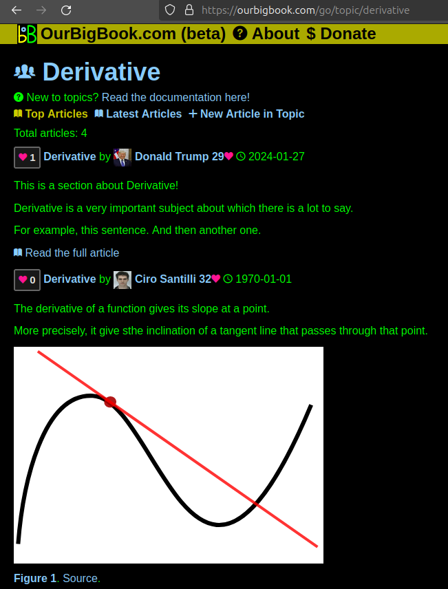
Figure 1. Screenshot of the "Derivative" topic page. View it live at: ourbigbook.com/go/topic/derivativeVideo 2. OurBigBook Web topics demo. Source. - local editing: you can store all your personal knowledge base content locally in a plaintext markup format that can be edited locally and published either:This way you can be sure that even if OurBigBook.com were to go down one day (which we have no plans to do as it is quite cheap to host!), your content will still be perfectly readable as a static site.
- to OurBigBook.com to get awesome multi-user features like topics and likes
- as HTML files to a static website, which you can host yourself for free on many external providers like GitHub Pages, and remain in full control

Figure 2. You can publish local OurBigBook lightweight markup files to either OurBigBook.com or as a static website.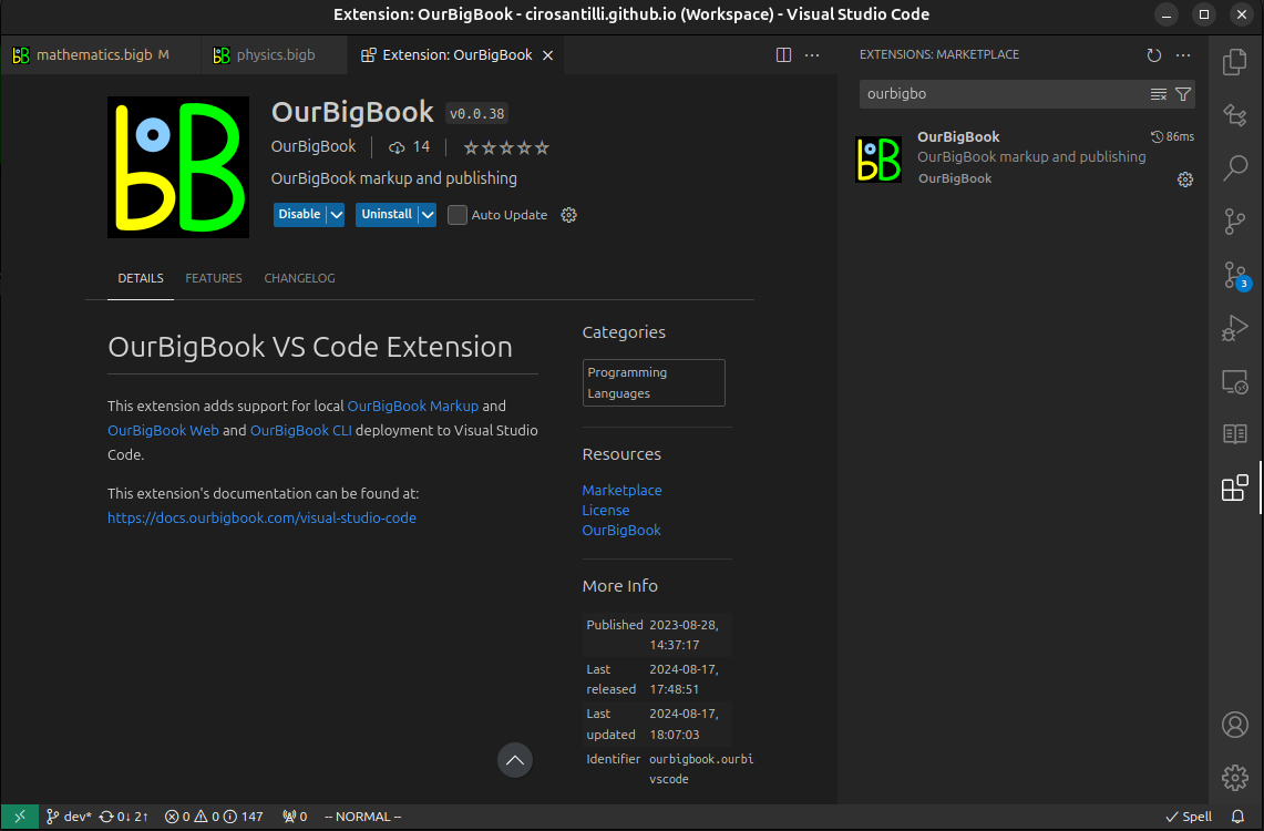
Figure 3. Visual Studio Code extension installation.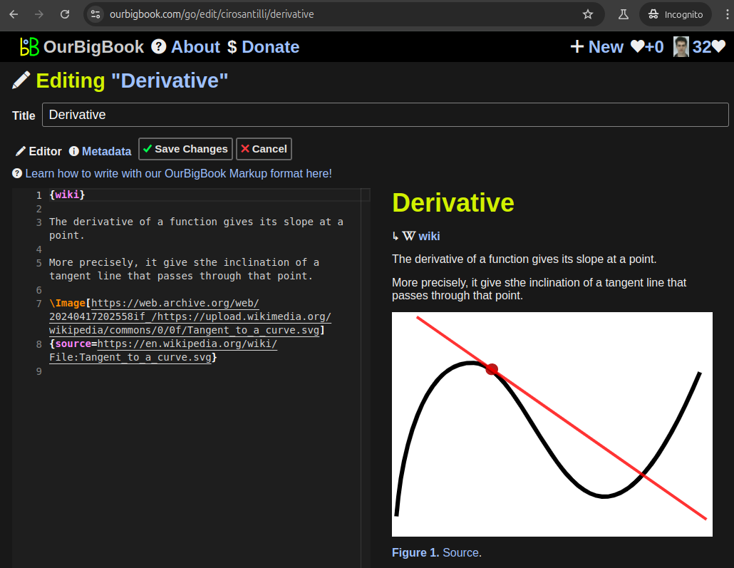
Figure 5. . You can also edit articles on the Web editor without installing anything locally. Video 3. Edit locally and publish demo. Source. This shows editing OurBigBook Markup and publishing it using the Visual Studio Code extension. 
- Infinitely deep tables of contents:
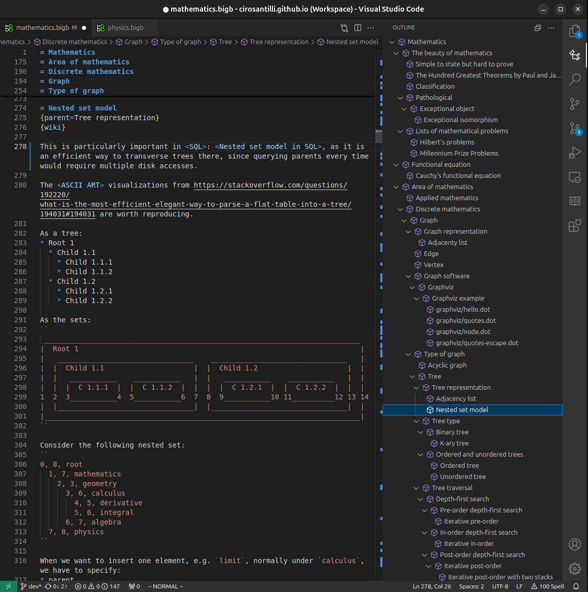
All our software is open source and hosted at: github.com/ourbigbook/ourbigbook
Further documentation can be found at: docs.ourbigbook.com
Feel free to reach our to us for any help or suggestions: docs.ourbigbook.com/#contact