A biochemical receptor is a protein that recognizes a signal and couples its detection to a biological effect. Its binding partner is a ligand; recognition depends on compatible shape and chemical interactions at a binding site within the protein structure. Specificity is selective recognition, not a guarantee that only one ligand can bind. Binding also need not activate the receptor: a competing ligand may occupy it while preventing activation by another ligand. See Alberts et al., Molecular Biology of the Cell, 4th edition: protein binding and specificity.
Compare a cell-surface receptor, accessible to an extracellular ligand, with an intracellular receptor, whose ligand must reach the interior. A cell-surface receptor can detect a hydrophilic ligand without transporting it through the cell membrane. In contrast, some membrane-permeable signals reach intracellular receptors that regulate gene expression. Location and downstream coupling are separate distinctions: an ionotropic receptor directly gates an ion channel, whereas a metabotropic receptor works through intracellular signal transduction. See Clark, Douglas and Choi, Biology 2e, 9.1: receptor locations and mechanisms.
Receptor binding occupancy quantifies recognition separately from response. A dissociation constant measures binding in a specified equilibrium model; it does not by itself predict the cellular effect. This separation is useful when comparing signals that bind equally well but produce different responses, or when interpreting a binding experiment alongside a functional measurement.
An inhibitory receptor activated by glycine; blockade contributes to strychnine poisoning.
A receptor responding to serotonin. Subtypes differ in signalling and distribution, so increasing transmitter availability can affect several mechanisms at once.
A serotonin receptor subtype implicated in particular local circuits, including some threat-related extended-amygdala pathways.
A serotonin receptor that can act as a raphe autoreceptor or a receptor on non-serotonergic target cells. Developmental and acute functions must be distinguished.
A family of soluble abscisic acid receptors. ABA-bound receptor complexes inhibit selected clade-A protein phosphatase 2C enzymes, allowing ABA-responsive kinases to remain active.
A methyl-accepting chemotaxis protein is a bacterial chemoreceptor whose signalling and adaptation are coupled to receptor modification. Receptor arrays interact with CheW and CheA to regulate downstream phosphorylation.
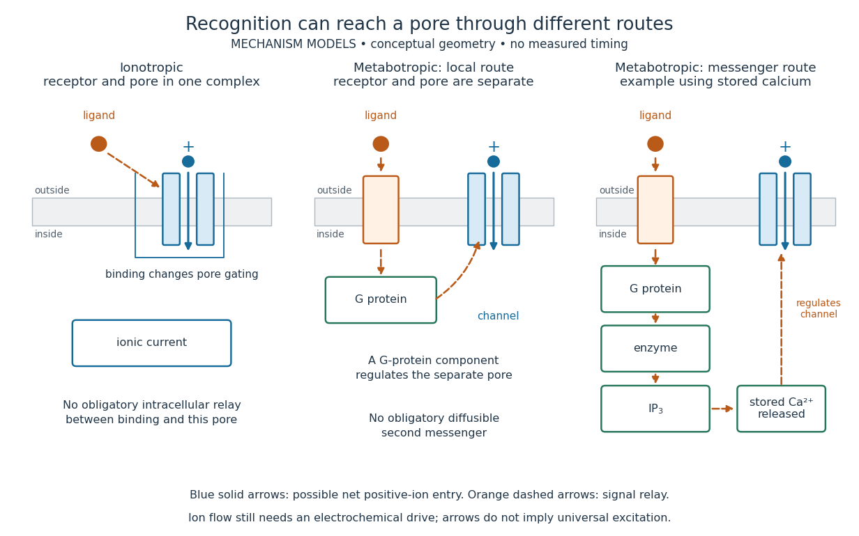
An ionotropic receptor is a receptor in which binding of a ligand directly gates an ion channel within the same protein complex. The binding site detects the chemical signal; the pore supplies a path across the cell membrane. At a chemical synapse, a neurotransmitter therefore changes synaptic conductance without an obligatory intervening G protein or second messenger. [ref]
A familiar example is the nicotinic acetylcholine receptor of skeletal muscle: acetylcholine binding opens a pore passing cations, including sodium and potassium. The muscarinic acetylcholine receptor instead uses intracellular signal transduction; the same neurotransmitter can engage different mechanisms. Direct gating does not mean that an ionotropic receptor pumps ions: electrochemical gradients drive passage through the open ion channel. Nor does “ionotropic” specify excitation. The reversal potential, initial membrane potential and other conductances determine whether the effect favors an action potential, causes hyperpolarization, or supplies shunting inhibition. [ref]
A concrete electrical approximation separates opening from drive. Let identical ion channels each have open-channel electrical conductance and opening probability . At fixed membrane potential , with common reversal potential , the expected value of inward-positive electric current isFor stipulated values , , , and , the mean open count is , the population electrical conductance is and . One open ion channel carries . These are illustrative values, not measurements. At , the net electric current vanishes despite open ion channels; neither binding nor opening alone determines the readout. The approximation omits changes in ion concentrations and voltage-dependent open-channel electrical conductance.
A motivating experiment made the elementary electrical events visible. As Bert Sakmann recounts, chronically denervated frog muscle fibers had acetylcholine-sensitive receptors spread beyond the original nerve contact. Mild enzyme treatment exposed the cell membrane; a polished glass pipette sealed over a small patch to reduce background noise. Two intracellular electrodes held the local membrane potential through voltage clamp, while the patch pipette recorded tiny electric currents. Applying suberyldicholine, a ligand that activates the acetylcholine receptor and prolongs openings, revealed abrupt, approximately rectangular current events. Their amplitudes agreed reasonably with estimates from whole-membrane current fluctuations. This established discrete opening and closing events; it did not make every ligand response instantaneous. Background noise and finite recording bandwidth concealed sufficiently small or brief events.[ref]
Recording an intact membrane patch alone does not exclude intracellular relays. Tank, Huganir, Greengard and Webb supplied a complementary isolation test in 1983: they purified nicotinic acetylcholine receptors from an electric ray's electric organ, incorporated them into artificial lipid membranes, and fused small vesicles into larger vesicles by freezing and thawing. Sealed glass-pipette recordings from isolated membrane patches resolved functional ion channels with properties resembling those in muscle. Removing the intact cytoplasm and its signaling machinery supports the sufficiency of the receptor complex for channel function; an artificial lipid environment still cannot reproduce every native regulatory interaction.[ref]
Direct gating supports millisecond communication at many chemical synapses. Response duration also depends on neurotransmitter clearance, binding and channel-state transitions. Continued ligand exposure can prolong activity. Classify an ionotropic receptor by its direct connection between recognition and pore gating, rather than a universal duration cutoff. [ref]
An ionotropic receptor activated by glutamate and related agonists. Subunit composition affects its properties; a role in one form of plasticity is not a complete explanation of a memory process.
A GABA-A receptor is an ionotropic GABA receptor whose pore primarily supports anion conductance. Its effect on membrane voltage depends on the relevant ion gradients as well as opening probability.
A nicotinic acetylcholine receptor is a pentameric cation-selective channel gated by acetylcholine. Extracellular binding interfaces communicate with a pore lined by M2 helices; transmitter recognition and ion selectivity are different properties.
A metabotropic receptor couples ligand recognition to intracellular signal transduction, which can regulate a separate ion channel, alter enzyme activity or change gene expression. In the usual neurotransmitter context, metabotropic receptors are G protein-coupled receptors: receptor activation is relayed by a G protein. There need not be a change in membrane potential at all. [ref]
Two routes should be distinguished. An activated G protein can regulate an ion channel locally at the cell membrane, without an obligatory diffusible second messenger. Alternatively, it can regulate an enzyme that changes the availability of a second messenger, such as cAMP or inositol trisphosphate. Downstream proteins then change channel opening, phosphorylation or other cellular activities. The receptor, G protein, second messenger and final target are distinct components; an activated G protein is a relay protein, rather than itself a small-molecule second messenger.[ref]
General Principles of Cell Communication. Figure 15-15
Do not turn this useful chemical synapse classification into the claim that every receptor is a GPCR, or that every indirect response must pass through a G protein. For example, the insulin receptor is an enzyme-linked cell-surface receptor whose intracellular catalytic activity initiates protein phosphorylation. Intracellular receptors supply another category. “Metabotropic” commonly names the GPCR branch in neuroscience; the broader idea of biochemical coupling does not make all non-channel receptors structurally identical. [ref]
Separating recognition from execution allows signal amplification and branching. A persistently activated receptor can activate several relay proteins; a regulated enzyme can produce many second messenger molecules. As an explicitly invented count example, suppose one activated receptor activates four relay proteins during one interval, and each relay activates a distinct enzyme that produces fifteen second messenger molecules during the next interval. With sufficient substrates and no loss, this gives molecules. Those counts do not specify a physiological gain or response duration. A directly gated ion channel also passes many ions while open, so amplification is not an exclusive property of metabotropic receptors: always name the input and output being compared. [ref]
A particularly instructive electrophysiology experiment separated a chemical trigger from the ion channel carrying its eventual electric current. In 1987, Hirono, Ito and Sugiyama injected messenger RNA from rat brain into frog egg cells, then tested acquired responses to neurotensin and muscarinic acetylcholine stimulation. Under voltage clamp at , neurotensin elicited transient, oscillatory inward electric currents after a delay of several tens of seconds. This delay refers to that response and preparation. Reversal potential measurements implicated increased permeability to chloride anions, rather than a pore specific to the applied ligand. Both neurotensin and acetylcholine responses were inhibited by pertussis toxin, which interferes with susceptible G proteins, and by intracellular EGTA, which binds free calcium. Injecting inositol trisphosphate mimicked the responses; a large prior dose of inositol trisphosphate reduced subsequent responses to either extracellular signal. These interventions implicate a shared intracellular relay and calcium-dependent channel opening.[ref]
The informative comparison with the nicotinic acetylcholine receptor experiment is mechanistic: an extracellular ligand can produce an inward electric current either by directly gating the pore or by mobilizing an intracellular second messenger and stored calcium that activate another pore. Intracellular inositol trisphosphate bypasses reception, whereas EGTA tests the need for free calcium. However, neither inhibition nor mimicry alone identifies every molecular connection: a perturbation can affect several targets, and an injected mediator can activate a pathway without proving that it is the only natural route. The egg cell's endogenous machinery and the mixed messenger RNA preparation also limit extrapolation to an intact neuron. The experiment establishes functional coupling in its preparation, rather than the exact molecular composition or timing of every metabotropic receptor.
Even G protein-dependent channel regulation need not involve a lengthy second messenger cascade. Pfaffinger and colleagues reported in 1985 that acetylcholine-evoked potassium channel activation in embryonic chick atrial cells required intracellular GTP, a guanine-containing nucleotide, and was abolished by pertussis-toxin pretreatment. Changes in cyclic nucleotides, including cAMP, did not account for the channel response. The report discusses intrinsic activation delays of roughly –, illustrating relatively rapid metabotropic receptor action. These observations support a G protein coupling step without cyclic-nucleotide second messengers; they do not by themselves exclude every possible intermediary or prove the specific subunit-channel interaction.[ref]
A small timing model makes the extra steps visible without inventing an experimental trace. Let jump from zero to one at and remain on. For a direct-gating model output , one relay variable , and its downstream output , take zero initial conditions andAll outputs are normalized dimensionless variables; they are neither recorded electric currents nor fitted concentrations. With illustrative time constants , and , their exponential function solutions for areInitially , while because its relay must first build up. Both outputs eventually approach one. Equal normalized final values do not imply equal biological amplitudes or absence of signal amplification; normalization deliberately removes that question.
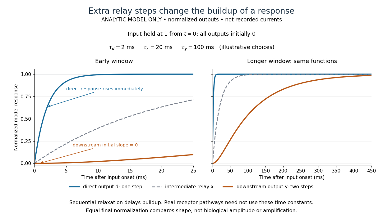
The model's useful feature is the delayed buildup created by sequential relaxation, not its arbitrary numbers. Actual kinetics also depend on ligand delivery, relay deactivation, neurotransmitter clearance and the final target. A local G protein route can act promptly, while an ionotropic receptor can remain active during continued exposure or feed a longer-lasting intracellular response. The beauty of metabotropic receptor coupling is its flexibility: the same recognition event can adjust electrical responsiveness and other cellular tasks through different targets. To classify an observed response, combine timing with molecular identity and controlled perturbations.
A G protein-coupled receptor is a cell-surface receptor that relays activation through a G protein on the intracellular side of the cell membrane. The activated G protein can regulate an enzyme or ion channel, so a GPCR supplies a concrete metabotropic receptor example: ligand recognition and the eventual response are connected through distinct components. Different G proteins couple to different targets. See Alberts et al., Molecular Biology of the Cell, 4th edition: signaling through G-protein-linked cell-surface receptors.
A protein family involved in regulation and signalling of activated receptors; recruitment can alter receptor coupling and trafficking.
The capacity of a signalling system to produce a maximal response without occupancy or activation of all receptors. It makes observed maximal response an unreliable direct measure of intrinsic ligand efficacy.
Preferential activation of one receptor-linked response relative to another, defined against a reference ligand and appropriate system controls. Different amplification or receptor reserve can mimic apparent bias.[ref]
An opioid receptor ligand, also called oliceridine, studied for differences between G protein activation and arrestin recruitment. Apparent signalling preference must be distinguished from low intrinsic efficacy.
A ligand that inhibits receptor activation by occupying the binding site used by an activating ligand.
The beta-2 adrenergic receptor is a G protein-coupled receptor whose ligand-dependent conformational populations have been studied by NMR spectroscopy and electron paramagnetic resonance.[ref]
Structural insights into the dynamic process of β2-adrenergic receptor signaling by Aashish Manglik and others. pp. 1101–1111, ligand and intracellular-partner effects on receptor dynamics
A cell-surface receptor detects an extracellular signal at the cell membrane. Its changed state affects the intracellular side, coupling reception to signal transduction or directly to an ion channel's opening.
An inhibitory immune regulator whose blockade can enhance T-cell responses while also increasing risk of immune-mediated damage.
An engineered receptor combining an antigen-binding region with intracellular signalling modules. Conventional designs recognise accessible cell-surface targets without peptide–HLA presentation.
The receptor complex through which a T cell recognises antigen, conventionally a peptide presented by a compatible MHC molecule.
An inhibitory receptor on immune cells whose engagement can reduce activity during antigen stimulation. Blocking it does not replace missing antigen recognition or presentation.
A receptor involved in embryonic patterning and immunity. In the early Drosophila melanogaster embryo, spatially restricted activation by Spätzle promotes nuclear Dorsal protein.
A receptor tyrosine kinase combines extracellular ligand recognition with an intracellular protein kinase protein domain. A membrane-spanning segment connects these parts across the cell membrane: the signal changes the intracellular machinery without requiring the ligand to enter the cytosol. The protein kinase transfers a phosphate group from ATP to selected tyrosine residues on proteins, including the receptor itself.[ref]
Signaling through Enzyme-Linked Cell-Surface Receptors. “Activated Receptor Tyrosine Kinases Phosphorylate Themselves”: extracellular binding, intracellular catalytic domains and ATP-dependent phosphate transfer
This architecture makes a reception event useful in two ways: it can change catalytic activity and assemble a local group of signaling proteins. Trans-autophosphorylation can create phosphotyrosine residues recognized by SH2 domains. Recruitment positions a downstream enzyme or relay protein near its partners. Neither every modified site nor every recruited protein has the same effect; association is a specific molecular connection, rather than a universal command to activate everything nearby.[ref]
Signaling through Enzyme-Linked Cell-Surface Receptors. “Phosphorylated Tyrosines Serve as Docking Sites For Proteins With SH2 Domains”, Figure 15-53: signaling enzymes, adaptors and negative regulators
Compare the epidermal growth factor receptor with the insulin receptor. Their common topology does not impose one activation geometry or make assembly equivalent to activity. The receptor phosphorylation assay consequently needs more than evidence that two protein chains associate.
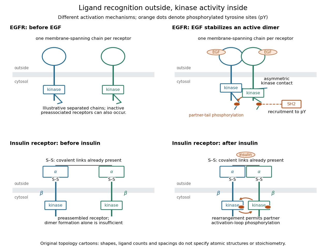
Receptor tyrosine kinase architecture and distinct activation mechanisms
. Original topology cartoons contrasting EGFR with the preassembled insulin receptor. Orange dots mark phosphotyrosine residues; S–S marks protein disulfide bonds. The EGFR panel separates asymmetric catalytic contact from partner-tail phosphorylation. Shapes, distances and drawn ligand counts are explanatory, rather than atomic structures or binding stoichiometry. Details.A member of the ErbB receptor family. Different ligands and receptor combinations have distinct signalling properties.
Platelet-derived growth factor receptor, a receptor tyrosine kinase activated by PDGF. Its characteristic ligand-induced phosphotyrosines require an appropriate detection reagent.
A receptor for fibroblast growth factors. Its outputs include, but are not restricted to, MEK and ERK activity.
A receptor important in early mouse lineage responses and primitive-streak morphogenesis. Its effects on fate and movement require separate measurements.
In an illustrative EGFR activation route, EGF stabilizes an associated pair of receptor chains. Asymmetric contact between their protein kinase protein domains activates one catalytic partner without requiring its activation-loop phosphorylation. Subsequent phosphorylation of cytoplasmic tails creates recruitment sites. Preassociated inactive EGFR populations can also occur: the cartoon's separated starting chains are not a universal initial condition.[ref]
Cell signaling by receptor-tyrosine kinases by Mark A Lemmon and Joseph Schlessinger. Lemmon and Schlessinger, “Mechanisms of Receptor Activation” and “Allosteric activation of TKDs”, Figure 2F
The mature insulin receptor already contains two extracellular chains and two membrane-spanning chains, joined through protein disulfide bonds, before insulin binds. Insulin induces rearrangement of this assembly. Partner-mediated phosphorylation of the inhibitory activation loop permits increased protein kinase activity. Ligand-induced creation of a previously absent dimer is therefore an unsuitable universal explanation.[ref]
Cell signaling by receptor-tyrosine kinases by Mark A Lemmon and Joseph Schlessinger. “Mechanisms of Receptor Activation” and “TKD autoinhibition by the activation loop: insulin and FGF receptors”, Figure 2E
An insulin receptor substrate is a docking protein phosphorylated by the activated insulin receptor. Its phosphotyrosine residues recruit downstream signaling proteins, including SH2 domain-containing components of phosphoinositide 3-kinase. This connects reception to the pathway described under insulin, rather than making the insulin receptor substrate another extracellular ligand.[ref]
Cell signaling by receptor-tyrosine kinases by Mark A Lemmon and Joseph Schlessinger. “Linking RTK activation to cell signaling”: IRS docking proteins and Figure 3A
A receptor phosphorylation assay asks whether, and how much, a specified receptor is modified by phosphorylation under defined conditions. It is distinct from a binding assay for receptor binding occupancy, a test of protein kinase activity on a supplied substrate, and a test of downstream signal transduction. Those measurements answer related but different questions.
For a Western blot implementation, compare matched samples with ligand omitted or added, detect a specified phosphorylated epitope, and separately detect total receptor abundance. Preserve the sampled state by stopping further enzyme reactions during sample preparation; otherwise protein kinase or protein phosphatase activity after sampling can change the result. A general anti-phosphotyrosine residue antibody recognizes many modified proteins; a site-selective antibody gives a narrower readout, but still needs Western blot antibody specificity controls.
Reason through the controls before interpreting the difference. Loss of the proposed signal after receptor-specific gene deletion supports protein identity. Treatment with a protein phosphatase, alongside preservation of the total-receptor signal, tests phosphate-dependent recognition. Western blot loading controls and Western blot transfer controls address sample recovery; a usable detection range addresses Western blot response saturation. Lower signal after perturbing protein kinase activity supports dependence on that perturbation, but alternative targets and altered receptor abundance can complicate the inference. These are proposed assay comparisons, not outcomes attributed to the historical membrane experiment.
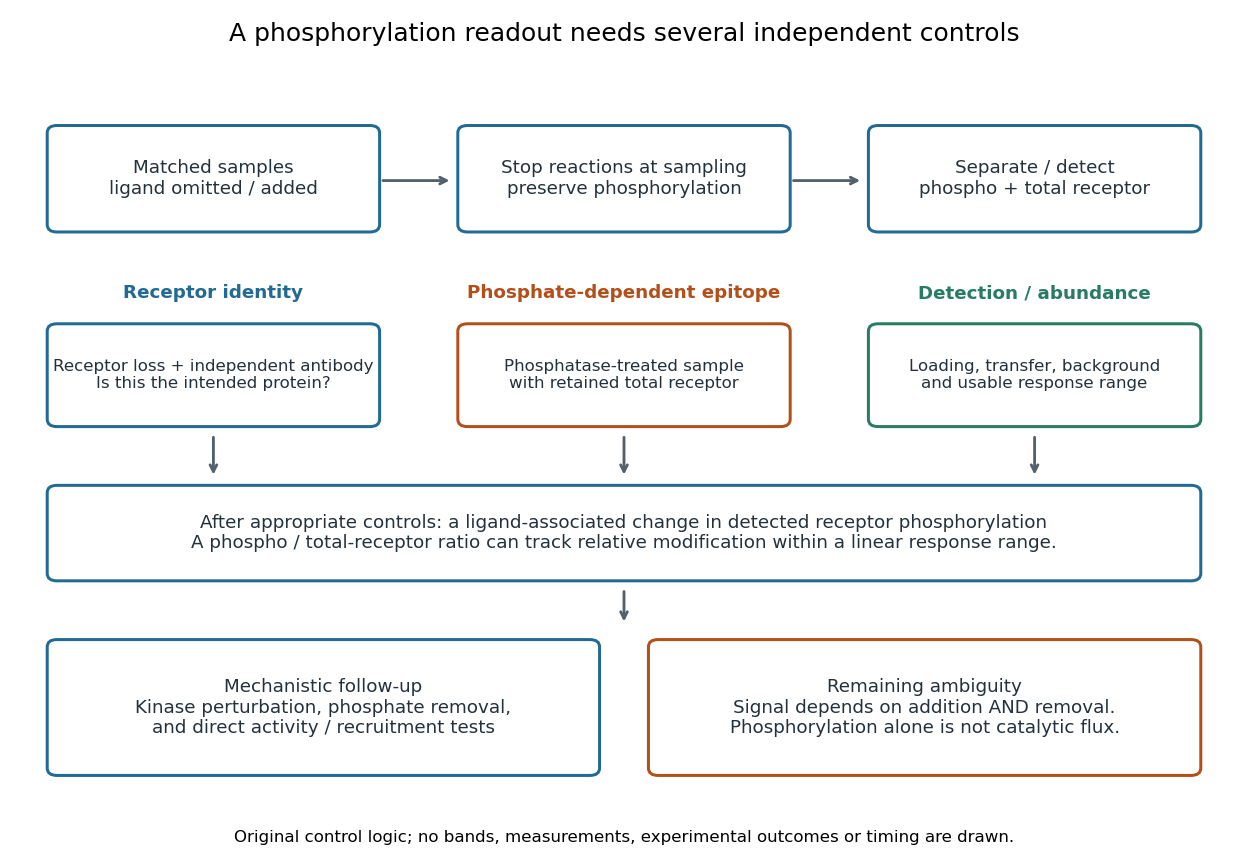
An original idealized measurement model explains the normalization. Let be the retained number of receptor molecules, the fraction carrying the selected modified epitope, and fixed detection gains. After subtracting their respective backgrounds, suppose the phosphorylated and total-receptor signals lie in a linear response range:Dividing by total receptor cancels under these assumptions. It differs from total-protein Western blot normalization, whose denominator includes many other proteins. For invented values , samples with and give and , but both give . Twice the phospho signal here means twice the retained receptor amount, with no change in fractional modification. Unequal recovery, changed epitope accessibility, nonzero uncorrected background or Western blot response saturation invalidates the cancellation. Unknown relative gains prevent reading an absolute modified fraction directly from the ratio.
A motivating experiment used isolated membranes from A-431 biological cells, which supplied abundant EGFR. Separate membrane aliquots were incubated at with ATP carrying radioactive phosphorus in its terminal phosphate group, plus magnesium or manganese ions. Radioactivity in material recovered by chemical acid-induced precipitation supplied the readout. Adding EGF beforehand enhanced incorporation relative to the unstimulated preparation; this placed a ligand-dependent biochemical response in a cell-free membrane system.[ref]
Epidermal Growth Factor by Stanley Cohen. Cohen, Epidermal Growth Factor, printed p. 339: A-431 membrane aliquots, terminally labeled ATP, divalent ions and trichloroacetic acid-insoluble incorporation
The follow-up comparisons separated increased addition from suppressed removal: phosphate removal was rapid but unaffected by EGF, and anti-EGF antibody reversed activation by removing the ligand. These observations support reversible regulation of membrane-associated protein kinase activity.[ref]
Epidermal Growth Factor by Stanley Cohen. printed p. 341, numbered observations 1–4: rapid activation, dephosphorylation, lack of soluble kinase release and anti-EGF reversal
Total incorporation alone cannot identify which protein or amino acid residue received the label, prove intrinsic receptor catalysis, or predict an intact biological cell's response.The same account records an instructive identification error: receptor phosphorylation was initially assigned to the wrong amino acid residue because released modified amino acids migrated together in the electrophoretic electric field. Reanalysis identified phosphotyrosine residues. Retained radioactivity is evidence about the assay's separation and detection, not unique chemical identity.[ref]
Epidermal Growth Factor by Stanley Cohen. printed p. 341: co-migration in the electrophoretic system and subsequent identification of tyrosine phosphorylation
Protein kinase activity adds modification, while protein tyrosine phosphatase activity can remove it. An original one-site model shows why a receptor phosphorylation assay cannot determine either process from a single modified fraction. Let be the fraction of a fixed receptor population carrying one selected phosphotyrosine residue. Stipulate constant effective first-order rate constants , with ATP availability absorbed into , and no receptor synthesis, degradation or trafficking:Here is the steady state fraction and the time constant. The ordinary differential equation has the exponential function solutionThe balance equation and a single relaxation step give this result; it is an illustrative model, rather than a fit or a claim about every RTK.
With stipulated rate constants , and . Doubling both rate constants leaves but changes to . The balanced phosphate turnover per site,doubles from to . Identical steady state modification therefore permits different catalytic fluxes. Removing the addition term instead gives ; this illustrates why a perturbation that stops protein kinase activity can expose removal dynamics, subject to the model's unchanged-removal assumption. A maintained steady state with ongoing ATP consumption is not chemical equilibrium.
An intracellular receptor recognizes a ligand inside a biological cell, including in the cytoplasm or cell nucleus. Some intracellular receptors regulate transcription after binding a membrane-permeable signal; an intracellular receptor does not universally imply direct control of DNA. See the receptor-location reference under receptor.
A ligand-regulated protein family whose members often act as DNA-binding regulators of transcription. Subcellular distribution and activation mechanisms vary between members.
A nuclear receptor responding to glucocorticoid ligands, with regulated localization and recruitment of transcriptional partners.
Receptor binding occupancy is the fraction of binding sites occupied by a ligand. Consider reversible, noncooperative binding at chemical equilibrium, with homogeneous independent sites: every site has the same binding properties, and binding at one site does not change binding at another. Let denote free ligand, an unoccupied receptor site and the bound complex. In this dilute concentration model,Square brackets denote concentrations; is the total site concentration. Here the dissociation constant has concentration units. Use free ligand in excess so binding negligibly depletes it; only under that approximation may the supplied total ligand concentration substitute for . See Analysis of Receptor–Ligand Interactions: equilibrium binding and saturation.
For an illustrative dissociation constant , where , free ligand concentrations , and give , and , respectively. At , exactly half the sites are occupied in this model. Raising tenfold from to raises occupancy from to about , rather than tenfold: the finite pool of sites causes saturation.
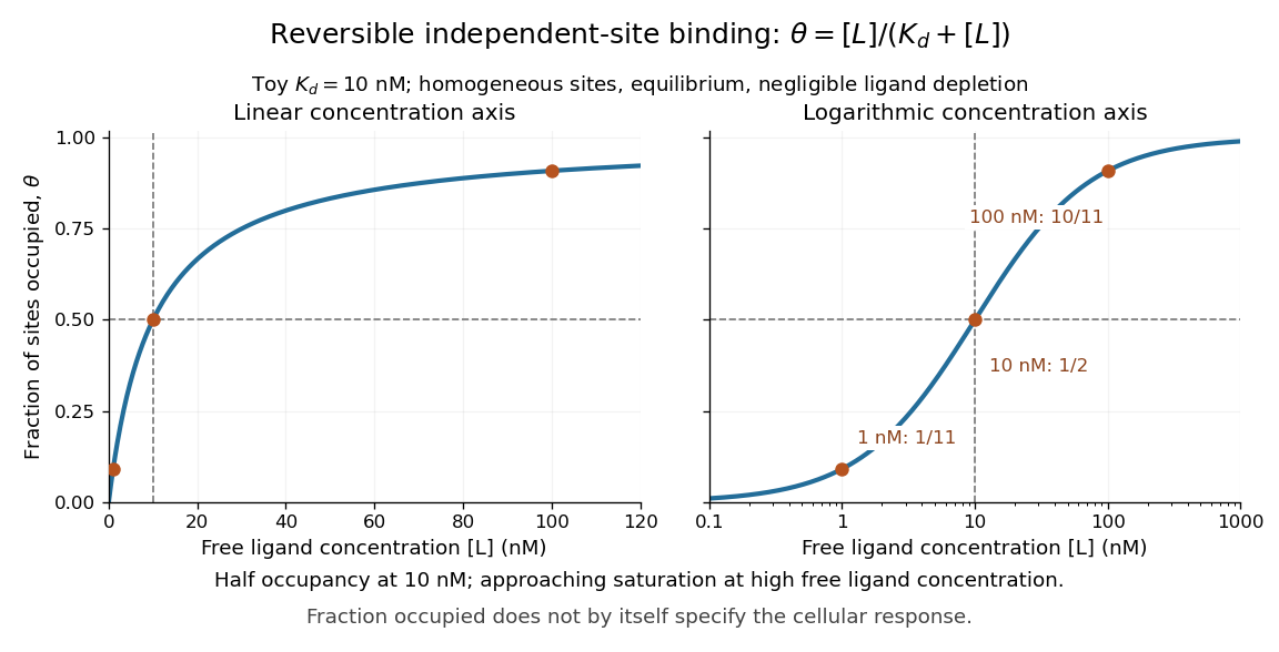
A short derivation uses the law of mass action: at chemical equilibrium, association balances dissociation, giving . Substitute this into and cancel to obtain the displayed formula. The limiting values follow directly: zero free ligand gives zero occupancy, and increasing far above drives occupancy toward one.
Receptor binding occupancy is not automatically the fraction of maximal response. Signal amplification, differing effects of bound ligands, and downstream saturation can change the relationship. Consequently, specifies half occupancy here; it does not universally specify either half-maximal or maximal cellular response. These numbers describe a toy binding model, not measured responses of every receptor. See Higham and Colquhoun, 2024: the affinity–efficacy problem.



