Topics (384k) Articles (398k) Users (410) Discussions (242) Comments (383) Files (13k) New article

Retinol is a retinoid with an alcohol end group and molecular formula . A general structural description does not specify the geometry of every double bond: all-trans-retinol and 11-cis-retinol distinguish particular configurations of the same framework.[ref]
That distinction matters because molecular formula and molecular geometry answer different questions. Two samples can have the same elemental composition while containing different proportions of retinol isomers. Their identical nominal molecular masses do not make those samples chemically interchangeable.
The alcohol group also distinguishes retinol from retinyl esters. Lecithin retinol acyltransferase transfers an acyl group onto the alcohol oxygen, producing an ester form that participates in storage and retinoid metabolism.[ref]
For a simple amount-to-mass example, the average molar mass gives for of retinol. This calculation specifies total chemical amount, without identifying its isomer distribution or biological availability.[ref]
A retinyl ester is an ester formed at the alcohol group of retinol. The retinyl portion is attached through oxygen to an acyl group; different attached groups produce different members of the class. The name therefore does not identify one unique molecular formula or molar mass.[ref]
All-trans-retinyl ester in ChEBI. CHEBI:63410, generic structure and retinyl-ester classification
Write a retinol alcohol schematically as , where Ret represents its carbon framework. The corresponding ester has the arrangement . Varying changes the attached acyl group without requiring a change in the retinyl framework. This is a structural comparison, not a claim that a free carboxylic acid supplies every biological esterification reaction.
For example, lecithin retinol acyltransferase uses phosphatidylcholine as an acyl donor. Its products include retinyl esters that can be stored or used in subsequent retinoid pathways.[ref]
Lecithin retinol acyltransferase in IUBMB Enzyme Nomenclature. reaction and comments, donor and storage products
An assay of total ester therefore needs an additional identity measurement if the question concerns a particular ester, geometric isomer or downstream substrate.cis-Resveratrol is the resveratrol stereoisomer with the two aryl groups on the same side of the central double bond, corresponding to the configuration. It has the same neutral molecular formula, , and formula-based molar mass as trans-Resveratrol, but a different spatial arrangement.[ref]
The isomers need different optical response factors. Brent C. Trela and Andrew L. Waterhouse measured an ethanol absorption maximum at with molar absorption coefficient for the cis form; their stability tests also depended on light exposure and pH.[ref]
Resveratrol: Isomeric Molar Absorptivities and Stability by Brent C. Trela and Andrew L. Waterhouse. 1996, Abstract, cis-isomer maximum and stability
At that maximum, an original solution in a path has ideal absorbanceThis is lower than the trans-isomer's corresponding example at its own maximum, but the two maxima occur at different wavelengths. Dividing their peak coefficients does not supply the cis/trans response ratio at one common detector wavelength.
For a mixture at a chosen wavelength , an additive Beer-Lambert law model isOne reading generally cannot identify both concentrations. Two wavelengths help only if their response rows are independent and the coefficients are appropriate to the solution. Multivariate calibration makes that requirement explicit; physical separation by chromatography supplies another route. Neither equal molecular formulas nor a single colour measurement establishes isomeric purity.
trans-Resveratrol is the resveratrol stereoisomer with the two aryl groups on opposite sides of the central double bond, corresponding to the configuration. It retains the three hydroxy groups and neutral molecular formula of resveratrol. ChEBI specifies the double-bond geometry explicitly, unlike the geometry-unspecified parent record.[ref]
Resveratrol in ChEBI. CHEBI:45713, definition, formula and stereochemical structure; CHEBI:27881, parent
The geometry affects the optical measurement. Brent C. Trela and Andrew L. Waterhouse reported an ethanol absorption maximum at and a molar absorption coefficient of for this isomer. They tested chemical stability under different light and pH conditions.[ref]
Resveratrol: Isomeric Molar Absorptivities and Stability by Brent C. Trela and Andrew L. Waterhouse. 1996, Abstract, trans-isomer maximum and stability observations
For an original Beer-Lambert law calculation under those stated spectral conditions, in a path givesThe calculation assumes a suitable homogeneous solution of the specified isomer, correctly subtracted background and the stated wavelength and solvent. A different solvent, wavelength or isomer mixture requires the corresponding optical model.
The trans and cis forms have the same molar mass, so weighing alone cannot determine their proportions. Illumination can also change a preparation after it has been weighed. Separating the species by chromatography and using appropriate wavelength-dependent response factors addresses a different question from merely measuring total formula-based mass. This chemical identity does not, by itself, establish a biological benefit.
Airway resistance quantifies the pressure difference needed to drive a specified volumetric flow rate through the conducting airways. For a steady linear relation with flow positive toward the alveoli,The units are pressure divided by volume per time, for example . A stipulated pressure drop of 1 cm water at gives in those units. The pressure references matter: mouth-to-alveolar pressure concerns the flow path, whereas transpulmonary pressure also describes elastic loading of the lung.
An instructive idealization is the Hagen-Poiseuille equation. For a rigid cylindrical tube carrying steady laminar flow of a fluid with dynamic viscosity ,Reducing its radius to 80% while holding length and dynamic viscosity fixed multiplies resistance by . Real airways form a branching, deformable network and need not carry fully developed laminar flow. The tube equation explains a sensitivity, not a universal law for every lung measurement.
Network structure also matters. For identical linear branches in parallel, pressure drop is shared and their volumetric flow rates add, giving . Many narrow distal branches can therefore have a very different combined resistance from one isolated narrow tube. Radius alone is insufficient without the arrangement and number of pathways.
Secant versus differential airway resistance distinguishes two values when the relation is nonlinear. For an original model , the finite ratio is , while its local slope is away from zero. Report the operating flow and which definition is used.
A 1959 primary study combined mouth pressure with an intra-esophageal balloon and a differential pressure transducer to investigate resistance and compliance.[ref]
Airway Resistance and Lung Compliance by Ole Storstein and others. Methods: balloon pressure, mouthpiece connection and Sanborn transducer
The mouth-to-esophageal difference is not a pure airway pressure drop: its elastic component must be separated. Simultaneous pressure, volume and flow information is what makes this mechanical distinction experimentally accessible.Spirometry measures the volume of air moved into or out of the respiratory system as a function of time. A spirometer records displaced gas directly or derives volume by integrating volumetric flow rate. The resulting trace describes gas movement at the mouth; it does not independently determine alveolar pressure, pleural pressure or lung compliance.[ref]
Standardization of Spirometry 2019 Update by Brian L. Graham and others. Introduction and Equipment: volume and flow measurements
Two important outputs of a forced expiration following maximal inspiration are forced vital capacity, the total volume expelled in that maneuver, and forced expiratory volume in one second, the volume expelled during its first second. Their ratio compares early emptying with the eventual expelled volume.[ref]
Standardization of Spirometry 2019 Update by Brian L. Graham and others. FVC Maneuver and definitions of FVC and FEV1
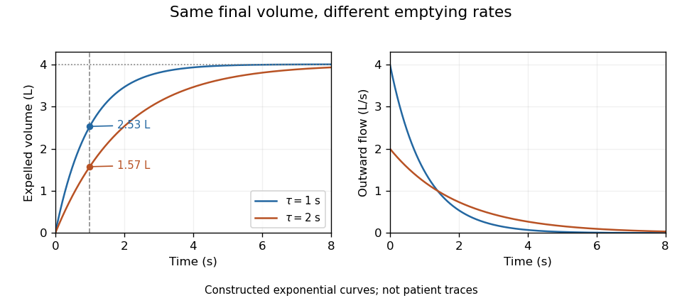
Equal final volumes can accompany substantially different emptying rates.An original mathematical example sets expelled volumewhere the numerical time and time constant are in seconds. Both and give an asymptotic expelled volume of 4 L. Their first-second volumes are instead L and L. The corresponding ratios to the limiting volume are approximately 0.632 and 0.393. These smooth toy curves explain the measurements; they are not measured traces or diagnostic reference limits.
Spirometry measures volume differences. The gas left after maximal expiration is residual lung volume, which has not crossed the instrument. Consequently a volume trace alone does not determine total lung capacity: different absolute starting volumes can produce the same expelled-volume trace. This is the absolute-volume ambiguity in spirometry.
Timing, effort, leaks and early termination can alter what a trace represents. Distinguish an instrumentally repeatable signal from a maneuver that answers the intended physiological question. The primary technical statement treats equipment quality assurance and maneuver acceptability separately.[ref]
Standardization of Spirometry 2019 Update by Brian L. Graham and others. Device Quality Assurance and FVC Maneuver acceptability sections
Respiratory-system compliance is the change in volume per change in elastic pressure across the passive lung and chest wall together. For a local static relation,Lung compliance alone instead uses transpulmonary pressure, . The whole-system and lung-only quantities are different even when they refer to the same volume change.
At that shared volume, the elastic pressure increments add. With component compliances and ,The reciprocals are component stiffnesses; respiratory-system elastance is . For positive component compliances, the combined value is smaller than either component. OpenStax describes opposing lung and wall recoil and their pressure references.[ref]
Anatomy and Physiology 2e by J. Gordon Betts and others. §22.3, pleural pressure and thoracic wall compliance
An original example uses and . The combined compliance is about 0.0667 in the same units. An added 0.50 L requires 2.5 cm water of lung elastic pressure and 5 cm water of wall pressure, totaling 7.5 cm water. Attributing that total entirely to the lung would underestimate lung-only compliance threefold.
In a passive ventilated-breath measurement, a zero-flow plateau airway pressure can be compared with the appropriately measured end-expiratory pressure. Under the stated equilibrium and no-effort assumptions, an interval estimate isFor stipulated L, and cm water, this is again about 0.0667. The baseline must account for the relevant positive end-expiratory pressure; a pressure measured during nonzero flow also contains a resistive contribution. The primary pilot study explicitly compares pressure-based respiratory-system estimates under passive ventilation.[ref]
Static and Dynamic Measurements of Compliance and Driving Pressure: A Pilot Study by Pierre Tawfik and others. Introduction, Methods A–D; Study Procedures
State whether the number is a local slope or an interval ratio, which pressure references were used, and whether muscle activity and flow were excluded. Separating lung and wall contributions requires internal pressure information beyond the whole-system volume response.
A seminal vesicle is a paired accessory gland that contributes fluid to semen. Its secretion includes fructose and other substances that help form the environment in which sperm are transported. The gland's duct joins the sperm-carrying pathway near the prostate.[ref]
Human Reproductive Anatomy and Gametogenesis. Section 43.3, male reproductive structures and accessory glands
Despite its name, a seminal vesicle is not primarily a storage sac for sperm. Sperm production, maturation, movement through ducts and addition of accessory-gland fluid are distinct contributions to reproduction. This distinction also explains why semen volume and sperm count describe different quantities: much of the ejaculated fluid is glandular secretion rather than sperm cells themselves.
A fallopian tube is one of the paired ducts connecting the region near an ovary to the uterus. Its ovarian end opens into the peritoneal cavity rather than forming a sealed attachment to the ovary. Fimbriae help receive the released oocyte, which is transported along the tube by ciliary activity and smooth-muscle movement.[ref]
Human Reproductive Anatomy and Gametogenesis. Section 43.3, female reproductive structures and oviducts
The tube is therefore an active transport environment, not a passive storage vessel. It can bring gametes into the same region and transport an early embryo toward the uterus. These tasks distinguish ovarian release, fertilization and uterine implantation: they are different events with different locations, even when they occur in a connected sequence.
Reference-standard uncertainty in relative potency is the contribution of a shared activity reference to uncertainty in results calibrated against it. It may arise through preparation, dilution, stability or transfer of the reference material. The contribution can remain common to many specimen results even when their biological readouts are independently repeated.
For an original calibration model, let , where is reference activity on a specified concentration basis and is measured relative biological potency. First-order propagation givesThe covariance term matters when reference preparation or calibration information also enters the fitted ratio. The GUM explicitly retains correlations arising from common standards and reference data.[ref]
Suppose is independent of all specimen ratio estimates, but is shared unchanged between them. For independent and ,This identity is exact under the stated independence assumptions. The reference contribution is not independent just because the two specimens occupy different wells or are assayed on different days.
An original numerical model uses reference mean 100 activity units per millilitre with standard uncertainty 5, and independent ratio estimates with mean 0.8 and standard deviation 0.04. The first-order standard uncertainty of one calibrated result near 80 units per millilitre is . Averaging four independent ratio estimates against the same reference gives approximately , not .
For this model the exact variance of the average of calibrated results isThe small extra 0.04 term comes from multiplying two uncertain quantities. As grows, standard uncertainty approaches 4 activity units per millilitre. This common-reference floor in potency averaging is a consequence of the shared calibration, rather than an unexplained failure of repeatability.
A reference's assigned unitage can itself be conventional. A 2017 WHO collaborative-study report proposed separate assigned bioactivity units and explained that first-standard unitage is set arbitrarily, while replacement standards are calibrated against the first.[ref]
Report on a Collaborative Study for Proposed Candidate 1st International Standard for the biological activities of Rituximab by Sandra Prior and others. printed p. 33, Appendix 3, response concerning assigned unitage
Uncertainty in realizing, preparing or transferring that convention is distinct from uncertainty in a stipulated number. Specify which quantity the uncertainty budget represents.A ratio of two results using exactly the same multiplicative reference has , so that common factor cancels algebraically. A comparison involving different reference lots need not have the same cancellation. Retain the calibration lineage and shared terms before combining results or interpreting a change in calibrated potency.
A binary selection mask records which pixels belong to a chosen subset of an image. On a finite pixel grid , it is the indicator functionIt can represent an area region of interest, the result of image thresholding or a combination of selection rules. The mask describes membership; its entries need not equal the underlying image intensities.
For a selected pixel count , the count and selected arithmetic mean areConsider the original arraysThe selected values are 0, 20 and 40, so and . The selected zero-valued pixel still contributes to the denominator. Replacing excluded pixels by zeros and averaging all six entries would instead give : that asks a different question. An empty mask has , so this selected-mean formula is undefined.
For rectangular pixels with calibrated dimensions and , the complete-pixel area from a calibrated pixel count is . These are stipulated array values and cell dimensions, not a measurement of an object.
A stored mask can use another two-value encoding. ImageJ's Create Mask command stores 255 inside an area selection and 0 outside, corresponding to in this model.[ref]
ImageJ User Guide by Tiago Ferreira and Wayne Rasband. IJ1.46r, section27.12.10, Create Mask
Summing gives 765 in the example, rather than the selected count 3. Convert the encoding or count selected entries before interpreting a sum as an area.Two normalized masks and combine by set intersection through . Their complement within the specified grid is . Record the foreground-background polarity, coordinate alignment and grid extent when transferring a mask between images.
Hemolysis is the breakdown of red blood cells. In a membrane-rupture experiment, release of hemoglobin into the surrounding liquid can supply a measurable readout. The process and the signal used to detect it are different things: interpreting the measurement requires a stated reference and background.[ref]
For an original calibrated-release model, assume absorbance is proportional to released hemoglobin and compare equally loaded samples at the same final volume. Let denote sample absorbance, the blank and an otherwise matched complete-release reference. DefineWith invented readings , and , this gives . It measures 30 percent of the corrected reference signal under the stated assumptions. It does not automatically count individual lysed cells if their hemoglobin contents or release behaviors differ.
The complement system can be studied through a hemolytic response. A 1965 component-isolation study assigned a serum protein to complement C5 and reported that its lytic function depended on complement C6. Such a functional assay tests the joint reaction, while a protein-abundance measurement asks how much of a component is present.[ref]
Isolation of beta 1F-globulin from human serum and its characterization as the fifth component of complement by U. R. Nilsson and others. Abstract: component assignment and C6-dependent hemolytic function
The growth hormone receptor is a membrane receptor that recognizes growth hormone and couples its binding to intracellular signaling through the associated tyrosine kinase JAK2. The receptor binds the extracellular hormone, while JAK2 supplies a catalytic signaling activity on the cytoplasmic side. Binding and kinase activation are related but distinct events.[ref]
Mechanism of activation of protein kinase JAK2 by the growth hormone receptor by Andrew J. Brooks and others. Abstract: receptor-bound JAK2 and proposed activation mechanism
A 1992 crystal structure resolved one growth hormone molecule bound to two receptor extracellular domains at 2.8-angstrom resolution. The hormone contacted the two receptor molecules through different surfaces. This establishes an informative stoichiometry and geometry, but the isolated extracellular complex does not by itself show all movements or phosphorylation reactions in a living membrane.[ref]
Later experiments tested the geometry inside receptor dimers. A 2014 study used Förster resonance energy transfer to monitor the relative positions of receptor motifs that bind JAK2, together with engineered receptors and molecular modeling. The resulting model linked receptor rearrangement to changes in how kinase and inhibitory pseudokinase regions interact.[ref]
Mechanism of activation of protein kinase JAK2 by the growth hormone receptor by Andrew J. Brooks and others. Abstract: FRET, controlled receptor geometry and domain-docking model
An important consequence is that measuring hormone occupancy alone cannot specify the signaling output. Two receptor configurations can have bound ligand yet position their intracellular partners differently. Experiments that separately measure binding, geometry and downstream kinase activity are therefore more informative than assuming that every occupied receptor is equally active.
A cytokine receptor recognizes a cytokine and couples its binding to changes in cellular behavior. Receptor identity, ligand recognition and the intracellular signaling machinery are distinct parts of the mechanism. The term covers receptors with different structures; the well-studied cytokine-receptor superfamily includes members that recruit associated kinases rather than supplying their own kinase domain.
The erythropoietin receptor provides a concrete example. Erythropoietin regulates the proliferation and differentiation of erythroid cells, yet its receptor lacks an intrinsic kinase domain. A 1993 study identified association with JAK2 and activation-associated phosphorylation after erythropoietin stimulation. This connected extracellular recognition with a separate intracellular protein kinase.[ref]
JAK2 associates with the erythropoietin receptor and is tyrosine phosphorylated and activated following stimulation with erythropoietin by B. A. Witthuhn and others. Abstract: receptor-superfamily membership, missing kinase domain and JAK2 response
For an experimental reasoning example, compare receptor binding with a downstream phosphorylation measurement. Preserved binding but loss of signaling after disruption of an associated kinase would distinguish recognition from intracellular coupling. Reduced response alone would not identify which step failed: receptor abundance, ligand binding and kinase function require separate measurements.
This distinction also helps interpret other members, such as the growth hormone receptor, whose activation has been studied through receptor rearrangement and associated JAK2 signaling.[ref]
Mechanism of activation of protein kinase JAK2 by the growth hormone receptor by Andrew J. Brooks and others. 2014 abstract: receptor rearrangement and JAK2 activation model
A mechanism established for one receptor should not be assumed for every receptor that recognizes a cytokine.Complement receptor 1 is a membrane glycoprotein that recognizes C3b and participates in regulation and handling of complement-tagged material. It is distinct from complement receptor 3, the Mac-1 complex associated with recognition of iC3b. Naming a receptor by its complement ligand does not make all complement-receptor proteins or all cleaved complement fragments equivalent.[ref]
Identification of the membrane glycoprotein that is the C3b receptor of the human erythrocyte, polymorphonuclear leukocyte, B lymphocyte, and monocyte by D. T. Fearon. JEM 152,20–30(1980), abstract: membrane gp205 and C3b-receptor identification.
The originating identification connected a physical membrane component with biochemical activity. D. T. Fearon investigated an erythrocyte membrane glycoprotein, termed gp205, that could reduce activity of the alternative-pathway C3 convertase by displacing Bb from the complex. It also promoted cleavage-inactivation of C3b in the presence of the C3b-inactivating enzyme. These activities motivated testing whether the same membrane component was the C3b receptor on several blood-cell classes.[ref]
Identification of the membrane glycoprotein that is the C3b receptor of the human erythrocyte, polymorphonuclear leukocyte, B lymphocyte, and monocyte by D. T. Fearon. Abstract: convertase decay, inactivation cofactor activity and erythrocyte/leukocyte receptor identity.
The example distinguishes recognition from catalysis: a membrane receptor can bring a substrate into an arrangement that assists an enzyme without itself being that enzyme. Likewise, observing less complement activation after adding a membrane preparation does not alone identify the responsible protein. Purification and independent binding/function evidence link an inhibitory activity to a particular molecular component. The receptor's presence on red blood cells also shows why a complement-binding cell need not be a phagocyte.
A chemokine receptor is a cell-surface receptor that recognizes a chemokine. Conventional chemokine receptors couple ligand recognition to G protein signaling, often helping leukocytes respond to spatial chemical cues. A chemokine receptor should not be confused with the secreted chemokine that binds it. The wider family includes atypical members whose signaling and ligand-handling properties require separate description.[ref]
https://www.guidetopharmacology.org/GRAC/FamilyIntroductionForward?familyId=14. IUPHAR/BPS originating receptor-family introduction: chemokines and seven-transmembrane receptor signaling
An important experimental example is a receptor being used for a purpose beyond its normal signaling role. H. Deng and colleagues reported that the receptor then called CC-CKR-5, now CCR5, provided a principal entry cofactor for the envelope proteins of primary macrophage-tropic HIV-1 strains. CD4 alone did not account for the entry requirements, and the result differed from the cofactor used by the tested T-cell-line-adapted viruses.[ref]
This illustrates why binding, signaling and viral entry must be distinguished. A cell might express CD4 yet lack a compatible additional entry factor; conversely, detecting a chemokine receptor does not demonstrate infection. A receptor-expression comparison can therefore test an entry requirement without establishing that every virus strain uses the same receptor. In migration experiments, receptor expression likewise does not by itself measure the direction, speed or persistence of cell movement.
Pinned article: Introduction to the OurBigBook Project
Welcome to the OurBigBook Project! Our goal is to create the perfect publishing platform for STEM subjects, and get university-level students to write the best free STEM tutorials ever.
Everyone is welcome to create an account and play with the site: ourbigbook.com/go/register. We belive that students themselves can write amazing tutorials, but teachers are welcome too. You can write about anything you want, it doesn't have to be STEM or even educational. Silly test content is very welcome and you won't be penalized in any way. Just keep it legal!
Intro to OurBigBook
. Source. We have two killer features:
- topics: topics group articles by different users with the same title, e.g. here is the topic for the "Fundamental Theorem of Calculus" ourbigbook.com/go/topic/fundamental-theorem-of-calculusArticles of different users are sorted by upvote within each article page. This feature is a bit like:
- a Wikipedia where each user can have their own version of each article
- a Q&A website like Stack Overflow, where multiple people can give their views on a given topic, and the best ones are sorted by upvote. Except you don't need to wait for someone to ask first, and any topic goes, no matter how narrow or broad
This feature makes it possible for readers to find better explanations of any topic created by other writers. And it allows writers to create an explanation in a place that readers might actually find it.
Figure 1. Screenshot of the "Derivative" topic page. View it live at: ourbigbook.com/go/topic/derivativeVideo 2. OurBigBook Web topics demo. Source. - local editing: you can store all your personal knowledge base content locally in a plaintext markup format that can be edited locally and published either:This way you can be sure that even if OurBigBook.com were to go down one day (which we have no plans to do as it is quite cheap to host!), your content will still be perfectly readable as a static site.
- to OurBigBook.com to get awesome multi-user features like topics and likes
- as HTML files to a static website, which you can host yourself for free on many external providers like GitHub Pages, and remain in full control

Figure 2. You can publish local OurBigBook lightweight markup files to either OurBigBook.com or as a static website.
Figure 3. Visual Studio Code extension installation.
Figure 5. . You can also edit articles on the Web editor without installing anything locally. Video 3. Edit locally and publish demo. Source. This shows editing OurBigBook Markup and publishing it using the Visual Studio Code extension. 
- Infinitely deep tables of contents:



All our software is open source and hosted at: github.com/ourbigbook/ourbigbook
Further documentation can be found at: docs.ourbigbook.com
Feel free to reach our to us for any help or suggestions: docs.ourbigbook.com/#contact