Topics (384k) Articles (398k) Users (410) Discussions (242) Comments (383) Files (13k) New article

The peptidyl transferase center is the catalytic region of the large ribosomal subunit where a growing peptide is transferred between transfer RNAs during translation. An amino group on the incoming aminoacyl-tRNA attacks the ester linking the existing chain to the P-site tRNA. A new peptide bond forms, leaving the extended chain attached to the A-site tRNA. Thus the reaction transfers a chain between tRNAs rather than joining two free amino acids in solution.[ref]
The structural basis of ribosome activity in peptide bond synthesis by Poul Nissen and others. Science 289, 920–930: substrate-analogue complexes and location of peptide-bond synthesis
For example, if the P-site tRNA carries a three-residue chain and the A-site tRNA carries one amino acid, the product is a four-residue chain on the A-site tRNA plus a deacylated P-site tRNA. Subsequent ribosomal movement places that peptidyl-tRNA for another round. The peptidyl transferase center performs the bond-forming step; decoding and translocation are coordinated but distinct functions of the ribosome.
A decisive structural experiment placed substrate analogues in crystals of an archaeal large subunit. The analogues occupied a cavity formed by conserved ribosomal RNA; in those structures the nearest protein side-chain atoms were about 18 angstroms from the bond-forming position. This strongly supported an RNA-based catalytic center and the description of the ribosome as a ribozyme.[ref]
The structural basis of ribosome activity in peptide bond synthesis by Poul Nissen and others. Abstract and Figures 4–7, RNA enclosure and protein-distance observation; original scan pages.ucsd.edu/~msereno/170/readings/16-RiboCatalysis.pdf
The result identifies the catalytic environment, but a static structure alone does not determine every proton-transfer step or every kinetic contribution of neighboring groups. Structural placement, chemical perturbations and reaction-rate measurements answer complementary mechanistic questions.
The small ribosomal subunit is the part of a ribosome that organizes messenger RNA and transfer RNA during decoding in translation. A codon in the messenger RNA pairs with an appropriate anticodon in a transfer RNA. The large ribosomal subunit supplies the site for joining the delivered amino acids into the growing protein. Both ribosomal subunits are required for the complete translating machine.[ref]
Molecular Biology of the Cell, 4th edition by Bruce Alberts and others. From RNA to Protein, ribosomes and the roles of their two subunits
In bacteria, the small ribosomal subunit is conventionally called 30S. It contains ribosomal RNA and ribosomal proteins. The S labels refer to sedimentation coefficients, not additive masses: a bacterial 30S small ribosomal subunit and 50S large ribosomal subunit form a 70S ribosome. Their combined shape and resistance to motion affect sedimentation, so ordinary addition of the labels is inappropriate.[ref]
Molecular Biology of the Cell, 4th edition by Bruce Alberts and others. From RNA to Protein, ribosome size and subunit composition
An invented messenger RNA example illustrates why positioning matters. Grouping the sequence
AUGGCUAAU in one reading frame gives AUG|GCU|AAU. Starting one nucleotide later instead gives UGG|CUA|.... The sequence has not changed, but the codons presented for decoding have. The small ribosomal subunit participates in organizing the messenger RNA and transfer RNA interactions needed to maintain the appropriate reading frame.A major structural advance was the 2000 determination of the bacterial 30S ribosomal subunit structure by B. T. Wimberly and collaborators using X-ray crystallography. Their model, refined to resolution, gave a molecular framework for interpreting biochemical results about decoding and ribosome assembly.[ref]
Structure of the 30S ribosomal subunit by B. T. Wimberly and others. Nature 407, 327–339; Abstract, structure determination and functional interpretation
A static structure establishes spatial organization; understanding the steps of translation additionally requires evidence about the moving, interacting complexes.The large ribosomal subunit is the ribosomal subunit containing the peptidyl transferase center, where peptide bonds are formed during biological translation. It contains ribosomal RNA and ribosomal proteins, and provides a ribosomal exit tunnel through which the growing polypeptide passes. The small ribosomal subunit organizes decoding of messenger RNA; synthesis requires coordination between both parts of the ribosome.[ref]
For a reaction example, let one peptidyl-tRNA carry a chain of four amino acid residues and an incoming aminoacyl-tRNA carry one more. Peptidyl transfer produces a five-residue chain attached to the incoming transfer RNA. The existing chain is transferred and extended; the reaction does not join five free amino acids all at once. Subsequent movement of the ribosome prepares the complex for another elongation cycle.
A 2000 structural experiment examined an archaeal large ribosomal subunit and complexes containing two substrate analogues. The analogues contacted conserved 23S ribosomal RNA in the catalytic region, with no protein side-chain atoms within about 18 angstroms of the bond-forming position. This supported the identification of the ribosome as a ribozyme. The same study found that both RNA and proteins contribute to the ribosomal exit tunnel.[ref]
The structural basis of ribosome activity in peptide bond synthesis by Poul Nissen and others. Abstract: Haloarcula marismortui structures, analogue contacts and exit-tunnel composition
The structural evidence identifies the reaction environment; it does not make every chemical step or proton-transfer mechanism directly visible.A 30S initiation complex is an assembly on the bacterial 30S ribosomal subunit that positions a messenger RNA and an initiator transfer RNA for the start of biological translation. Correct codon–anticodon pairing establishes the starting reading frame. Bacterial translation initiation factors guide physiological assembly; experimentally simplified complexes need not contain every component present during the cellular pathway.[ref]
Escherichia coli Ribosomal Protein S1 Unfolds Structured mRNAs Onto the Ribosome for Active Translation Initiation by Mélodie Duval and others. Introduction, formation of the initial codon–anticodon interaction; Figure5 and Toeprinting methods, simplified initiation complexes
A useful conceptual example contrasts two states of the same message. In one, messenger RNA is attached to the small ribosomal subunit but its start codon remains inaccessible. In the other, a change in RNA secondary structure permits the start codon to engage the initiator. Counting attached RNA alone would not distinguish these states.
Toeprinting helps resolve the distinction by locating where a ribosomal complex stops primer extension. In the S1 study, a stop associated with an RNA pseudoknot differed from the downstream toeprint associated with productive start-site positioning. Comparing subunits lacking ribosomal protein S1 with reconstituted subunits connected the observed pattern to S1-dependent RNA handling.[ref]
Escherichia coli Ribosomal Protein S1 Unfolds Structured mRNAs Onto the Ribosome for Active Translation Initiation by Mélodie Duval and others. Figure5 and FigureS2, binding versus accommodated complexes and toeprint positions
The complex is an initiation state on a small ribosomal subunit, rather than an already elongating complete ribosome. Joining the large ribosomal subunit and proceeding through initiation changes the molecular assembly and the reactions it can perform.
The ribosomal P site, or peptidyl site, is the ribosome binding position associated with the transfer RNA carrying the growing peptide before a peptide-transfer step. During initiation, the initiator transfer RNA is placed there directly, paired with the start codon of the messenger RNA. Its decoding contacts involve the small ribosomal subunit, while its acceptor end extends toward the peptidyl transferase center in the large ribosomal subunit.[ref]
Large-Scale Movements of IF3 and tRNA during Bacterial Translation Initiation by Tanweer Hussain and others. Highlights and Introduction, initiator-tRNA accommodation in the P site
For an original chain-length example, let a P-site peptidyl-tRNA carry four amino acid residues, and let an aminoacyl-tRNA at the ribosomal A site carry one. Forming the next peptide bond transfers the four-residue chain onto the incoming residue: the five-residue product is initially attached to the A-site carrier, leaving the P-site carrier deacylated. Subsequent movement positions the carriers for another cycle.[ref]
The ribosomal A site, or aminoacyl site, is the ribosome position that accepts an incoming aminoacyl-tRNA during biological translation. Its anticodon is tested against a messenger RNA codon at the small ribosomal subunit, while its amino-acid-bearing end reaches the peptidyl transferase center of the large ribosomal subunit. The A site therefore spans coordinated decoding and chemical positions rather than being a hole in only one subunit.[ref]
For an original chain-transfer example, a peptidyl-tRNA at the ribosomal P site carries eight amino acid residues, and an A-site aminoacyl-tRNA carries one. Peptidyl transfer produces a nine-residue chain attached to the A-site carrier. Movement of the ribosome then relocates the carriers and presents a new codon for the next incoming transfer RNA. Entry, bond formation and movement are distinct stages.
Occupancy also depends on the stage of biological translation. During bacterial initiation, bacterial initiation factor IF1 binds in the small-subunit A site instead of an elongating aminoacyl-tRNA. A primary X-ray crystallography study found IF1 occluding that position and changing nearby 16S ribosomal RNA bases.[ref]
Crystal structure of an initiation factor bound to the 30S ribosomal subunit by A. P. Carter and others. Abstract, A-site occlusion and A1492/A1493 displacement
This explains why observing a bound molecule at the A site does not by itself identify an elongation-ready complex.A 30S preinitiation complex is an early bacterial translation initiation assembly on the 30S ribosomal subunit, before the initiating messenger RNA–transfer RNA arrangement is fully organized for subsequent steps. The name can cover an ensemble of intermediate states: recruitment of bacterial translation initiation factors, messenger RNA and initiator transfer RNA does not imply that every component already occupies its final productive position.[ref]
Real-time assembly landscape of bacterial 30S translation initiation complex by Pohl Milón and others. Abstract, early30S PIC and ligand recruitment
A kinetic study found a favored pathway in which bacterial initiation factor IF2 and bacterial initiation factor IF3 associate before bacterial initiation factor IF1 stabilizes the assembly. Messenger RNA can join at different stages. This is a favored assembly route, rather than a rule that every individual binding event follows one universal order.[ref]
Real-time assembly landscape of bacterial 30S translation initiation complex by Pohl Milón and others. Abstract, factor ordering and mRNA binding
Complementary cryo-electron microscopy resolved eleven structural classes of bacterial initiation assemblies. Movements of bacterial initiation factor IF3 and initiator transfer RNA accompanied accommodation at the ribosomal P site for start codon recognition.[ref]
Large-Scale Movements of IF3 and tRNA during Bacterial Translation Initiation by Tanweer Hussain and others. Abstract and Highlights, eleven reconstructions and conformational changes
Such classes reveal spatially different states; their existence alone does not assign transition rates.For an original population example, stipulate 100 small-subunit assemblies: ten lack the tested factors, 25 have factors but no message, 45 also have the message but lack the correctly positioned initiating carrier, and 20 meet the chosen positioning criterion. A message-binding assay counts 65 assemblies, while the stricter positional criterion counts 20. Among the message-bound assemblies, the latter fraction is . The invented counts show why composition, binding and productive geometry require distinct readouts, rather than one generic label of assembled.
The RNA oligonucleotide association coefficient measures the overlap of two catalogs produced through ribosomal RNA oligonucleotide cataloguing, weighting the retained shared fragments by their nucleotide content. In the 1977 comparison, catalogued sequences had at least six residues, andwhere count nucleotide residues represented in the two eligible catalogs and counts those represented in their shared portion. It is a catalog-overlap statistic, not the percentage identity of a complete sequence alignment.[ref]
Phylogenetic structure of the prokaryotic domain: the primary kingdoms by Carl R. Woese and George E. Fox. p.5089, Table1 legend: association coefficient, nucleotide counts and hexamer-or-larger cutoff
An original example uses distinct fragment labels with specified lengths. CatalogA contains ; catalogB contains . The subscripts denote nucleotide counts, and differently named fragments are different sequences. ThusCounting only matched labels would instead give two shared labels out of three per catalog, producing . The difference exposes the effect of weighting: a shared eight-residue fragment contributes more than a shared six-residue fragment.
For a nonzero denominator, gives . A useful equivalent form isThe numerator in the subtracted fraction represents the catalog material outside the overlap under this counting model. If both catalogs are empty, the original formula has denominator zero; assigning a value would require an extra convention.
The coefficient is also sensitive to catalog coverage. Add the same new six-residue fragment to both example catalogs: the totals become30 and26, with20 shared, so the coefficient rises to . Add it only toA instead, and the coefficient is . These constructed changes alter the statistic without specifying any new evolutionary history.
The original ribosomal RNA oligonucleotide cataloguing experiments used comparisons of this kind as evidence for phylogenetics. Turning a similarity into a particular phylogenetic tree or a divergence date needs additional assumptions and evidence. The statistic itself records the specified molecular overlap.[ref]
Phylogenetic structure of the prokaryotic domain: the primary kingdoms by Carl R. Woese and George E. Fox. Abstract and Table1: molecular comparison and phylogenetic interpretation
A small nuclear ribonucleoprotein is a ribonucleoprotein particle containing a small nuclear RNA assembled with proteins. Several such particles cooperate in the spliceosome, which removes introns from precursor messenger RNA. U1 snRNP is a concrete example: its U1 small nuclear RNA helps recognize a splice donor site, while its proteins stabilize and organize the particle.[ref]
Crystal structure of human spliceosomal U1 snRNP at 5.5 Å resolution by Daniel A. Pomeranz Krummel and others. Introduction, spliceosomal particles and U1 composition
The distinction between component and assembly matters. An isolated small nuclear RNA is not the entire small nuclear ribonucleoprotein. Human U1 contains an RNA and ten proteins, including seven different Sm proteins. Nor is one U1 particle by itself the complete spliceosome.[ref]
Crystal structure of human spliceosomal U1 snRNP at 5.5 Å resolution by Daniel A. Pomeranz Krummel and others. Abstract and Introduction, U1 RNA, seven Sm proteins and three U1-specific proteins
A structural experiment rebuilt a functional U1 core from separately prepared components and determined a X-ray crystallography map. Labels containing heavy atoms helped identify which protein occupied which density. The crystallized particle was engineered and lacked one native U1 protein; modeling a complete particle required additional structural information.[ref]
Crystal structure of human spliceosomal U1 snRNP at 5.5 Å resolution by Daniel A. Pomeranz Krummel and others. Structure determination, Figures1–4 and model in Figure5
For an original completeness example, detecting an RNA together with one of its expected proteins establishes co-recovery of those components. It does not establish that all ten proteins occupy each recovered particle. Comparing component inventories with a functional RNA splicing assay tests composition and activity separately.
Ribonuclease H cleaves the RNA component of an RNA-DNA hybrid, a paired structure containing one RNA strand and one DNA strand. Its substrate recognition therefore concerns both chemical composition and pairing, rather than merely the presence of RNA anywhere in a sample.[ref]
Ribonuclease H in IUBMB Enzyme Nomenclature. EC 3.1.26.4, endonucleolytic cleavage and hybrid substrate
A 1973 calf-thymus study purified the enzyme more than three thousandfold and tested several substrates. It cleaved an artificial DNA/RNA polymer pair and a hybrid made with phage DNA, while the tested ribosomal RNA was not attacked. Activity also required a suitable metal ion.[ref]
Purification and properties of ribonuclease H of calf thymus by J. G. Stavrianopoulos and others. Abstract, purification, substrates and metal-dependent activation
The comparison illustrates a specificity test. Detecting cleavage in one preparation would show activity, while testing alternative substrates helps establish what the activity recognizes. Conversely, failure to cleave a hybrid under conditions lacking an activating ion would not establish that the hybrid is intrinsically resistant. Protein purification, substrate controls and assay conditions jointly support the molecular interpretation; the behavior of one preparation should not be assumed to specify every member of the broader enzyme family.
Ribonuclease T1 is a guanosine-selective ribonuclease: it cleaves susceptible phosphodiester bonds on the 3′ side of guanosine residues in single-stranded RNA. Its two-stage chemistry first creates an RNA cyclic phosphate end and a 5′-hydroxyl end, then converts the cyclic end to a 3′-phosphate end by hydrolysis.[ref]
The current classification is EC 4.6.1.24, a lyase entry. The former EC 3.1.27.3 entry was transferred in 2020 because the primary cleavage step is a phosphorus–oxygen lyase reaction. Merely describing the final overall reaction as hydrolysis hides that first step.[ref]
An invented sequence example makes its selectivity concrete. Read
ACGUAAGCU from 5′ to 3′ and suppose all eligible internal bonds are accessible and completely cleaved. Cutting after each G gives ACG|UAAG|CU: fragments of three, four and two nucleotides, respectively. The nine nucleotides are redistributed among three fragments rather than disappearing. For a linear chain, distinct internal cuts produce fragments.The letters alone do not guarantee that a folded RNA exposes every candidate bond. Sequence preference and physical accessibility jointly affect a cleavage pattern; the toy calculation explicitly assumes complete accessibility.
This selectivity also made ribonuclease T1 useful in historical sequence comparisons. A 1977 study characterized 16S ribosomal RNA from ten methane-producing microbial species through the fragments produced by T1 digestion, then compared the resulting catalogs.[ref]
Classification of methanogenic bacteria by 16S ribosomal RNA characterization by George E. Fox and others. Abstract, T1 digestion and comparative oligonucleotide characterization
Selective cleavage converted a large molecule into a collection of experimentally comparable sequence pieces.RNase R is a bacterial exoribonuclease that degrades RNA from the3′ end toward the5′ end. It hydrolyzes successive phosphodiester bonds rather than requiring an internal cut at each step. Its ability to digest structured RNA distinguishes it from enzymes restricted mainly to accessible single-stranded regions.[ref]
The direction matters. For a sufficiently accessible linear RNA whose final three nucleotides are written5′–…GCA–3′, degradation encounters the terminal A before the preceding C. This notation illustrates direction, not a claim that an isolated three-nucleotide fragment is an efficient substrate. An intact circular RNA has no free end for this entry route; a strand break creates ends and changes the substrate's topology.
A2016 mechanistic study compared designed single-stranded and duplex RNA substrates with altered enzyme variants. It examined the S1 RNA-binding domain, intrinsic RNA helicase activity, ATP binding and conformational changes measured through tryptophan fluorescence. Those comparisons separated RNA binding, strand separation and phosphodiester cleavage rather than treating disappearance of an RNA band as a complete mechanistic explanation.[ref]
How RNase R Degrades Structured RNA: Role of the Helicase Activity and the S1 Domain by Sk Tofajjen Hossain and others. Abstract and Figures1–3: designed substrates, binding assays and conformational observations
An exoribonuclease is an enzyme that removes nucleotide units successively from an end of an RNA chain. The prefix exo describes end-directed action, in contrast to cleavage at an internal position. The attacked end must be specified: RNase R acts3′→5′, whereas Xrn1 acts5′→3′.[ref]
How RNase R Degrades Structured RNA: Role of the Helicase Activity and the S1 Domain by Sk Tofajjen Hossain and others. Abstract: RNase R3′ exoribonuclease
[ref]Structural and biochemical studies of the 5′→3′ exoribonuclease Xrn1 by Jeong Ho Chang and others. Abstract and Introduction: Xrn-family5′→3′ activity
End removal has a different inventory from an internal cut. In an idealized 100-nucleotide chain, 20 successive single-nucleotide removal events leave an 80-nucleotide chain. One internal cut can split that same original chain into two fragments while retaining all 100 nucleotides in those fragments. A change in fragment size therefore needs interpretation alongside product amounts and the type of cleavage.
Direction is also distinct from processivity, the persistence of successive reactions during one binding encounter. Binding to an end, removing a nucleotide and remaining associated are separable parts of an enzyme's behavior. End accessibility, RNA structure and terminal chemistry can all affect which substrates are degraded.
The 2011 structural and biochemical study of Xrn1 illustrates why the whole protein matters. Crystal structures of a defined construct were combined with mutational tests; regions remote from the active site helped stabilize its catalytic architecture. Thus a lost degradation signal after deleting a region need not mean that the deleted region itself contains the bond-cleaving chemistry.[ref]
Structural and biochemical studies of the 5′→3′ exoribonuclease Xrn1 by Jeong Ho Chang and others. Abstract and Results: E178Q construct and roles of domainsD1–D4
Lentivirus is a genus within Retroviridae that includes HIV-1 and feline immunodeficiency virus. Members infect several mammalian host groups. Their virions characteristically have an internal core that appears bar-like or cone-shaped in structural observations.[ref]
Lentivirus report by the International Committee on Taxonomy of Viruses. Distinguishing features, Virion and Biology
The name covers a shared evolutionary grouping rather than one human disease. Primate lentiviruses use CD4 together with a chemokine receptor for recognition and entry; that statement is narrower than claiming that every member of the genus uses the same host receptor.[ref]
Lentivirus report by the International Committee on Taxonomy of Viruses. Distinguishing features
The genus illustrates complementary kinds of evidence. Electron microscopy reveals particle morphology, while genome comparisons examine sequence divergence and distinctive genetic features. The ICTV account describes species demarcation increasingly in terms of genome evidence, alongside historical criteria such as host range and antigenic properties.[ref]
Lentivirus report by the International Committee on Taxonomy of Viruses. Species demarcation criteria
A cone-shaped core can consequently be an informative structural observation without uniquely identifying a viral species. Conversely, sequence-based placement does not by itself measure the entry properties of a particle in a particular cell. Classification and functional characterization answer connected but different scientific questions.
Orthoretrovirinae is the subfamily of Retroviridae containing Lentivirus and several other retroviral genera. It includes human immunodeficiency virus and feline immunodeficiency virus. The family placement describes a replication strategy; it does not imply one host range or one disease outcome for all members.[ref]
Orthoretrovirinae report by the International Committee on Taxonomy of Viruses. Subfamily classification and Distinguishing features
A typical virion packages two copies of its RNA genome. After entry and release of the viral core into a host cell, reverse transcription produces DNA that can become an integrated provirus. Proteolytic processing of a viral polyprotein also releases functional components during maturation.[ref]
Orthoretrovirinae report by the International Committee on Taxonomy of Viruses. Distinguishing features, RNA copies, reverse-transcription timing and protease processing
[ref]Retroviridae report by the International Committee on Taxonomy of Viruses. Table 1, replication and integration
An invented counting example clarifies the two-copy description. If each packaged RNA molecule has 10,000 nucleotides, the pair contains 20,000 RNA nucleotides in total. The length of one genome copy is still 10,000 nucleotides. A corresponding 10,000-base-pair double-stranded DNA molecule contains 20,000 DNA nucleotides, but its length is conventionally reported in base pairs. Copy count, nucleotide count and base-pair length answer different questions.
The subfamily's genera are distinguished in part by phylogenetic analysis of conserved reverse-transcriptase regions. A shared enzyme mechanism can coexist with differences in accessory proteins, host distribution and cellular targets. Observing the shared mechanism therefore provides a classification clue while leaving those other biological properties to be determined.[ref]
Orthoretrovirinae report by the International Committee on Taxonomy of Viruses. Genus demarcation criteria and differences among members
Spumaretrovirinae is a subfamily of Retroviridae that includes the foamy viruses. The name refers to a characteristic foamy appearance that many isolates produce in cell culture. That visible effect is one observation within a classification also supported by molecular features and phylogenetic analysis.[ref]
Retroviridae report by the International Committee on Taxonomy of Viruses. Derivation of names, Spumaretrovirinae; Relationships within the family
A notable feature concerns the timing of reverse transcription. Released virions can already contain substantial reverse-transcribed DNA. In Orthoretrovirinae, the usual packaged genome is instead two RNA copies and reverse transcription follows entry into a new host cell. Both groups use a DNA stage in their replication cycles.[ref]
Spumaretrovirinae report by the International Committee on Taxonomy of Viruses. Distinguishing features, reverse-transcribed DNA in virions
[ref]Orthoretrovirinae report by the International Committee on Taxonomy of Viruses. Distinguishing features, RNA packaging and post-entry reverse transcription
The ICTV account also describes cytoplasmic capsid assembly and a genome of roughly 11.6 kilobases for a typical spumavirus. Its viral enzyme precursor is expressed from a spliced transcript, rather than the same fused precursor arrangement emphasized for Orthoretrovirinae.[ref]
Spumaretrovirinae report by the International Committee on Taxonomy of Viruses. Distinguishing features, assembly, typical genome and Pro/Pol expression
The timing distinction gives a useful experimental question: at which point in the cycle is a particular nucleic-acid form present? Detecting DNA in released particles and detecting newly made DNA after entry describe different stages. A single positive measurement, without identifying the sampled stage and material, cannot reconstruct when conversion occurred. Molecular form, location and sequence in time are complementary evidence.
Pinned article: Introduction to the OurBigBook Project
Welcome to the OurBigBook Project! Our goal is to create the perfect publishing platform for STEM subjects, and get university-level students to write the best free STEM tutorials ever.
Everyone is welcome to create an account and play with the site: ourbigbook.com/go/register. We belive that students themselves can write amazing tutorials, but teachers are welcome too. You can write about anything you want, it doesn't have to be STEM or even educational. Silly test content is very welcome and you won't be penalized in any way. Just keep it legal!
Intro to OurBigBook
. Source. We have two killer features:
- topics: topics group articles by different users with the same title, e.g. here is the topic for the "Fundamental Theorem of Calculus" ourbigbook.com/go/topic/fundamental-theorem-of-calculusArticles of different users are sorted by upvote within each article page. This feature is a bit like:
- a Wikipedia where each user can have their own version of each article
- a Q&A website like Stack Overflow, where multiple people can give their views on a given topic, and the best ones are sorted by upvote. Except you don't need to wait for someone to ask first, and any topic goes, no matter how narrow or broad
This feature makes it possible for readers to find better explanations of any topic created by other writers. And it allows writers to create an explanation in a place that readers might actually find it.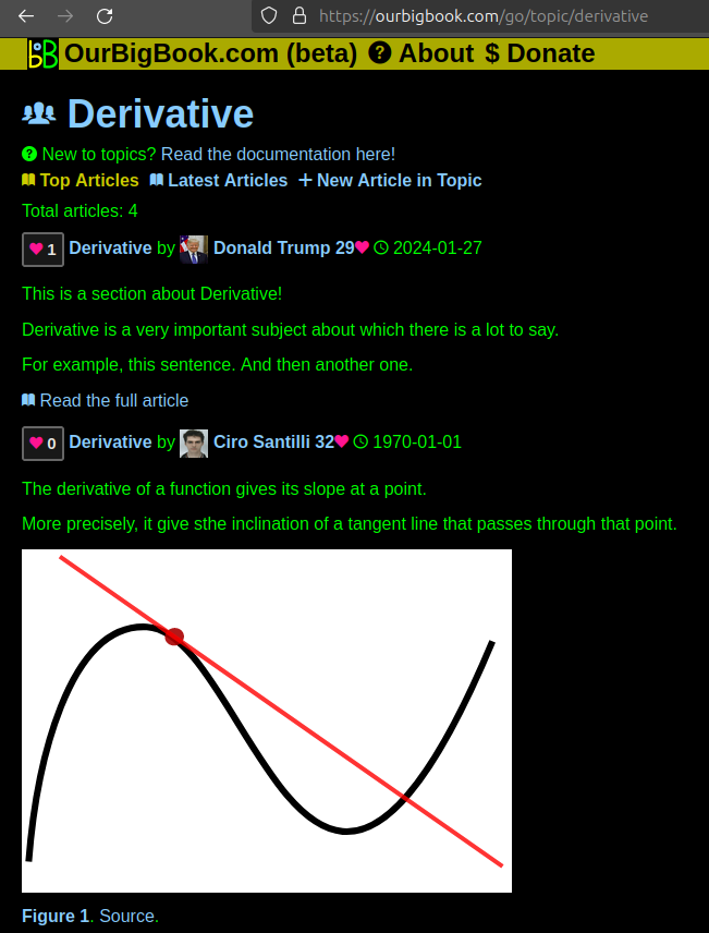
Figure 1. Screenshot of the "Derivative" topic page. View it live at: ourbigbook.com/go/topic/derivativeVideo 2. OurBigBook Web topics demo. Source. - local editing: you can store all your personal knowledge base content locally in a plaintext markup format that can be edited locally and published either:This way you can be sure that even if OurBigBook.com were to go down one day (which we have no plans to do as it is quite cheap to host!), your content will still be perfectly readable as a static site.
- to OurBigBook.com to get awesome multi-user features like topics and likes
- as HTML files to a static website, which you can host yourself for free on many external providers like GitHub Pages, and remain in full control

Figure 2. You can publish local OurBigBook lightweight markup files to either OurBigBook.com or as a static website.
Figure 3. Visual Studio Code extension installation.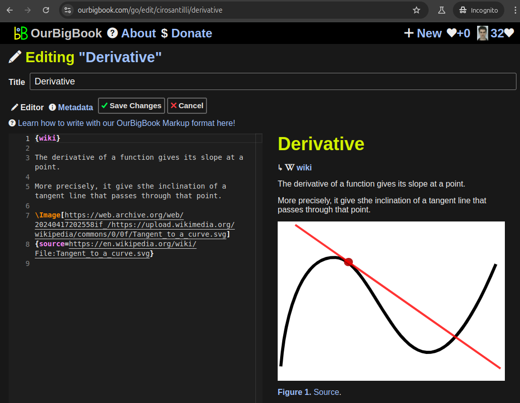
Figure 5. . You can also edit articles on the Web editor without installing anything locally. Video 3. Edit locally and publish demo. Source. This shows editing OurBigBook Markup and publishing it using the Visual Studio Code extension. 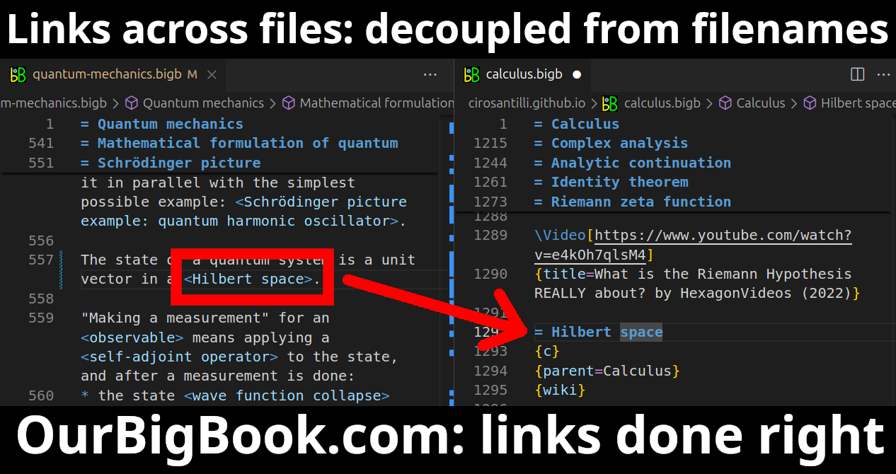
- Infinitely deep tables of contents:
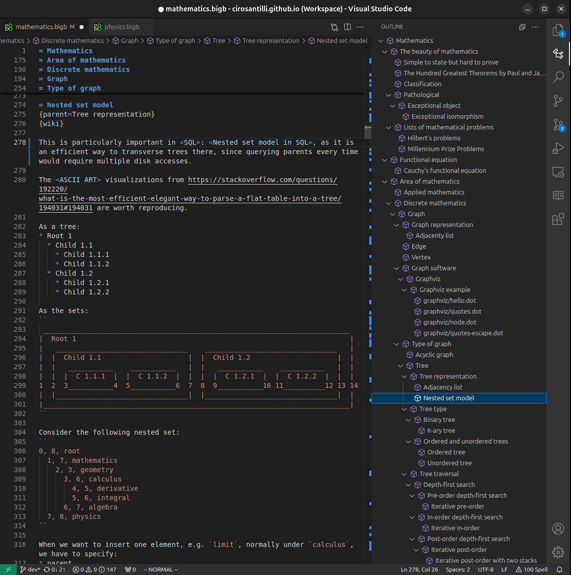
All our software is open source and hosted at: github.com/ourbigbook/ourbigbook
Further documentation can be found at: docs.ourbigbook.com
Feel free to reach our to us for any help or suggestions: docs.ourbigbook.com/#contact