Topics (384k) Articles (398k) Users (410) Discussions (242) Comments (383) Files (13k) New article

Oxyhemoglobin is oxygen-bound hemoglobin. Binding is reversible: an available haem site can acquire or release an oxygen molecule as conditions change. A fully occupied adult hemoglobin tetramer carries four oxygen molecules, one per haem site.[ref]
Hemoglobin by Radiometer. Structure and function; additional Oxygen saturation parameter page, Physiological background
Oxygenation describes ligand occupancy and should not be confused with the ferric haem state defining methemoglobin.A quantitative description must specify its denominator. The manufacturer's functional oxygen saturation iswhere HHb denotes unoxygenated, oxygen-capable hemoglobin. A fractional oxyhemoglobin quantity instead divides by total hemoglobin, including specified non-oxygen-carrying derivatives.[ref]
Hemoglobin by Radiometer. Additional Oxygen saturation parameter page, definition of functional hemoglobin; total-hemoglobin definition
The concentrations must use a consistent haem-equivalent convention.For an original four-component example, suppose those equivalent concentrations are 80 units oxyhemoglobin, 5 units deoxyhemoglobin, 10 units carboxyhemoglobin and 5 units methemoglobin. Then functional saturation is , whereas the oxyhemoglobin fraction of the full 100-unit total is . These describe the same hypothetical mixture with different denominators. Neither percentage alone states the total oxygen amount in a blood volume.
A separate amount calculation assumes 2 micromoles of tetramers, all with four functional sites, at mean occupancy . Bound oxygen is thenAny oxygen dissolved outside hemoglobin is an additional pool. Concentration, binding fraction and gas partial pressure are related through a physical model, rather than being interchangeable measurements.
Primary spectral work measured oxyhemoglobin alongside other derivatives, providing coefficient data for distinguishing their contributions to absorbance.[ref]
Absorption spectra of human fetal and adult oxyhemoglobin, de-oxyhemoglobin, carboxyhemoglobin, and methemoglobin by W. G. Zijlstra and others. Abstract, 450–1000 nm derivative spectra
This connects molecular oxygenation with a measurable optical signal while retaining the distinction between one derivative and the whole sample.Carboxyhemoglobin is hemoglobin with carbon monoxide bound at a haem site. Carbon monoxide competes with oxygen for binding; a CO-occupied site is unavailable for oxygen while CO remains bound.[ref]
Hemoglobin by Radiometer. Additional manufacturer parameter page, Carboxyhemoglobin, What is COHb?
This differs from methemoglobin, where the defining change is oxidation of haem iron. Binding carbon monoxide is not itself the same operation as converting an available ferrous site into a ferric site.An original independent-site equilibrium model illustrates competition. Let oxygen and carbon-monoxide binding weights be and , with values defined on the same pressure basis. For empty, oxygen-bound and CO-bound states,With stipulated and no carbon monoxide, the oxygen-bound fraction is . Adding a competing weight changes it to and gives CO the same fraction. These weights are hypothetical; this model omits the cooperative and conformational behavior of real hemoglobin.
The calculation explains why knowing the oxygen partial pressure alone does not identify the amount bound to hemoglobin. Available-site count, competing ligands and binding properties also enter. Even a high occupied fraction among the remaining oxygen-capable sites does not imply that all haem sites are oxygenated.
Co-oximetry separates carboxyhemoglobin from other derivatives using their spectral responses. The 1991 fetal/adult hemoglobin study quantified derivative absorptivities and emphasized that even small spectral differences affect multicomponent analysis.[ref]
Absorption spectra of human fetal and adult oxyhemoglobin, de-oxyhemoglobin, carboxyhemoglobin, and methemoglobin by W. G. Zijlstra and others. Abstract
Identifying a derivative therefore requires a suitable measurement model; total hemoglobin concentration alone leaves its ligand composition unresolved.Methemoglobin is a hemoglobin derivative with oxidized ferric iron at a haem site. The ferric site cannot perform the usual reversible oxygen binding of an available ferrous site. The distinction is one of oxidation state, not merely whether an oxygen molecule currently occupies the site.[ref]
Hemoglobin by Radiometer. Additional manufacturer parameter page, Methemoglobin, What is MetHb?
An adult hemoglobin tetramer contains four haem sites, so site occupancy and the state of the whole tetramer must not be silently conflated.Deoxyhemoglobin retains the capacity to bind oxygen at its available sites. A ferric site in methemoglobin instead requires chemical reduction to restore the corresponding ferrous state. Formation of oxyhemoglobin is therefore different from accumulating ferric haem. Changes in the surrounding protein also influence the properties of remaining sites; a site-count calculation alone is not a complete cooperative-binding model.
For an original capacity calculation, suppose a sample contains 100 micromoles of haem sites, with 20 micromoles ferric and the other 80 capable of reversible oxygen binding. The latter can bind at most 80 micromoles of oxygen in a one-molecule-per-site model. If 60 micromoles are oxygenated, the oxygenated fraction of all haem sites is , while the occupied fraction of the stipulated capable sites is . A denominator change produces different percentages without changing the sample.
Experimentally, spectrophotometry distinguishes derivatives through calibrated absorption spectra. A 1991 primary study measured human fetal and adult oxyhemoglobin, unoxygenated hemoglobin, carboxyhemoglobin and methemoglobin spectra over 450–1000 nm.[ref]
Absorption spectra of human fetal and adult oxyhemoglobin, de-oxyhemoglobin, carboxyhemoglobin, and methemoglobin by W. G. Zijlstra and others. Abstract, measured millimolar absorptivities
The relevant inverse problem has the formwhere path length , derivative concentrations and their calibrated absorption coefficients determine the modeled absorbance. Co-oximetry uses multiple spectral measurements to estimate such derivative fractions. A single colour observation cannot generally separate several unknown components.Deoxyhemoglobin is the unoxygenated form of hemoglobin whose available haem sites can reversibly bind oxygen. Releasing oxygen produces a binding-state change, rather than the ferric iron state defining methemoglobin. Carboxyhemoglobin differs again because a competing carbon monoxide ligand occupies a binding site. These distinctions matter when interpreting the components of an optical measurement.
In the instrument convention for functional oxygen saturation, HHb denotes the unoxygenated, oxygen-capable component. Its concentration appears with oxyhemoglobin in the denominator:The HHb label is not an instruction to treat deoxygenation as addition of a hydrogen molecule. Functional saturation also uses a different denominator from fractional oxyhemoglobin referenced to total hemoglobin.[ref]
Oxyhemoglobin by Radiometer. FO2Hb versus oxygen saturation, definition of the functional denominator
An original independent-site model illustrates a further counting distinction. Let each of four sites have oxygen-occupancy probability , independently. The probability that a tetramer has exactly occupied sites isAt , half the sites are empty on average, while only of tetramers have all four sites empty. A haem-equivalent unoxygenated fraction therefore need not equal the fraction of wholly unliganded tetramers. Real hemoglobin binding is cooperative, so independence here is a stated teaching assumption rather than a physiological model.
Deoxyhemoglobin and oxyhemoglobin have distinguishable absorption spectra. A primary study measured the absorptivities of these and other human hemoglobin derivatives across 450–1000 nm, including fetal/adult differences relevant to multicomponent analysis.[ref]
Absorption spectra of human fetal and adult oxyhemoglobin, de-oxyhemoglobin, carboxyhemoglobin, and methemoglobin by W. G. Zijlstra and others. Abstract
A controlled oxygenation comparison can therefore follow changing spectral contributions without inferring oxygen amount from apparent colour alone. The measured path length, concentrations and calibrated component spectra remain necessary inputs.Fetal hemoglobin, commonly HbF, is a hemoglobin form built from two alpha-globin and two gamma-globin subunits, written . The major adult form, hemoglobin A, instead has the combination. Fetal identity concerns subunit composition; oxyhemoglobin and deoxyhemoglobin concern oxygen occupancy. The same fetal molecule can change its oxygenation state without becoming adult hemoglobin.
Primary structural evidence is available in the human fetal deoxyhemoglobin crystal structure 1FDH. Its experimental record identifies an alpha/gamma tetramer determined by X-ray diffraction at resolution.[ref]
Structure of human foetal deoxyhaemoglobin by J. A. Frier and M. F. Perutz. RCSB PDB 1FDH, Experimental Data Snapshot, Biological Assembly and Macromolecules
The related paper connects structural differences with weaker interaction with 2,3-Bisphosphoglycerate, an effector relevant to oxygen-binding behavior.[ref]Structure of human foetal deoxyhaemoglobin by J. A. Frier and M. F. Perutz. Indexed primary publisher abstract, diphosphoglycerate-site comparison
A structure constrains a mechanism, while an affinity curve requires a functional measurement under specified conditions.An original Hill equation model illustrates what higher oxygen affinity means. Let fractional occupancy at oxygen partial pressure beWith the same stipulated , compare midpoints and 30 in arbitrary pressure units. At , their occupancies areThe lower midpoint gives greater occupancy at the same pressure. It does not imply that the protein carries more than one oxygen molecule per available haem site. These midpoints and the exponent are teaching parameters, not measured fetal/adult reference values.
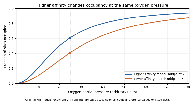
Original Hill models showing how a lower binding midpoint raises occupancy at the same oxygen pressure; parameters are illustrative
. Details. Subunit identity also matters analytically. A primary 1991 experiment measured fetal and adult hemoglobin derivative spectra over 450–1000 nm and found differences relevant to multicomponent analysis.[ref]
Absorption spectra of human fetal and adult oxyhemoglobin, de-oxyhemoglobin, carboxyhemoglobin, and methemoglobin by W. G. Zijlstra and others. Abstract
An optical model that distinguishes oxygenation states should therefore also justify which protein-form spectra it uses. Oxygen occupancy, subunit composition and total protein concentration are different quantities.Hemoglobin A, commonly HbA, is the major adult human hemoglobin assembly made of two alpha-globin and two beta-globin chains, .[ref] It is one hemoglobin form rather than a name for every hemoglobin molecule present in an adult. Fetal hemoglobin has a different non-alpha-chain composition.
Human deoxyhemoglobin structure 4HHB is a primary structural example. The associated study refined the model at using room-temperature diffraction data recorded on film with a synchrotron X-ray source.[ref]
The crystal structure of human deoxyhaemoglobin at 1.74 A resolution by G. Fermi and others. Primary abstract and RCSB Experimental Data
This connects the abstract notation with experimentally determined chain and haem organization.Composition and oxygenation are independent descriptors. Hemoglobin A can occur in oxygenated or deoxygenated states while retaining its alpha/beta identity. Each intact tetramer contains four haem sites, so a concentration expressed per tetramer differs by a factor of four from a haem-equivalent concentration.
For an original accounting example, take a pure preparation at 1.00 g/L and use a stipulated nominal tetramer molar mass of . Thenand haem-site concentration is about . If all sites are functional and mean oxygen occupancy is 0.80, bound oxygen concentration is about . Oxygen dissolved outside the protein is an additional pool. The assumptions are explicit teaching conditions, not a physiological reference range.
A primary spectral study measured both fetal and adult hemoglobin derivatives and found differences relevant to multicomponent analysis.[ref]
Absorption spectra of human fetal and adult oxyhemoglobin, de-oxyhemoglobin, carboxyhemoglobin, and methemoglobin by W. G. Zijlstra and others. Abstract
A measurement model should therefore state which protein-form and oxygenation-state spectra it uses, alongside the basis of the reported concentration.The sinoatrial node is a group of specialized cardiac cells that normally initiates the electrical rhythm of the heart. Its pacemaker cells gradually undergo depolarization between action potentials instead of remaining at a stable resting voltage. Their activity spreads into surrounding atrial cardiac muscle through electrical coupling. [ref]
Anatomy and Physiology 2e by J. Gordon Betts and others. Section 19.2, Autorhythmicity of Cardiac Cells and the Sinoatrial Node
The rhythm can arise without a separate motor nerve triggering every beat. Autonomic inputs modulate its rate, while conduction through other cardiac structures organizes the order of activation. Pacemaker activity, propagation and mechanical contraction are therefore different stages: recording the first does not by itself prove that every later stage proceeds normally.
The right ventricle is the heart chamber receiving blood from the right atrium through the tricuspid valve and ejecting it through the pulmonary valve into the pulmonary trunk.[ref]
Anatomy and Physiology 2e by J. Gordon Betts and others. Section 19.1, Chambers and Circulation through the Heart
Its output enters the pulmonary circulation rather than passing directly into the aorta.The chamber's volume changes through the cardiac cycle. Define stroke volume as end-diastolic minus end-systolic volume:The second ratio is the right ventricular ejection fraction. In an original example with and , stroke volume is and ejection fraction . At beats per minute, multiplying stroke volume by rate gives .
That last conversion represents net forward output only under its stated assumptions. Significant backward or alternate flow can separate chamber-volume displacement from the amount delivered onward. Nor does an ejection fraction alone provide the chamber's absolute output: chambers with different initial volumes can share the same fraction.
In a steady serial-circulation model without shunts or sustained storage, right- and left-sided net outputs must balance. A persistent difference would accumulate blood somewhere in the system. This conservation statement does not require the two ventricles to generate equal pressure or perform identical mechanical work. Anatomy, volume displacement, forward flow and pressure are distinct but connected observations.
The coronary sinus is a major venous collecting channel of the heart that opens into the right atrium. It receives blood from cardiac veins, including the great cardiac vein, middle cardiac vein and small cardiac vein.[ref]
Anatomy and Physiology 2e by J. Gordon Betts and others. Section 19.1, Coronary Veins and Figure 19.15
It belongs to the venous return pathway, distinct from the coronary arteries supplying the myocardium.The collecting-channel arrangement explains its use in a regional balance. Incoming flows combine before the sample or outflow observation is taken. In a steady model with negligible local storage,If represented inflows are and , the sinus outflow is . This is a chosen continuity example, not a normal flow value.
A measurement at the coronary sinus does not automatically represent every myocardial route. If another modeled pathway returns directly to a chamber, the total venous return from the represented heart is , while the sinus observation remains . Treating the latter as the whole total would miss about in this example.
Likewise, venous oxygen content sampled from a collecting channel is a mixture weighted by its contributing flows. It should be paired with flow and arterial measurements representing compatible tissue when calculating coronary oxygen uptake. Knowing the sampling site is part of defining the biological quantity, not simply a label attached after measurement.
The right atrium is the heart chamber receiving blood from the superior vena cava, inferior vena cava and coronary sinus. Its principal outlet is the tricuspid valve, leading to the right ventricle.[ref]
Anatomy and Physiology 2e by J. Gordon Betts and others. §19.1, Right Atrium
This places it between systemic venous return and the pump supplying the pulmonary circulation.The right atrium is both a junction and a chamber that can store blood. For a simple volume balance,where sums the incoming blood flow rates and is net outward flow through the tricuspid valve. If a constructed inflow exceeds outflow by L/min for min, atrial volume increases by 50 mL. Such an imbalance cannot persist indefinitely in a fixed-volume steady-state model. Over a repeating cardiac cycle, the time-averaged inflow and outflow must balance if there is no net accumulation.
The joining streams can have different venous oxygen content. Their location in one chamber does not guarantee instantaneous perfect mixing at every sample position. Calculating mixed venous oxygen content requires a representative mixture, with the entering blood flow rates supplying the weights. This connects an anatomical chamber to the sampling assumptions behind the Fick principle.
The tricuspid valve is the right atrioventricular heart valve, between the right atrium and right ventricle. It permits filling from the right atrium and limits backward blood flow during ventricular contraction.[ref]
The leaflets are supported by chordae tendineae attached to papillary muscles. Rising ventricular pressure brings the leaflets together; tension in the supporting apparatus helps prevent their displacement into the right atrium. The papillary muscles do not need to pull the leaflets open to initiate filling.[ref]
Anatomy and Physiology 2e by J. Gordon Betts and others. §19.1, Right Ventricle and Heart Valve Structure and Function
A useful mechanical analogy is a pressure-loaded flap restrained by tethers. The pressure difference supplies a load, while the tethers limit how far the flap can move. The analogy distinguishes the role of the chordae tendineae from the action of a motorized door.
The valve also determines how to interpret right ventricular ejection fraction. Suppose a constructed ventricular contraction reduces cavity volume by 80 mL, but 20 mL of that loss returns through the tricuspid valve into the right atrium. Only 60 mL remains for forward transport through the pulmonary valve, if other transfers are negligible. The measured cavity-volume change is still 80 mL. An ejection fraction calculated from chamber volumes therefore does not by itself measure the useful forward stroke volume of the pulmonary circulation.
The pulmonary valve is the heart valve between the right ventricle and pulmonary trunk. During forward ventricular ejection it allows blood to enter the pulmonary circulation; closure limits return from the pulmonary trunk into the relaxing right ventricle.[ref]
Its pocket-like cusps respond to the changing pressure difference. Unlike the tricuspid valve, the pulmonary valve has no attached chordae tendineae or papillary muscles.[ref]
Anatomy and Physiology 2e by J. Gordon Betts and others. §19.1, Heart Valve Structure and Function, pulmonary semilunar valve
Thus the same direction-control task is achieved by different supporting structures at the chamber inlet and outlet.A deliberately idealized valve model sets forward blood flow to zero when the downstream pressure exceeds the upstream pressure, and permits positive flow when ventricular pressure exceeds trunk pressure. This model captures direction but omits the inertia and deformation of real cusps. It is useful for checking the sign of a flow term, not for predicting an exact opening time.
For example, if a constructed cycle sends 80 mL forward and 10 mL backward across the pulmonary valve, net transport is 70 mL. Counting only the positive part of blood flow overestimates net delivery. This is why stroke volume inferred from changing chamber volume and net forward flow in the pulmonary trunk require compatible assumptions about valve function.
The interventricular septum is the partition separating the cavities of the right ventricle and left ventricle. It is internal to the heart, unlike the external anterior interventricular sulcus and posterior interventricular sulcus. The distinction can be seen by comparing an intact surface view with an opened view of the ventricular chambers.[ref]
An intact partition lets the two ventricular pumps operate at different blood pressure values without a direct open route between their cavities. An elementary physical model makes the role clear: if a hypothetical opening connected two chambers, a pressure gradient across that opening could drive an additional blood flow. Conservation would then require a term for transfer between chambers, in addition to the flow through their respective heart valves. This thought experiment explains why a partition is part of the circulation's topology, rather than merely a line on an anatomical drawing.
The interventricular septum also belongs to the mechanically connected ventricular structure. Consequently, measuring right ventricular ejection fraction and measuring left ventricle function describe two chambers in one coupled heart, rather than two unrelated pumps. For an anatomical identification exercise, first find the two cavities, then identify their common separating wall; locating a surface groove alone does not establish the wall's thickness or internal structure.
Pinned article: Introduction to the OurBigBook Project
Welcome to the OurBigBook Project! Our goal is to create the perfect publishing platform for STEM subjects, and get university-level students to write the best free STEM tutorials ever.
Everyone is welcome to create an account and play with the site: ourbigbook.com/go/register. We belive that students themselves can write amazing tutorials, but teachers are welcome too. You can write about anything you want, it doesn't have to be STEM or even educational. Silly test content is very welcome and you won't be penalized in any way. Just keep it legal!
Intro to OurBigBook
. Source. We have two killer features:
- topics: topics group articles by different users with the same title, e.g. here is the topic for the "Fundamental Theorem of Calculus" ourbigbook.com/go/topic/fundamental-theorem-of-calculusArticles of different users are sorted by upvote within each article page. This feature is a bit like:
- a Wikipedia where each user can have their own version of each article
- a Q&A website like Stack Overflow, where multiple people can give their views on a given topic, and the best ones are sorted by upvote. Except you don't need to wait for someone to ask first, and any topic goes, no matter how narrow or broad
This feature makes it possible for readers to find better explanations of any topic created by other writers. And it allows writers to create an explanation in a place that readers might actually find it.
Figure 1. Screenshot of the "Derivative" topic page. View it live at: ourbigbook.com/go/topic/derivativeVideo 2. OurBigBook Web topics demo. Source. - local editing: you can store all your personal knowledge base content locally in a plaintext markup format that can be edited locally and published either:This way you can be sure that even if OurBigBook.com were to go down one day (which we have no plans to do as it is quite cheap to host!), your content will still be perfectly readable as a static site.
- to OurBigBook.com to get awesome multi-user features like topics and likes
- as HTML files to a static website, which you can host yourself for free on many external providers like GitHub Pages, and remain in full control

Figure 2. You can publish local OurBigBook lightweight markup files to either OurBigBook.com or as a static website.
Figure 3. Visual Studio Code extension installation.
Figure 5. . You can also edit articles on the Web editor without installing anything locally. Video 3. Edit locally and publish demo. Source. This shows editing OurBigBook Markup and publishing it using the Visual Studio Code extension. 
- Infinitely deep tables of contents:



All our software is open source and hosted at: github.com/ourbigbook/ourbigbook
Further documentation can be found at: docs.ourbigbook.com
Feel free to reach our to us for any help or suggestions: docs.ourbigbook.com/#contact