Protein targeting directs a protein to its cellular destination. Synthesis in the cytosol does not imply that the protein works there.
For entry into the endoplasmic reticulum, distinguish three questions: does the nascent chain reach the membrane, does part of it cross into the ER lumen, and does a segment remain embedded as a transmembrane protein? Cotranslational protein translocation couples these events to biological translation. A signal peptide can initiate delivery without specifying the eventual destination of the mature protein.
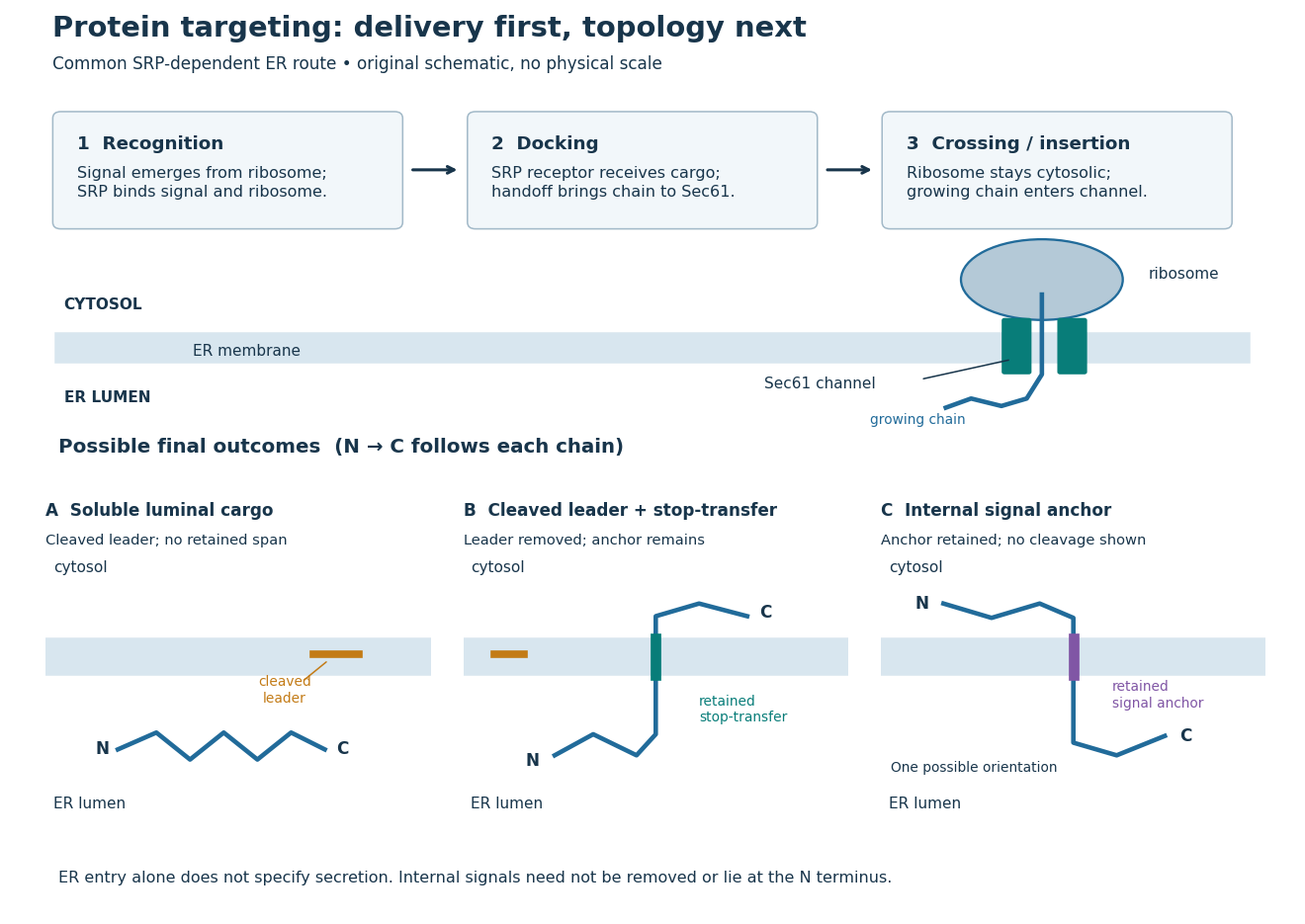
Protein targeting to the endoplasmic reticulum: delivery and three possible outcomes
. Original pathway and membrane topology schematic. The upper row separates SRP recognition, membrane docking and passage through a translocon. Lower cartoons stipulate a soluble cargo, a cleaved signal peptide plus retained stop-transfer sequence, and an uncleaved internal signal anchor. Each drawn terminus is labeled; the internal-anchor orientation is one possibility. Shapes and lengths are illustrative, with no molecular coordinates or measured timings. Details.
This distinction matters experimentally. Finding a protein in a membrane-containing centrifugation pellet could reflect surface binding or protein aggregation. The microsomal protein import experiment instead asks whether an intact membrane prevents an external protease from reaching the product. That tests accessibility across a barrier, rather than proximity to it.
Delivery of a protein from its producing cell to the extracellular environment, enabling communication and extracellular functions.
A signal peptide is a short targeting segment of a newly made polypeptide chain. A familiar ER signal sequence lies near the N-terminus and is removed during entry; Günter Blobel and Bernhard Dobberstein's 1975 experiments linked a larger immunoglobulin light chain precursor to a processed product.[ref]
Transfer of Proteins across Membranes I by Günter Blobel and Bernhard Dobberstein. Transfer of proteins across membranes. I, Introduction p. 836; Figures 5 and 14; Discussion pp. 845–847
Cleavage is a separate question from recognition. Not every targeting signal is a cleaved signal peptide: an internal signal anchor can remain part of a transmembrane protein. Conversely, loss of a segment does not by itself prove membrane crossing. Compare the independent size and accessibility readouts in the microsomal protein import experiment.
A signal peptidase removes a cleavable signal peptide; a suitable cleavage site and access are required.[ref]
The Endoplasmic Reticulum. Molecular Biology of the Cell, 4th edition by Bruce Alberts and others; “The ER Signal Sequence Is Removed from Most Soluble Proteins After Translocation”: adjacent cleavage-site requirement
For the stipulated -amino acid residue precursor under ER signal sequence, removal of the first amino acid residues leaves . Now stipulate that positions – of that precursor are instead retained as a stop-transfer sequence. Their positions in the mature chain become –, because the numbering origin changes when the first positions are removed. The mature chain still has amino acid residues, but its membrane-spanning segment is retained. A second hypothetical chain with an uncleaved internal signal anchor loses no positions at all. These are conditional bookkeeping examples, not predictions from invented biological sequences.
A signal anchor combines targeting or initiation of membrane passage with a retained membrane-spanning segment. Its orientation determines which flanking part enters the ER lumen; a single internal signal can yield either orientation.[ref]
Membrane Protein Insertion at the Endoplasmic Reticulum by Sichen Shao and Ramanujan S. Hegde. Shao and Hegde, 2011; “Cotranslational Membrane Protein Topogenesis”, Figure 5
In the figure's stipulated internal-anchor example, the N-terminus remains in the cytosol and the C-terminus enters the ER lumen. Reversing that orientation would reverse which end is protected from an externally added protease. Merely counting one membrane-spanning segment cannot choose between the two.
A stop-transfer sequence interrupts ongoing passage of a polypeptide chain across a membrane and retains a membrane-spanning segment. Protein targeting, insertion and subsequent sorting involve distinguishable sequence instructions.[ref]
Intracellular protein topogenesis by Günter Blobel. Blobel, Intracellular protein topogenesis, 1980; abstract: signal, stop-transfer, sorting and insertion sequences
In the figure's cleaved-leader example, assume the N-terminus of the mature chain has entered the ER lumen, a stop-transfer sequence remains in the membrane, and the remaining C-terminus is made on the cytosol side. The outcome differs from releasing the entire mature chain into the ER lumen. The latter is soluble cargo, potentially retained there or transported onward; entry alone does not establish secretion.
Organelle protein import brings externally synthesized proteins into an organelle. Many plastid proteins made from nuclear genes carry targeting information recognized by machinery that transports them across the envelope membranes. This delivery enables nuclear genes to supply functions inside a plastid, but the presence of targeting information is not itself evidence that a gene was acquired through endosymbiotic gene transfer. [ref]
Origin and Evolution of Plastids and Photosynthesis in Eukaryotes by Geoffrey I. McFadden. “How to tame your symbiont”: targeting, membrane passage and host-derived proteins
For the endoplasmic reticulum, cotranslational protein translocation illustrates how organelle protein import can begin before a polypeptide chain is complete. Import into the ER lumen and insertion as a transmembrane protein are different outcomes; neither alone establishes subsequent secretion from the biological cell.
A test of whether a protein crosses a specified organelle boundary, with controls distinguishing import from surface binding or membrane rupture.
Cotranslational protein translocation is passage of a growing polypeptide chain across a membrane during biological translation. SRP recognizes an exposed hydrophobic ER signal sequence. The SRP receptor enables handoff to the Sec61 complex. Biological translation can slow during delivery; the ribosome remains on the cytosol side.[ref]
Membrane Protein Insertion at the Endoplasmic Reticulum by Sichen Shao and Ramanujan S. Hegde. Shao and Hegde, Membrane Protein Insertion at the Endoplasmic Reticulum, 2011; “Cotranslational Targeting”, Figure 3
The useful engineering feature is coordination: a targeting instruction can become accessible while the rest of its cargo is still being made. Recognition, docking and crossing remain distinct operations. A defect in any one can prevent successful import even when the protein primary structure contains an appropriate signal.
This route is not universal. Many tail-anchored proteins use delivery after biological translation, and their membrane-spanning segment lies near the C-terminus. Thus protein targeting signals need not be at the N-terminus.[ref]
Membrane Protein Insertion at the Endoplasmic Reticulum by Sichen Shao and Ramanujan S. Hegde. Shao and Hegde, 2011; “Posttranslational Membrane Protein Topogenesis”, Figure 6
The signal recognition particle is an RNA–protein targeting complex linking a ribosome-associated nascent chain to the SRP receptor.[ref]
Membrane Protein Insertion at the Endoplasmic Reticulum by Sichen Shao and Ramanujan S. Hegde. Shao and Hegde, 2011; “Cotranslational Targeting”, Figure 3
The bacterial homolog of the SRP54 protein, containing a GTPase involved in cotranslational membrane targeting.
The conserved eukaryotic SRP subunit containing a signal-sequence-binding region and a GTPase domain; bacterial Ffh is its homolog.
The SRP receptor receives the SRP–ribosome–nascent chain complex for handoff to the translocon.[ref]
Membrane Protein Insertion at the Endoplasmic Reticulum by Sichen Shao and Ramanujan S. Hegde. Shao and Hegde, 2011; “Cotranslational Targeting”, Figure 3
A translocon is membrane machinery through which a polypeptide chain crosses or enters a membrane. In endoplasmic reticulum protein targeting, the Sec61 complex supplies the central channel.[ref]
Membrane Protein Insertion at the Endoplasmic Reticulum by Sichen Shao and Ramanujan S. Hegde. Shao and Hegde, 2011; “The Sec61 Translocon”, Figure 4
The Sec61 complex supports passage across the endoplasmic reticulum membrane and lateral entry of membrane-spanning segments into its lipid bilayer.[ref]
Membrane Protein Insertion at the Endoplasmic Reticulum by Sichen Shao and Ramanujan S. Hegde. Shao and Hegde, 2011; “The Sec61 Translocon”, Figure 4
In 1975, Günter Blobel and Bernhard Dobberstein combined cell-free translation with dog-pancreas microsomes. Density-gradient centrifugation isolated the membranes; removal of resident ribosomes allowed reconstitution with rabbit-reticulocyte ribosome subunits. Added messenger RNA encoded an immunoglobulin light chain; globin provided a comparison. Products made with or without membranes were challenged with proteases and analyzed by SDS-PAGE and autoradiography. Membranes present during synthesis yielded processed, protected light chain; globin remained susceptible. Adding membranes after precursor synthesis failed to reproduce import. These comparisons support selective passage during synthesis in this preparation.[ref]
The companion paper specifies radioactive amino acid incorporation, ice-cold trypsin plus chymotrypsin treatment, a slab-gel electrophoresis apparatus and dried-gel exposure to X-ray film. Product-size analysis distinguishes a preserved full-size product from residual labeled fragments.[ref]
Transfer of Proteins across Membranes I by Günter Blobel and Bernhard Dobberstein. Methods pp. 837–838: proteolysis, SDS-PAGE and autoradiography; Discussion p. 847
Protection was incomplete. The authors considered leaky vesicles, and warned that heat treatment could damage processing as well as ribosome attachment. Their experiments did not identify SRP or the Sec61 complex. Nor does failure of late membrane addition establish that all organelle protein import must occur during synthesis.[ref]
Transfer of Proteins across Membranes II by Günter Blobel and Bernhard Dobberstein. pp. 858–859: incomplete protection; p. 861: heat-inactivation caveat; p. 862: alternatives for failed processing
A protease protection assay asks whether a barrier prevents an added protease from reaching a protein. A membrane-disruption comparison tests whether protection depends on that barrier.
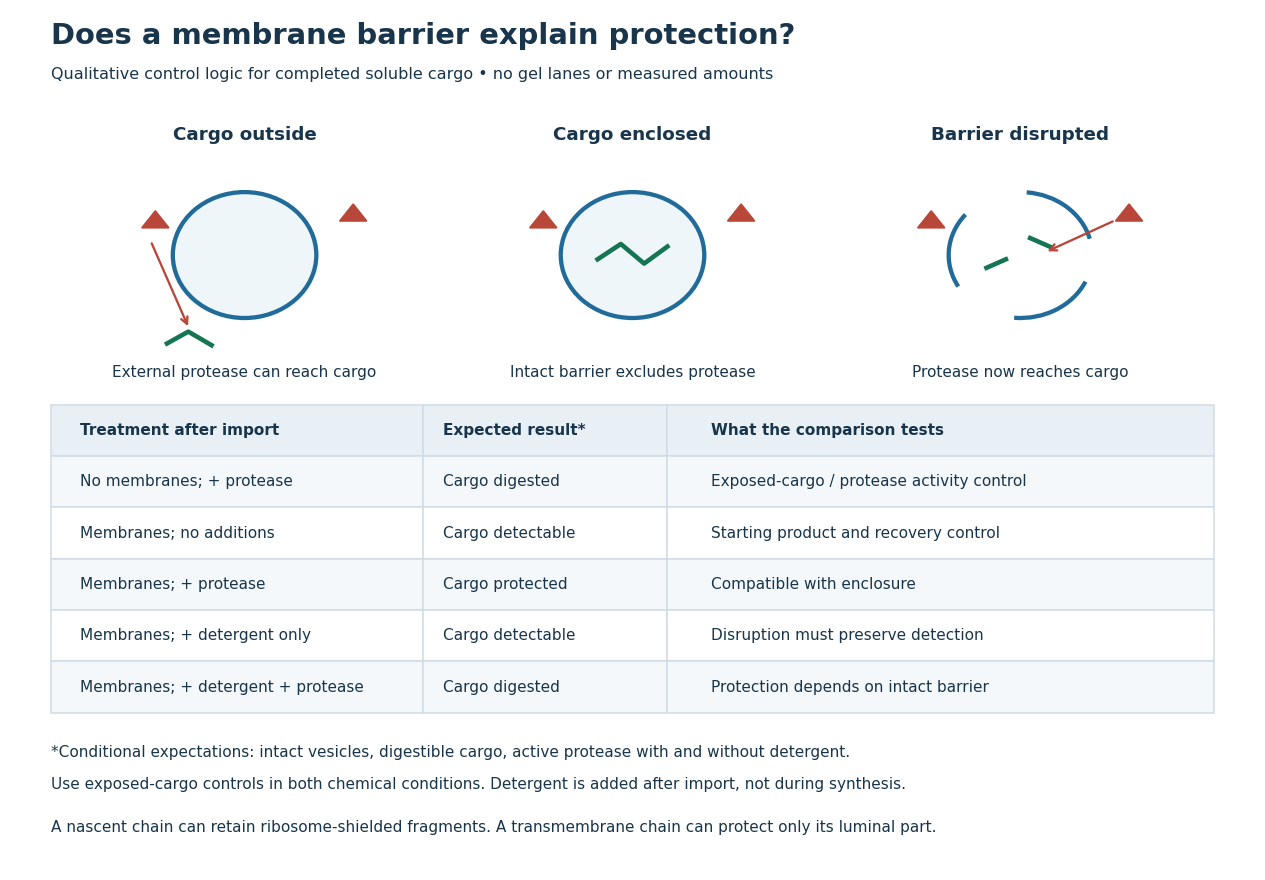
Protease protection assay: accessibility and control logic
. Original qualitative decision diagram for a completed soluble cargo. Icons and words describe expected outcomes under stated assumptions; they are not an autoradiography readout, copied historical lanes, measured survival fractions or simulated experimental intensities. Detergent is added after the import reaction. The historical nascent chain experiment also retained small ribosome-protected fragments, which this completed-cargo model deliberately distinguishes. Details.
The membrane-disruption test was performed by David D. Sabatini and Günter Blobel in 1970. Rat-liver microsomes purified by sucrose centrifugation incorporated radioactive leucine during cell-free translation. Samples received cold trypsin and chymotrypsin, with or without membrane-solubilizing detergents. Electron microscopy checked vesicle preservation; size-exclusion chromatography separated protected fragment sizes. Large membrane-associated fragments lost protection after disruption, whereas small ribosome-protected fragments persisted. An exposed ribosome preparation remained digestible when mixed with microsomes, checking that membranes did not simply inhibit the proteases.[ref]
Controlled Proteolysis of Nascent Polypeptides in Rat Liver Cell Fractions II by David D. Sabatini and Günter Blobel. Controlled proteolysis of nascent polypeptides in rat liver cell fractions. II, Methods p. 147; activity control p. 148; Figure 5C–D and text p. 152; Discussion pp. 155–156
To read the qualitative diagram, first compare untreated aliquots: they establish that detectable cargo exists before digestion. Next compare external protease with intact versus disrupted membranes. Persistence only while the barrier remains intact supports enclosure, provided the exposed-cargo control confirms active protease in both chemical conditions. The detergent-only aliquot checks that disruption itself does not destroy recovery or detection. Adding membranes after synthesis tests a different alternative: capture of a completed product rather than entry during biological translation.
This is an argument from controlled accessibility. Binding predicts membrane association but generally leaves an exposed cargo digestible. Enclosure predicts protection that depends on membrane integrity. However, protein aggregation, intrinsic resistance to proteolysis, inactive protease or undetected small fragments can obscure this distinction. For a transmembrane protein, expect protection of the enclosed portion rather than necessarily the full chain. Combine fragment size, compartment controls and membrane topology; a protected signal alone does not uniquely specify the whole protein's location.
Articles by others on the same topic
There are currently no matching articles.