A chloroplast is a photosynthetic plastid in plant and algae biological cells. The green-plant chloroplast modeled here has a chloroplast envelope surrounding chloroplast stroma and internal thylakoids. Light-dependent reactions occur at the thylakoid membrane; the Calvin cycle operates in the chloroplast stroma.[ref]
8.1 Overview of Photosynthesis. Biology 2e, 8.1, Basic Photosynthetic Structures and The Two Parts of Photosynthesis
Cyanobacteria perform oxygenic photosynthesis without chloroplasts: the prokaryotic biological cell itself contains the photosynthetic machinery. Thus chloroplast identifies an organelle, while oxygenic photosynthesis identifies a process. Endosymbiotic theory explains their historical connection; organelle protein import explains how many nuclear-encoded proteins reach the modern organelle.
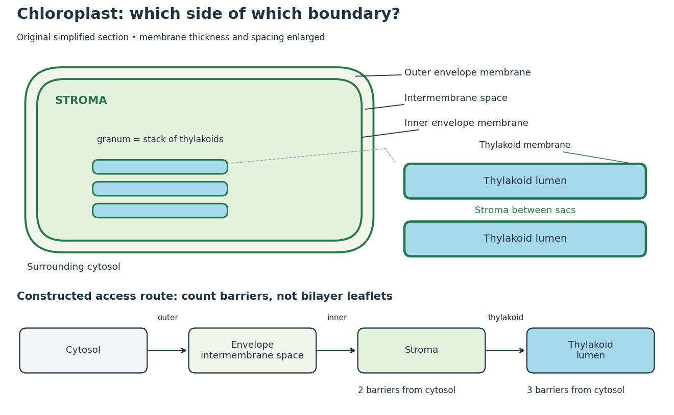
Chloroplast compartments and boundary counting
. Original simplified section and compartment-access model, not a micrograph or scale drawing. The enlargement separates thylakoid lumen from the chloroplast stroma between stacked sacs. The route counts crossings of membrane barriers, not molecular leaflets; actual permeability and transport mechanisms are omitted. Details.
The useful feature is selective separation: enzymes and substrates can share an organelle without sharing a compartment. For a constructed accessibility example, put a soluble enzyme in chloroplast stroma and its substrate in thylakoid lumen. Sharing the same chloroplast does not permit direct contact; contact requires a transport or boundary-disruption step. Placing the substrate between adjacent thylakoids instead puts it on the stromal side. The visual stack alone cannot establish chemical accessibility.
For the simplest boundary-counting model, represent surrounding cytosol, chloroplast intermembrane space, chloroplast stroma and thylakoid lumen by four vertices in a chain. Its three edges represent the outer envelope, inner envelope and thylakoid membrane, respectively. A route from cytosol to chloroplast stroma crosses two barriers; extending it into thylakoid lumen adds a third. This follows by listing the edges along the chain. Counting the two leaflets of each lipid bilayer separately would answer a different question. These counts describe this idealized geometry, not the number of protein targeting signals or the transport itinerary of every protein.
The light-dependent reactions article develops ATP, NADPH and electrochemical gradient coupling, and the Calvin cycle article develops carbon fixation bookkeeping. Their different locations make the chloroplast interesting as an organized chemical workspace. Light-independent chemical reactions still depend on supplied carriers and regulation; the name does not prescribe nighttime operation.
Production and regulation of functional RNAs and proteins from the chloroplast genome. The location of the regulated process and the genomic origin of its regulator are different properties.
A nuclear-encoded plastid factor implicated in coordination of plastid state with nuclear gene expression. It should not be identified with every exported plastid stress signal.[ref]
Regulation of nuclear gene expression in response to the state of an organelle. The identity and route of a signal require experimental evidence beyond correlation of nuclear and organellar changes.
Regulation directed from the nuclear system toward an organelle, including control of imported regulators. It contrasts with retrograde signaling in direction, not necessarily in timescale.
A nuclear-specified plastid RNA polymerase of the phage-type family, with promoter preferences distinct from plastid-encoded RNA polymerase.
A bacterial-type plastid RNA polymerase with core subunits encoded by the plastid genome. Nuclear-encoded sigma factors and associated components mean that its name does not imply autonomous regulation.
The double boundary formed by the outer chloroplast envelope membrane and inner chloroplast envelope membrane.[ref]
Separates surrounding cytosol from chloroplast intermembrane space.
Together with the inner chloroplast envelope membrane, it defines the boundary of the chloroplast. Crossing the envelope and entering the thylakoid lumen are different transport problems because the thylakoid system supplies an additional membrane barrier. [ref]
Molecular Biology of the Cell, 4th edition by Bruce Alberts and others. Chapters 12, 18 and 19, compartments, division and cell junctions
Separates chloroplast intermembrane space from chloroplast stroma.
Its selective transport systems help maintain the distinct composition of the chloroplast stroma. The thylakoid membrane is a separate internal membrane, so an imported molecule reaching the stroma has not automatically reached the thylakoid lumen. [ref]
Molecular Biology of the Cell, 4th edition by Bruce Alberts and others. Chapters 12, 18 and 19, compartments, division and cell junctions
The space between the two chloroplast envelope membranes.
It is topologically distinct from both the chloroplast stroma and the thylakoid lumen. Naming the compartments prevents a transport measurement across one barrier from being mistaken for transport across the entire chloroplast membrane system. [ref]
Molecular Biology of the Cell, 4th edition by Bruce Alberts and others. Chapters 12, 18 and 19, compartments, division and cell junctions
The chloroplast stroma is the fluid compartment inside the inner chloroplast envelope membrane and outside thylakoids, containing Calvin cycle enzymes.[ref]
A chloroplast stroma location is therefore more informative than an unspecified “inside the chloroplast” location. In the boundary model above, entering chloroplast stroma has not yet entered thylakoid lumen. Protein targeting must distinguish these destinations; a fraction containing chloroplasts does not establish which internal compartment contains a detected protein.
A thylakoid is a membrane-enclosed sac; its thylakoid membrane contains chlorophyll and light-dependent reactions machinery. A stack is a granum.[ref]
The membrane and its enclosed fluid answer different location questions. A soluble protein in thylakoid lumen is inside the sac; a membrane protein is embedded in its boundary and can expose different parts on the two sides. Membrane topology and organelle protein import provide the reusable language for those distinctions.
The fluid interior enclosed by the thylakoid membrane.
A stack of thylakoids.
Stacking places many thylakoid membranes in a compact region of a chloroplast. The membranes remain part of a connected internal system; the stack is a structural arrangement, whereas the thylakoid lumen is the enclosed aqueous compartment. [ref]
Molecular Biology of the Cell, 4th edition by Bruce Alberts and others. Chapters 12, 18 and 19, compartments, division and cell junctions
The Hill reaction couples light-driven electron transfer from water to an added electron acceptor with molecular oxygen release in an isolated photosynthetic preparation. The chloroplast dye-reduction experiment uses DCPIP.[ref]
Investigating the light dependent reaction in photosynthesis at Practical Biology. Investigating the light dependent reaction in photosynthesis, introductory and Teaching notes: artificial acceptor assay
In 1937, Robin Hill's Oxygen Evolved by Isolated Chloroplasts described a spectroscopic molecular oxygen measurement in illuminated isolated chloroplasts: oxygenation of muscle hemoglobin provided the indicator. This was an oxygen-binding readout. The publicly accessible first paragraph establishes that method; it does not specify enough preparation or control details to reconstruct the entire historical experiment here.[ref]
Oxygen Evolved by Isolated Chloroplasts by Robin Hill. Hill, Nature 139, 881–882 (22 May 1937), public first-paragraph preview: hemoglobin oxygenation and spectroscopic saturation measurement
The experimental idea is powerful because isolation asks how much of photosynthesis remains functional outside an intact biological cell. Breaking a chloroplast envelope does not logically imply destroying every internal thylakoid membrane. Conversely, finding an active preparation cannot establish that its chloroplast envelope remains intact. Activity and structural integrity require different evidence.
The Practical Biology protocol uses cold sucrose in buffer solution, ground green leaves, muslin filtration, centrifugation and cold pellet resuspension. A lamp illuminates extract plus DCPIP from –; a red-filter colorimeter monitors blue-color loss.[ref]
Investigating the light dependent reaction in photosynthesis at Practical Biology. Apparatus and Chemicals; Technical note 2; Procedure a–k; Sample results, Using a bench centrifuge: EEL colorimeter with red filter
Compare extract plus dye in darkness, illuminated dye without extract, extract without dye, and supernatant plus dye: controls for light, extract dependence, background color and incomplete separation. Green supernatant can remain active; a centrifugation pellet establishes neither purity nor intact chloroplasts.[ref]
Investigating the light dependent reaction in photosynthesis at Practical Biology. Procedure i–k, five-tube table; Teaching notes; Questions 1–3 and answers: control interpretations and green-supernatant limitation
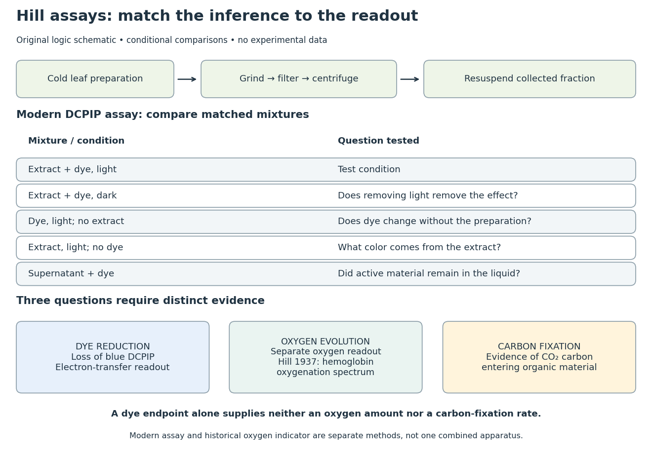
Hill reaction: preparation, comparisons and distinct readouts
. Original experimental-logic schematic. Control outcomes are conditional interpretations, not invented observations. The modern dye assay follows the inspected Practical Biology protocol; the separate historical branch denotes hemoglobin oxygenation. No measured time course, apparatus photograph or published figure is reproduced. Details.
Here is an original logical check of the controls. Suppose only the illuminated extract-plus-dye condition loses blue color, while the dark mixture and illuminated dye-only mixture retain it. Removing light or removing extract each removes the effect in those comparisons. That supports a joint requirement under the tested conditions. If the dye-only mixture also loses blue color, the proposed attribution to extract has failed its control; a faster change in the extract mixture would require a quantitative comparison before attribution. These are conditional cases, not experimental results.
Even the favorable case leaves three distinct questions. Dye reduction demonstrates delivery of electrons to an artificial electron acceptor. An independently calibrated molecular oxygen readout is needed to measure molecular oxygen evolution. Demonstrating carbon fixation additionally requires evidence that carbon dioxide becomes incorporated into organic material. None of the latter quantities is supplied by a blue-color endpoint alone. An absorbance change also needs background and scattering controls before it can be treated as dye concentration; Beer-Lambert law states the relevant assumptions.
In particular, two hypothetical preparations can give the same dye endpoint while differing in whether their stromal Calvin cycle is functional. Observing only the dye discards that distinction. The counterexample establishes why dye decolorization cannot alone prove complete photosynthesis, carbon fixation, or an intact chloroplast. Temperature, extract loading and illumination must also be matched when comparing rates: a lamp can alter both light exposure and heating.
Articles by others on the same topic
There are currently no matching articles.