Protein cell biology studies how proteins function within biological cells: where they are delivered, which partners they encounter, and how these interactions affect cellular processes. A protein's protein primary structure and abundance do not by themselves specify its location or available partners. RNA-binding proteins, G proteins and protein targeting illustrate different aspects of that problem.
The microsomal protein import experiment makes location experimentally testable. In 1975, Günter Blobel and Bernhard Dobberstein combined cell-free translation components with dog-pancreas microsomes. An immunoglobulin light chain product made while suitable membranes were present became processed and resistant to external proteolysis, whereas globin did not enter the same protected compartment. Adding membranes only after synthesis did not reproduce the effect in this preparation.[ref]
Transfer of Proteins across Membranes II by Günter Blobel and Bernhard Dobberstein. Abstract and pp.856–862, light-chain/globin comparisons and timing of membrane addition
The important observation is controlled accessibility, not just recovery near a membrane. A protein can bind a vesicle's outside without crossing it; enclosure can protect it from an external protease. Protease protection assays therefore complement measurements of total product and membrane topology.
For an original two-compartment example, suppose a fixed total of100 competent cargo molecules is divided between20 enclosed and80 exposed molecules. Redistributing the same total to70 enclosed and30 exposed changes accessibility even though total abundance stays100. If every enclosed molecule is protected and every exposed molecule is digested in the stipulated model, the protected fraction changes from0.20 to0.70. Real measurements also need checks for membrane integrity, protease activity and product recovery; the fractions alone do not identify every transport mechanism. This is why cellular organization belongs alongside the isolated molecule's chemistry.
A protein that binds RNA. Binding specificity can depend on nucleotide sequence, RNA secondary structure, chemical modification and other partners; recovery in an RNA pull-down alone does not establish direct recognition.
Recognition and positioning of a primary hairpin help the Drosha complex generate a shorter precursor. Later export and Dicer processing are additional stages, so loss of a mature microRNA can arise from defects at several different steps. [ref]
NCBI Gene by the National Library of Medicine. Homo sapiens, DGCR8 record, Summary: primary microRNA processing
An RNA-binding protein that collaborates with Nanos and other factors in sequence-specific repression of target messenger RNAs.
G3BP2 is an RNA-binding protein involved in the organization of stress granules. Together with related components, it helps couple RNA-protein interactions to formation of a condensed cytoplasmic assembly. The granule state depends on concentration, binding partners and stress context. Presence of G3BP2 in a granule supports participation, while testing a requirement for assembly needs depletion or a defined functional perturbation. [ref]
G3BP–Caprin1–USP10 complexes mediate stress granule condensation and associate with 40S subunits by Nancy Kedersha and others. 2016, G3BP-family interactions and stress-granule assembly
An RNA-associated regulator of stress-granule assembly. Requirements for assembly depend on stress and partner context.
Like G3BP2, it can organize interactions among RNA and proteins during stress-granule formation. Perturbing those interactions distinguishes a direct assembly requirement from mere co-localization in an already formed granule. [ref]
A Drosophila regulator whose RNA recognition supports sex-specific and autoregulatory splicing. It can act through competition at early recognition or through substrate-specific later spliceosomal interactions.
A common structured RNA-binding domain. Multiple motifs in a protein can recognize separated RNA segments and support geometry-dependent regulation.
A budding-yeast regulator that targets selected AU-rich transcripts for degradation during iron deficiency, reallocating resources away from iron-consuming processes.[ref]
Coordinated remodeling of cellular metabolism during iron deficiency through targeted mRNA degradation by Sergi Puig and others. Iron-deficiency-dependent target turnover
A family of microRNA-silencing effectors linking Argonaute-associated recognition to downstream repression and RNA-deadenylation machinery.[ref]
GW182 proteins directly recruit cytoplasmic deadenylase complexes to miRNA targets by Joerg E. Braun and others. Direct recruitment interactions
A bacteriophage RNA-recognition module used as a complement to MS2 coat protein for experimental RNA recruitment and imaging.
A bacteriophage protein recognizing a defined RNA hairpin, used as a fusion partner in RNA tethering assays and RNA imaging. Hairpin arrays and bound fusion proteins can themselves perturb RNA behaviour.
A member of a repeat-containing protein family with major roles in organellar RNA recognition and regulation. Different members can support distinct processing, editing, stability or translational functions.
A plastid RNA-binding protein that can protect a bound region against exonucleolytic decay from either direction, helping define stable transcript termini.[ref]
Zipcode-binding protein 1, also known as IGF2BP1. It recognizes a localization element in beta-actin messenger RNA and can repress its biological translation during transport.[ref]
An insulin-like growth factor 2 messenger RNA-binding protein family member. Stabilization and enhanced biological translation of modified targets have been reported; this does not identify every modification-dependent stabilizer as an IGF2BP protein.[ref]
A protein that binds a poly(A) tail and can influence messenger RNA stability and biological translation.
A protein family that binds small guide RNAs and participates in sequence-directed regulation. Guide complementarity can identify targets, but cleavage activity varies among family members and pathways.
A germline nuclear Caenorhabditis elegans Argonaute required for maintenance of some inherited small-RNA-dependent silencing states.
Endonucleolytic target cleavage directed by a small-RNA guide and a catalytically competent Argonaute. Cleavage typically falls opposite guide positions 10 and 11; not all Argonautes can slice.
An Argonaute-family branch binding piRNAs. Cytoplasmic cleavage and nuclear transcriptional silencing protect the germline in many animals, with important species and tissue differences.
The Caenorhabditis elegans Piwi-family protein associated with 21U piRNAs and germline surveillance of RNA expression.
Recognition by its associated small RNAs helps distinguish transcripts for germline surveillance. Establishing a persistent silenced state additionally involves downstream pathways, so initial recognition and long-term maintenance are different stages. [ref]
piRNAs Can Trigger a Multigenerational Epigenetic Memory in the Germline of C. elegans by Alyson Ashe and others. 2012, recognition and inherited silencing
A mouse Piwi-family protein associated with fetal male germ-cell transposon repression and de novo DNA methylation.
Its associated guide RNAs contribute sequence information for recognition of transposon-related material. Repression can involve both RNA-level and chromatin-associated outputs, depending on developmental stage and partner factors. [ref]
A piRNA Pathway Primed by Individual Transposons Is Linked to De Novo DNA Methylation in Mice by Alexei A. Aravin and others. 2008, germline piRNA pathway and methylation
Complementary cleavage relationships between small-RNA molecular populations can reinforce production of transposon-targeting guides. The resulting signature links sequence recognition to amplification rather than simply accumulating every small RNA indiscriminately. [ref]
A Drosophila Piwi-family protein participating in germline piRNA-mediated regulation, including ping-pong amplification.
It binds selected small RNAs and participates in transcript recognition and cleavage within a germline defence network. Its relationship with AGO3 helps amplify appropriate guide molecular populations, linking molecular specificity to transposon control. [ref]
An epigenetic role for maternally inherited piRNAs in transposon silencing by Julius Brennecke and others. 2008, inherited piRNAs and transposon repression
In Arabidopsis thaliana, it associates with small interfering RNAs that help target RNA-directed DNA methylation. Sequence complementarity supplies specificity while partner proteins connect recognition to a chromatin modification rather than cleavage of every matching transcript. [ref]
ARGONAUTE4 control of locus-specific siRNA accumulation and DNA and histone methylation by Daniel Zilberman and others. 2003, AGO4-dependent methylation and silencing
A heterogeneous nuclear ribonucleoprotein involved in RNA processing. Its binding can respond to modification-dependent accessibility of an RNA motif.[ref]
A polypyrimidine-tract-binding RNA-binding protein family member involved in precursor-RNA processing and other aspects of gene expression.
A polypyrimidine-tract-binding protein with four RNA recognition motifs. Its splicing effects depend on site position, RNA geometry and other proteins; it is not exclusively a repressor.
A member of a family of sequence-selective RNA-binding proteins involved in regulating alternative splicing.
Sequence-selective binding near an alternative exon can change recruitment or activity of the splicing machinery. The same binding motif can be associated with different outcomes depending on position and the surrounding regulatory context. [ref]
Molecular Biology of the Cell, 4th edition by Bruce Alberts and others. Chapter 7, RNA Processing and Alternative Splicing
A member of a neural RNA-binding protein family with position-dependent effects on alternative splicing. Binding-site position helps predict activation or repression of exon inclusion.[ref]
Protein targeting directs a protein to its cellular destination. Synthesis in the cytosol does not imply that the protein works there.
For entry into the endoplasmic reticulum, distinguish three questions: does the nascent chain reach the membrane, does part of it cross into the ER lumen, and does a segment remain embedded as a transmembrane protein? Cotranslational protein translocation couples these events to biological translation. A signal peptide can initiate delivery without specifying the eventual destination of the mature protein.
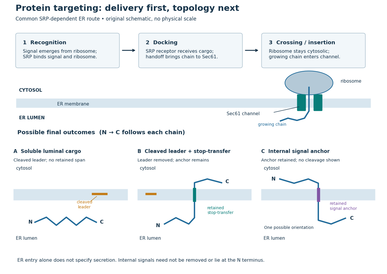
Protein targeting to the endoplasmic reticulum: delivery and three possible outcomes
. Original pathway and membrane topology schematic. The upper row separates SRP recognition, membrane docking and passage through a translocon. Lower cartoons stipulate a soluble cargo, a cleaved signal peptide plus retained stop-transfer sequence, and an uncleaved internal signal anchor. Each drawn terminus is labeled; the internal-anchor orientation is one possibility. Shapes and lengths are illustrative, with no molecular coordinates or measured timings. Details.
This distinction matters experimentally. Finding a protein in a membrane-containing centrifugation pellet could reflect surface binding or protein aggregation. The microsomal protein import experiment instead asks whether an intact membrane prevents an external protease from reaching the product. That tests accessibility across a barrier, rather than proximity to it.
Delivery of a protein from its producing cell to the extracellular environment, enabling communication and extracellular functions.
A signal peptide is a short targeting segment of a newly made polypeptide chain. A familiar ER signal sequence lies near the N-terminus and is removed during entry; Günter Blobel and Bernhard Dobberstein's 1975 experiments linked a larger immunoglobulin light chain precursor to a processed product.[ref]
Transfer of Proteins across Membranes I by Günter Blobel and Bernhard Dobberstein. Transfer of proteins across membranes. I, Introduction p. 836; Figures 5 and 14; Discussion pp. 845–847
Cleavage is a separate question from recognition. Not every targeting signal is a cleaved signal peptide: an internal signal anchor can remain part of a transmembrane protein. Conversely, loss of a segment does not by itself prove membrane crossing. Compare the independent size and accessibility readouts in the microsomal protein import experiment.
A signal peptidase removes a cleavable signal peptide; a suitable cleavage site and access are required.[ref]
The Endoplasmic Reticulum. Molecular Biology of the Cell, 4th edition by Bruce Alberts and others; “The ER Signal Sequence Is Removed from Most Soluble Proteins After Translocation”: adjacent cleavage-site requirement
For the stipulated -amino acid residue precursor under ER signal sequence, removal of the first amino acid residues leaves . Now stipulate that positions – of that precursor are instead retained as a stop-transfer sequence. Their positions in the mature chain become –, because the numbering origin changes when the first positions are removed. The mature chain still has amino acid residues, but its membrane-spanning segment is retained. A second hypothetical chain with an uncleaved internal signal anchor loses no positions at all. These are conditional bookkeeping examples, not predictions from invented biological sequences.
A signal anchor combines targeting or initiation of membrane passage with a retained membrane-spanning segment. Its orientation determines which flanking part enters the ER lumen; a single internal signal can yield either orientation.[ref]
Membrane Protein Insertion at the Endoplasmic Reticulum by Sichen Shao and Ramanujan S. Hegde. Shao and Hegde, 2011; “Cotranslational Membrane Protein Topogenesis”, Figure 5
In the figure's stipulated internal-anchor example, the N-terminus remains in the cytosol and the C-terminus enters the ER lumen. Reversing that orientation would reverse which end is protected from an externally added protease. Merely counting one membrane-spanning segment cannot choose between the two.
A stop-transfer sequence interrupts ongoing passage of a polypeptide chain across a membrane and retains a membrane-spanning segment. Protein targeting, insertion and subsequent sorting involve distinguishable sequence instructions.[ref]
Intracellular protein topogenesis by Günter Blobel. Blobel, Intracellular protein topogenesis, 1980; abstract: signal, stop-transfer, sorting and insertion sequences
In the figure's cleaved-leader example, assume the N-terminus of the mature chain has entered the ER lumen, a stop-transfer sequence remains in the membrane, and the remaining C-terminus is made on the cytosol side. The outcome differs from releasing the entire mature chain into the ER lumen. The latter is soluble cargo, potentially retained there or transported onward; entry alone does not establish secretion.
Organelle protein import brings externally synthesized proteins into an organelle. Many plastid proteins made from nuclear genes carry targeting information recognized by machinery that transports them across the envelope membranes. This delivery enables nuclear genes to supply functions inside a plastid, but the presence of targeting information is not itself evidence that a gene was acquired through endosymbiotic gene transfer. [ref]
Origin and Evolution of Plastids and Photosynthesis in Eukaryotes by Geoffrey I. McFadden. “How to tame your symbiont”: targeting, membrane passage and host-derived proteins
For the endoplasmic reticulum, cotranslational protein translocation illustrates how organelle protein import can begin before a polypeptide chain is complete. Import into the ER lumen and insertion as a transmembrane protein are different outcomes; neither alone establishes subsequent secretion from the biological cell.
A test of whether a protein crosses a specified organelle boundary, with controls distinguishing import from surface binding or membrane rupture.
Cotranslational protein translocation is passage of a growing polypeptide chain across a membrane during biological translation. SRP recognizes an exposed hydrophobic ER signal sequence. The SRP receptor enables handoff to the Sec61 complex. Biological translation can slow during delivery; the ribosome remains on the cytosol side.[ref]
Membrane Protein Insertion at the Endoplasmic Reticulum by Sichen Shao and Ramanujan S. Hegde. Shao and Hegde, Membrane Protein Insertion at the Endoplasmic Reticulum, 2011; “Cotranslational Targeting”, Figure 3
The useful engineering feature is coordination: a targeting instruction can become accessible while the rest of its cargo is still being made. Recognition, docking and crossing remain distinct operations. A defect in any one can prevent successful import even when the protein primary structure contains an appropriate signal.
This route is not universal. Many tail-anchored proteins use delivery after biological translation, and their membrane-spanning segment lies near the C-terminus. Thus protein targeting signals need not be at the N-terminus.[ref]
Membrane Protein Insertion at the Endoplasmic Reticulum by Sichen Shao and Ramanujan S. Hegde. Shao and Hegde, 2011; “Posttranslational Membrane Protein Topogenesis”, Figure 6
The signal recognition particle is an RNA–protein targeting complex linking a ribosome-associated nascent chain to the SRP receptor.[ref]
Membrane Protein Insertion at the Endoplasmic Reticulum by Sichen Shao and Ramanujan S. Hegde. Shao and Hegde, 2011; “Cotranslational Targeting”, Figure 3
The bacterial homolog of the SRP54 protein, containing a GTPase involved in cotranslational membrane targeting.
Its signal-recognition and GTPase functions connect a nascent hydrophobic segment with the bacterial SRP receptor FtsY. The pathway directs selected translating ribosomes toward the membrane before the complete protein has been synthesized. [ref]
Molecular Biology of the Cell, 4th edition by Bruce Alberts and others. Chapters 3, 6 and 15, protein function, translation and cell signalling
The conserved eukaryotic SRP subunit containing a signal-sequence-binding region and a GTPase domain; bacterial Ffh is its homolog.
The SRP receptor receives the SRP–ribosome–nascent chain complex for handoff to the translocon.[ref]
Membrane Protein Insertion at the Endoplasmic Reticulum by Sichen Shao and Ramanujan S. Hegde. Shao and Hegde, 2011; “Cotranslational Targeting”, Figure 3
Reciprocal regulation of the receptor and SRP GTPases couples recognition to transfer of the nascent chain toward a membrane translocation site. This illustrates a targeting cycle in which simple binding is only one stage of productive delivery. [ref]
Molecular Biology of the Cell, 4th edition by Bruce Alberts and others. Chapters 3, 6 and 15, protein function, translation and cell signalling
A translocon is membrane machinery through which a polypeptide chain crosses or enters a membrane. In endoplasmic reticulum protein targeting, the Sec61 complex supplies the central channel.[ref]
Membrane Protein Insertion at the Endoplasmic Reticulum by Sichen Shao and Ramanujan S. Hegde. Shao and Hegde, 2011; “The Sec61 Translocon”, Figure 4
The Sec61 complex supports passage across the endoplasmic reticulum membrane and lateral entry of membrane-spanning segments into its lipid bilayer.[ref]
Membrane Protein Insertion at the Endoplasmic Reticulum by Sichen Shao and Ramanujan S. Hegde. Shao and Hegde, 2011; “The Sec61 Translocon”, Figure 4
In 1975, Günter Blobel and Bernhard Dobberstein combined cell-free translation with dog-pancreas microsomes. Density-gradient centrifugation isolated the membranes; removal of resident ribosomes allowed reconstitution with rabbit-reticulocyte ribosome subunits. Added messenger RNA encoded an immunoglobulin light chain; globin provided a comparison. Products made with or without membranes were challenged with proteases and analyzed by SDS-PAGE and autoradiography. Membranes present during synthesis yielded processed, protected light chain; globin remained susceptible. Adding membranes after precursor synthesis failed to reproduce import. These comparisons support selective passage during synthesis in this preparation.[ref]
The companion paper specifies radioactive amino acid incorporation, ice-cold trypsin plus chymotrypsin treatment, a slab-gel electrophoresis apparatus and dried-gel exposure to X-ray film. Product-size analysis distinguishes a preserved full-size product from residual labeled fragments.[ref]
Transfer of Proteins across Membranes I by Günter Blobel and Bernhard Dobberstein. Methods pp. 837–838: proteolysis, SDS-PAGE and autoradiography; Discussion p. 847
Protection was incomplete. The authors considered leaky vesicles, and warned that heat treatment could damage processing as well as ribosome attachment. Their experiments did not identify SRP or the Sec61 complex. Nor does failure of late membrane addition establish that all organelle protein import must occur during synthesis.[ref]
Transfer of Proteins across Membranes II by Günter Blobel and Bernhard Dobberstein. pp. 858–859: incomplete protection; p. 861: heat-inactivation caveat; p. 862: alternatives for failed processing
A protease protection assay asks whether a barrier prevents an added protease from reaching a protein. A membrane-disruption comparison tests whether protection depends on that barrier.
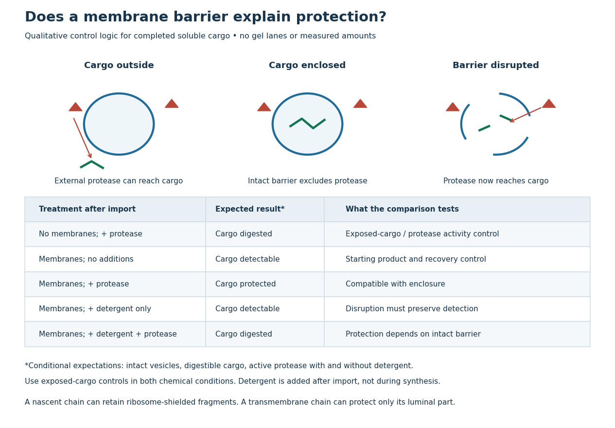
Protease protection assay: accessibility and control logic
. Original qualitative decision diagram for a completed soluble cargo. Icons and words describe expected outcomes under stated assumptions; they are not an autoradiography readout, copied historical lanes, measured survival fractions or simulated experimental intensities. Detergent is added after the import reaction. The historical nascent chain experiment also retained small ribosome-protected fragments, which this completed-cargo model deliberately distinguishes. Details.
The membrane-disruption test was performed by David D. Sabatini and Günter Blobel in 1970. Rat-liver microsomes purified by sucrose centrifugation incorporated radioactive leucine during cell-free translation. Samples received cold trypsin and chymotrypsin, with or without membrane-solubilizing detergents. Electron microscopy checked vesicle preservation; size-exclusion chromatography separated protected fragment sizes. Large membrane-associated fragments lost protection after disruption, whereas small ribosome-protected fragments persisted. An exposed ribosome preparation remained digestible when mixed with microsomes, checking that membranes did not simply inhibit the proteases.[ref]
Controlled Proteolysis of Nascent Polypeptides in Rat Liver Cell Fractions II by David D. Sabatini and Günter Blobel. Controlled proteolysis of nascent polypeptides in rat liver cell fractions. II, Methods p. 147; activity control p. 148; Figure 5C–D and text p. 152; Discussion pp. 155–156
To read the qualitative diagram, first compare untreated aliquots: they establish that detectable cargo exists before digestion. Next compare external protease with intact versus disrupted membranes. Persistence only while the barrier remains intact supports enclosure, provided the exposed-cargo control confirms active protease in both chemical conditions. The detergent-only aliquot checks that disruption itself does not destroy recovery or detection. Adding membranes after synthesis tests a different alternative: capture of a completed product rather than entry during biological translation.
This is an argument from controlled accessibility. Binding predicts membrane association but generally leaves an exposed cargo digestible. Enclosure predicts protection that depends on membrane integrity. However, protein aggregation, intrinsic resistance to proteolysis, inactive protease or undetected small fragments can obscure this distinction. For a transmembrane protein, expect protection of the enclosed portion rather than necessarily the full chain. Combine fragment size, compartment controls and membrane topology; a protected signal alone does not uniquely specify the whole protein's location.
A G protein is a protein switch whose activity depends on the bound guanine nucleotide. The three-subunit G proteins coupled to G protein-coupled receptors relay activation to other proteins; they are distinct from the receptors themselves. See the Alberts reference under G protein-coupled receptor.
A Gq protein is a heterotrimeric G protein whose alpha subunit couples activated G protein-coupled receptors to effectors including phospholipase C. GTP binding changes the alpha subunit's interactions; subsequent GTP hydrolysis helps terminate the signal. The beta and gamma subunits are components of the same signalling system, not parts of the receptor. [ref]
Molecular Biology of the Cell, 4th edition by Bruce Alberts and others. Chapter 15, Signaling through G-Protein-Linked Cell-Surface Receptors: phospholipase C and inositol phospholipids
The inhibitor YM-254890 supplies a molecular probe: structural work showed stabilization of a GDP-bound arrangement that impedes nucleotide exchange. Suppressing exchange is distinct from blocking the downstream enzyme directly, so an inhibitor response can help identify which stage of a signalling pathway is being controlled. [ref]
A small-molecule inhibitor that stabilizes a GDP-bound arrangement of Gαq and inhibits nucleotide exchange.[ref]
A small GTPase is a relatively small nucleotide-switch protein that cycles between GDP- and GTP-bound states. Ras family proteins illustrate signaling mechanisms distinct from the three-subunit organization of a heterotrimeric G protein.
Ran is a small GTPase involved in transport through the nuclear pore complex and in organization of microtubules. It switches between GDP- and GTP-bound states. The spatial organization of its regulators makes its nucleotide state informative about location, rather than simply reporting the total amount of the protein. [ref]
NCBI Gene by the National Library of Medicine. Human RAN, Gene ID 5901, Summary: nuclear transport, RCC1 and microtubule organization
In nuclear import, a high nuclear Ran-GTP activity promotes release of cargo from transport receptors, while the cytosolic side supports recycling through a different nucleotide state. This directional cycle depends on separated regulator activities and a supply of chemical energy. Equalizing all conditions on both sides would remove the same spatial cue even if the total Ran abundance stayed fixed. [ref]
Molecular Biology of the Cell, 4th edition by Bruce Alberts and others. Chapter 12, The Transport of Molecules between the Nucleus and the Cytosol: the Ran GTPase cycle
Nucleotide exchange changes recruitment of effectors that regulate the actin cytoskeleton. Spatially restricted activity can help select a protrusive region, connecting a biochemical switch to directional cell movement. [ref]
Molecular Biology of the Cell, 4th edition by Bruce Alberts and others. Chapter 16, Regulation of the Actin Cytoskeleton
A Rab GTPase is a small GTP-binding protein that regulates membrane traffic. GDP- and GTP-bound states differ in their interactions with effectors, while exchange factors and GTPase-activating proteins regulate the cycle. Different Rabs act at different trafficking stages, helping organize compartment identity, motor recruitment and carrier targeting. Rab8a and Rab11a illustrate related switches with different preferred routes and partners. [ref]
Rab Conversion as a Mechanism of Progression from Early to Late Endosomes by Jochen Rink and others. 2005, Rab5-to-Rab7 transition and endosomal identity
Rab8a is a Rab GTPase involved in delivery toward the cell surface and ciliary membrane organization. Rabin8 activates it through nucleotide exchange, linking upstream signals to effector recruitment. Its membrane location and nucleotide state together help determine productive interactions. A fluorescent Rab8a spot therefore reports localization without automatically specifying whether the switch is active or transport is proceeding. [ref]
NCBI Gene by the National Library of Medicine. Homo sapiens, RAB8A record, Summary and linked functional evidence
Rab11a is a Rab GTPase associated with recycling membrane traffic. It helps organize the return or redistribution of internalized components and contributes to polarized delivery in several cell types. Its effectors connect nucleotide state to targeting and motor systems. A recycled receptor can regain surface availability without being synthesized again, illustrating how trafficking changes cellular responsiveness on short timescales. [ref]
NCBI Gene by the National Library of Medicine. Homo sapiens, RAB11A record, Summary and linked functional evidence
A Rab-family regulator associated with late endosomal and degradative trafficking.
Its effectors help connect late endosomes to movement, targeting and fusion with degradative compartments. The transition from Rab5 to Rab7 illustrates maturation of membrane identity; a static marker image captures a state rather than the whole trafficking history. [ref]
Molecular Biology of the Cell, 4th edition by Bruce Alberts and others. Chapters 3, 6 and 15, protein function, translation and cell signalling
A Rab-family regulator associated with early endosomal membrane domains.
Active Rab5 recruits partners that organize early endosomes and membrane fusion. Replacement of Rab5-associated identity by Rab7-associated identity helps connect early uptake with later degradative trafficking, rather than requiring each endosome to retain one permanent molecular label. [ref]
Molecular Biology of the Cell, 4th edition by Bruce Alberts and others. Chapters 3, 6 and 15, protein function, translation and cell signalling
Replacement of an early Rab-regulated membrane identity by a later one, exemplified by Rab5 loss and Rab7 acquisition during endosome maturation.
An ARF GTPase is a small GTP-binding protein involved in membrane organization and coat recruitment. Nucleotide exchange promotes a membrane-associated active state that recruits selected effectors, while hydrolysis contributes to turnover of the assembly. ARF1 is a prominent regulator of coatomer recruitment at Golgi-associated membranes. Different family members and effectors act in different locations, so the family name does not specify one universal cargo route. [ref]
Molecular Biology of the Cell, 4th edition by Bruce Alberts and others. Chapter 13, Coat proteins and vesicle budding: ARF and COPI
Sar1 GTPase is a small GTP-binding protein that initiates COPII coat assembly at the endoplasmic reticulum. Its GTP-bound state associates with the membrane and recruits the Sec23-Sec24 complex, followed by the Sec13-Sec31 complex. Nucleotide cycling helps coordinate coat assembly and turnover. Reconstitution with defined components demonstrates a direct role in carrier formation rather than only an indirect consequence of cellular signalling. [ref]
A heterotrimeric G protein is a signaling assembly with , and subunits. Receptor-promoted nucleotide exchange on regulates interactions with and effectors; both signaling components can contribute downstream.
The associated β and γ subunits of a heterotrimeric G protein. Their interactions with downstream effectors transmit signals as well as support heterotrimer assembly.
The guanine-nucleotide-binding subunit of a heterotrimeric G protein. Nucleotide state changes its interactions with receptors, Gβγ and effectors.
A Gα family member that can inhibit adenylyl cyclase.
The GDP/GTP cycle changes its interaction with receptors, beta-gamma subunits and effectors. Inhibition of adenylyl cyclase is a common family property, while the resulting cellular response also depends on other targets and local cyclic AMP breakdown. [ref]
Molecular Biology of the Cell, 4th edition by Bruce Alberts and others. Chapters 3, 6 and 15, protein function, translation and cell signalling
A Gα family member that can stimulate adenylyl cyclase.
Receptor-promoted nucleotide exchange helps activate the switch, and GTP hydrolysis contributes to termination. Increased cyclic AMP then regulates effectors such as protein kinase A, turning a cell-surface recognition event into intracellular phosphorylation. [ref]
Molecular Biology of the Cell, 4th edition by Bruce Alberts and others. Chapters 3, 6 and 15, protein function, translation and cell signalling
A Gα family member that activates phospholipase C signalling.
The resulting cleavage of a membrane phosphoinositide generates inositol trisphosphate and diacylglycerol. These products can engage calcium release and kinase regulation in different cellular locations, so one receptor can initiate several coordinated downstream effects. [ref]
Molecular Biology of the Cell, 4th edition by Bruce Alberts and others. Chapters 3, 6 and 15, protein function, translation and cell signalling
Actin is a protein whose monomers assemble into filaments. An idealized actin filament has a bending modulus and persistence length; inferring its effective elastic modulus from those measurements additionally requires a cross-sectional model.
An individual soluble actin subunit. Total actin monomer concentration and concentration available for assembly differ when actin monomers are bound by sequestering molecules.
Binding that reduces the pool of actin monomers available for productive actin filament assembly. Existing actin filaments can then disappear through ordinary turnover; their loss does not establish direct severing.
A actin filament assembled from actin subunits. actin filament mass and actin filament number are distinct quantities; subunit exchange can continue while the total mass is approximately steady.
An actin-binding molecule used in fluorescent conjugates to visualize filamentous actin. Weak staining must be distinguished from background, and staining accessibility can affect inference about residual polymer.
Binding that restricts subunit exchange at a actin filament end. actin filament capping and actin monomer sequestration can both reduce bulk assembly but have different microscopic mechanisms.
Breakage of a actin filament into shorter pieces. This changes actin filament number and end number and should be distinguished from loss of subunits at pre-existing ends.
A bundle of actin filaments that can support directed intracellular transport. Absence of a visible cable does not exclude sparse short actin filaments below an imaging threshold.
A localized actin assembly in budding yeast, commonly associated with endocytosis. The lifetime of a visible patch and the lifetime of an individual constituent actin filament are different measurements.
Replacement and redistribution of subunits through assembly and disassembly. A persistent structure can be highly dynamic even when its appearance is stable.
Loss of subunits from actin filaments. Blocking replacement from soluble actin monomers can cause net actin filament loss without direct drug binding to the actin filament.
Assembly of actin subunits into actin filaments. An unseeded bulk reaction can show a actin nucleation lag, a growth phase and an approximately constant final polymer mass; equal endpoints need not imply equal kinetics.
A fluorescence readout of actin assembly using polymerization-dependent signal from a labelled actin fraction. Label effects, direct fluorescence changes, quenching and background must be controlled.[ref]
The free assembly-competent actin monomer concentration at which net addition at a specified actin filament end is zero in a specified model: . Different ends and nucleotide conditions can have different values.
Net addition of subunits at a actin filament end. A simple end model gives , with the sign changing at the relevant actin critical concentration.
Formation of a stable assembly from which an actin filament can grow. Lowering the concentration of assembly-competent actin monomer can affect actin nucleation much more strongly than addition to an existing end.
Myosin is a family of actin-associated motor proteins that couple ATP hydrolysis to mechanical work. Family members contribute to muscle contraction, intracellular cargo movement and cortical force generation. Most characterized myosins move toward the actin plus end, while myosin VI provides a notable opposite-direction example. Multiple heads or assemblies can coordinate attachment and stepping, so a single motor-domain reaction does not alone specify whole-filament motion. [ref]
Myosin VI is an actin-based motor that moves backwards by A. L. Wells and others. 1999, directionality assays
[ref]Myosin V Walks Hand-Over-Hand: Single Fluorophore Imaging with 1.5-nm Localization by Ahmet Yildiz and others. 2003, alternating-head stepping
The ability of a motor assembly to take repeated steps along actin before dissociating, often requiring coordination of multiple actin-binding heads.
A mechanically productive transition coupled to changes in nucleotide and actin binding. Load and isoform affect its detailed kinetics.
The neck-associated structural element that amplifies conformational changes in the motor domain into displacement.
Bound light chains help stabilize this elongated region. Changing lever-arm geometry can change step size without requiring a corresponding change in the distance of every chemical event at the ATP-binding site. [ref]
Molecular Biology of the Cell, 4th edition by Bruce Alberts and others. Chapters 3, 6 and 15, protein function, translation and cell signalling
The fraction of an ATPase cycle spent strongly bound to actin. It contributes to processivity and tension holding but does not alone determine cellular function.
An unconventional actin-based motor whose movement is toward the pointed end, opposite the direction of most characterized myosins.
A class of actin-based cargo motors; well-studied dimeric members coordinate their heads for processive stepping.
Coordination allows one head to remain associated while the other advances, supporting repeated motion along actin. Single-fluorophore localization experiments distinguish this alternating-head mechanism from a model in which the same head always leads. [ref]
Molecular Biology of the Cell, 4th edition by Bruce Alberts and others. Chapters 3, 6 and 15, protein function, translation and cell signalling
Myosin II participates in the actomyosin ring of typical animal cytokinesis.[ref]
The Events of M Phase. The Cell: A Molecular Approach, 2nd edition by Geoffrey M. Cooper; Chapter 14, The Events of M Phase, Cytokinesis and Figure 14.30
A proteasome is a protein complex that carries out proteolysis; unlike a lysosome, it has no surrounding lipid bilayer.
Ubiquitin is a small protein attached to other proteins in cellular sorting and regulation, including targeting many substrates to a proteasome.
Ubiquitination covalently attaches ubiquitin to a substrate, usually through successive action of a ubiquitin-activating enzyme, ubiquitin-conjugating enzyme and ubiquitin ligase. The modification can change degradation, localization or signalling; attachment alone does not specify which outcome occurs.
A ubiquitin ligase, or E3, promotes transfer of ubiquitin to a selected substrate within the ubiquitination system. Many E3s provide substrate recognition, while the detailed chemistry differs between E3 families. A ubiquitin-conjugating enzyme supplies activated ubiquitin. The resulting modification can signal protein degradation or other fates depending on substrate, linkage and context, so ubiquitination is not synonymous with immediate destruction. [ref]
Articles were limited to the first 100 out of 104 total.
Articles by others on the same topic
There are currently no matching articles.