Topics (384k) Articles (398k) Users (410) Discussions (242) Comments (383) Files (13k) New article

Phosphorus-32 is a radioactive isotope of phosphorus with 15 protons and 17 neutrons. It undergoes beta-minus decay to sulfur-32, preserving the mass number while increasing the atomic number by one. The cited laboratory evaluation gives a half-life of .[ref]
Phosphorus-32 in LNE-LNHB Table de Radionucleides. Page1, decay scheme and nuclear data; March2026 evaluation
Its radioactivity makes phosphorus-containing material experimentally traceable. In the 1952 Hershey-Chase experiment, phosphorus-32 labelled the DNA component of Bacteriophage T2. Mechanical agitation removed much less phosphorus label than sulfur label from infected bacteria, helping distinguish the material entering the cells from material remaining at the surface.[ref]
Independent functions of viral protein and nucleic acid in growth of bacteriophage by A. D. Hershey and Martha Chase. Abstract, points4–6: label separation and progeny comparison
The interpretation relies on the labelling and separation in this system; it is not a rule that all biological phosphorus belongs to DNA.For a closed sample changing only by radioactive decay, . After two quoted half-lives, about 28.546 days, the radioactivity is one quarter of its initial value. A falling signal can therefore reflect decay even without loss of labelled material from a vessel. Movement of material, chemical processing and detector response require additional measurements when interpreting a real tracer experiment.
A phosphate diester is a phosphoric ester with two organic groups linked through oxygen to the same phosphorus. Its neutral-acid representation is ; deprotonating the remaining O–H group gives without breaking either ester linkage.[ref]
Nomenclature of Phosphorus-Containing Compounds of Biochemical Importance by IUPAC and IUBMB. §4, phosphodiesters and the bridging phosphoric group
For an original connectivity example, let the two organic groups be methyl groups. The fragment contains one phosphorus center but two organic O–P connections. Two separate molecules of instead contain two phosphorus centers, each monoesterified. Counting phosphorus atoms alone therefore cannot determine the ester class.
A diester can bridge two different organic units or join two positions within one unit. In a cyclic nucleotide, the two attachments close a ring; they need not belong to two separate molecules.[ref]
Nomenclature of Phosphorus-Containing Compounds of Biochemical Importance by IUPAC and IUBMB. §4 comments(iii–iv) and Table3, polynucleotide names and cyclic phosphates
In DNA and RNA, successive sugar units are joined through phosphodiester bonds. Cleaving one ester linkage changes chain connectivity, which is why the same local chemistry can govern the integrity of a much longer nucleic acid.A phosphate triester is a phosphoric ester in which three organic groups are attached through oxygen to one phosphorus center. A general connectivity formula isUnlike a neutral-acid phosphate monoester or phosphate diester, this representation contains no remaining P–OH group. The organic groups may nevertheless contain their own ionizable sites; “triester” does not guarantee that every molecule in the class is electrically neutral.
Trimethyl phosphate supplies a concrete example: has formula . Its three methyl groups each contribute one carbon and three hydrogens, while the central unit and linking oxygens contribute one phosphorus and four oxygens.[ref]
Trimethyl phosphate in PubChem. CID10541, molecular formula, structure and SMILES
An original connectivity comparison replaces one group by . The resulting fragment, , is a diester because only two organic groups remain attached through oxygen. Merely removing a proton from that O–H group changes charge without restoring a third ester linkage.
The suffix counts esterification at one phosphorus center. Three separate phosphate monoesters elsewhere on a large molecule do not constitute one phosphate triester. This local counting rule helps distinguish a change in connectivity from a change in pH, protonation or total phosphorus content.
Phosphatidylglycerol is a glycerophospholipid whose usual membrane form has two fatty acid chains attached by ester linkages to a glycerol backbone, with a phosphate linking that backbone to a second glycerol. Its name describes a family: the fatty acid chains can vary. In the deprotonated form, the phosphate-containing head carries a net negative electric charge.[ref]
Phosphatidylglycerol in ChEBI. CHEBI:17517, structure and definition; CHEBI:60523, conjugate-base charge
The two chains and the charged head give phosphatidylglycerol different interactions with water and membrane-bound proteins. A useful controlled example comes from enzyme kinetics: J. L. van Rijn and colleagues activated bovine prothrombin using factor Xa on liposomes containing 20% phosphatidylglycerol and 80% phosphatidylcholine by mole fraction. Adding factor Va changed the apparent Michaelis constant from to . This compares the same stated membrane composition with and without a protein cofactor; it does not imply that adding phosphatidylglycerol alone universally multiplies the reaction rate by 55.[ref]
A phosphorothioate substitution replaces an oxygen of a phosphate group with sulfur. In a nucleic acid backbone, the usual experimental substitution replaces a non-bridging oxygen, while retaining the oxygen bridges connecting neighboring nucleotides. It changes backbone chemistry without deleting a nucleotide or changing its nucleobase sequence. The altered group is a phosphate analogue rather than a disulfide bond.
The two non-bridging positions matter. For a phosphate joining different nucleotide environments, replacing one oxygen or the other can give different configurations at phosphorus. Such phosphorothioate diastereomers can interact differently with an RNA-folding environment or a metal ion. The 2013 U6 metal rescue experiment tested both and substitutions at selected sites, rather than treating every sulfur replacement as equivalent.[ref]
RNA catalyses nuclear pre-mRNA splicing by Sebastian M. Fica and others. pp.229–230, Figure2a and site-specific substitution comparisons.
An original counting example shows the extra information required. At three independently stereogenic, substituted phosphates, two possible configurations at each site give configuration combinations. All eight can have the same nucleotide sequence and elemental formula. Measuring molecular mass alone therefore cannot tell which configuration combination was supplied. If every oxygen-to-sulfur replacement is made once at a selected site, each adds approximately unified atomic mass units; three add about , regardless of those configuration choices. These rounded atomic masses are illustrative, not exact isotope masses.
Replacing oxygen with sulfur also changes metal–ligand interactions. A loss of RNA activity in magnesium followed by preferential rescue with a different metal can test whether the original oxygen helped position a catalytic ion. It can also reflect altered folding or assembly, so the comparison needs controls. The substitution is a targeted chemical perturbation whose interpretation depends on position, configuration and the reaction being measured.
4-Nitrophenyl phosphate is a phosphate monoester in which a phosphate oxygen joins the aromatic group of 4-Nitrophenol. The neutral acid has formula ; a salt or hydrated reagent has a different complete formula. The “4” places the nitro group opposite the oxygen-linked substituent on the ring.[ref]
Alkaline phosphatase can hydrolyze 4-Nitrophenyl phosphate, releasing 4-Nitrophenol and inorganic phosphate. In the alkaline assay mixture the colored anionic product permits measurement with a spectrophotometer at . The manufacturer's calculation subtracts a blank and specifies a coefficient of under its stated conditions.[ref]
Diethanolamine Assay for the Enzymatic Assay of Alkaline Phosphatase by MilliporeSigma. §4, reaction; §§7.4–7.5, blank subtraction and coefficient
For an original endpoint example, suppose blank-corrected absorbance is through an optical path length of , with that coefficient applicable. The Beer-Lambert law givesThis is product. For an initial substrate concentration of , one-to-one conversion with no other product source would mean 40-percent conversion. These numbers are an illustrative model, not measured data from the cited assay.
The interpretation requires a suitable blank correction, known optical path length and stable measurement conditions. Changing pH changes the product's ionization and optical response. An endpoint measures accumulated product; estimating catalytic activity requires a time-dependent measurement and the corresponding calibration.
4-Nitrophenol is phenol with a nitro group opposite the hydroxy group on the benzene ring: positions 1 and 4. Its chemical formula is , and the alternative name p-nitrophenol describes the same substitution pattern.[ref]
Removing the phenolic proton produces its conjugate base, 4-Nitrophenolate. This change matters in optical enzyme assays: alkaline phosphatase can release 4-Nitrophenol from 4-Nitrophenyl phosphate, and the alkaline mixture favours the strongly absorbing anionic form. A spectrophotometer follows product-associated absorbance; an enzyme-free reference blank distinguishes the reaction signal from background.[ref]
Diethanolamine Assay for the Enzymatic Assay of Alkaline Phosphatase by MilliporeSigma. Reaction and §§7.4–7.5, 405 nm measurement and blank subtraction
A 1980 reference-material study measured a molar absorption coefficient of at and under its specified alkaline conditions.[ref]
High-purity 4-nitrophenol: purification, characterization, and specifications for use as a spectrophotometric reference material by G. N. Bowers Jr and others. Abstract, measured coefficient and final reaction conditions
For an illustrative blank-corrected absorbance and optical path under matching conditions, the Beer-Lambert law gives . Applying that coefficient at a different wavelength, pH or reaction composition needs validation: colour alone does not fix a concentration.The gradient first moment of an effective magnetic field gradient waveform is its time-weighted integral,Here effective includes the sign changes caused by refocusing pulses. Its units are when is in tesla per metre. The gradient zeroth moment is . For position with constant velocity, the accumulated nuclear magnetic resonance phase iswhere is the angular gyromagnetic ratio. Thus removes the dependence on initial position while can retain sensitivity to velocity, the basis of an idealized phase-contrast magnetic resonance imaging encoding.[ref]
A flow velocity zeugmatographic interlace for NMR imaging in humans by P. R. Moran. Original abstract; motion-sensitive phase encoding; equations here are an explicit constant-velocity derivation
For a teaching bipolar gradient, let the first lobe have signed area and time centroid , and the second have signed area and centroid . ThenEqual and opposite lobe areas cancel stationary-spin phase, but their time separation prevents cancellation of the constant-velocity phase. For and , the first moment is . These are illustrative waveform values, not measurements from the source.
The velocity encoding limit, commonly written , specifies the velocity scale represented by a phase difference of magnitude in a chosen phase-contrast magnetic resonance imaging encoding. With equal gradient zeroth moments and first-moment difference , an ideal constant-velocity model givesThe gyromagnetic ratio is angular, in radians per second per tesla, and is the difference of gradient first moments. A sign convention must be specified separately. A 1982 original paper described obtaining motion information through phase-sensitive nuclear magnetic resonance encoding.[ref]
A flow velocity zeugmatographic interlace for NMR imaging in humans by P. R. Moran. Abstract; phase encoding of flow information; displayed relations are the stated model
Choose the convention . If , then produces . But produces , which wraps to when phase is represented in . A reconstruction that ignores this aliasing would report . The limit describes an encoding ambiguity, not a physical maximum speed of the fluid.
For a fixed phase uncertainty in this simple model, propagation gives . Reducing improves velocity sensitivity under that assumption while narrowing the unambiguous range. For example, radians and give . Real measurements must also account for background phase, intravoxel variation and timing. Gated measurements of cerebrospinal fluid show why timing within the cardiac cycle matters even when net displacement is small.[ref]
The gradient zeroth moment is the signed time integral of an effective magnetic field gradient waveform along a specified encoding axis:Its units are tesla seconds per metre. Effective means that the sign convention accounts for any refocusing pulses in the nuclear magnetic resonance sequence; it is not necessarily the unsigned physical pulse area.
For a stationary spin at position , choose the phase convention . Then , where is the angular gyromagnetic ratio. A balanced waveform with cancels this position-dependent contribution within the model. It does not assert that every possible background contribution to phase vanishes.
As an original rectangular-pulse example, for has . Adding for cancels it. Equal areas, rather than equal pulse heights, produce the balance.
For a moving spin with , the remaining contribution can be , involving the gradient first moment. That separation explains how magnetic resonance velocity encoding can suppress sensitivity to stationary position while preserving motion information. Early phase-sensitive flow imaging supplied the experimental motivation for extracting such information from resonance phase.[ref]
A flow velocity zeugmatographic interlace for NMR imaging in humans by P. R. Moran. Primary abstract; displayed moment relations are explicit idealized derivations
A bipolar gradient is a magnetic field gradient waveform with lobes of opposite sign. In nuclear magnetic resonance, such waveforms can control sensitivity to position and motion. Opposite signs alone do not guarantee equal signed areas, and an applied waveform must be distinguished from its effective encoding waveform when refocusing pulses change phase evolution.
For an original counterexample, apply for and then for . The gradient zeroth moment isThe waveform is bipolar but unbalanced.
If the two durations are instead equal to , and the effective lobes are contiguous, from time 0 to and from to , then but the gradient first moment is . Thus cancellation of stationary-position phase can coexist with sensitivity to constant velocity. The sign follows this stated ordering.
Bipolar pulses also appear outside flow imaging. A 1989 stimulated echo study addressed background gradients that distort diffusion measurements. Alternating pulses could cancel a cross term under specified conditions; tests used proton signals from homogeneous and granular samples.[ref]
Pulsed field gradient stimulated echo methods for improved NMR diffusion measurements in heterogeneous systems by R. M. Cotts and others. Primary abstract: background-gradient reduction, conditional cross-term cancellation and measurements
This is a sequence-specific result, not a promise that every bipolar pair removes every imaging error.Drug distribution is movement of a drug between blood and other body compartments. Perfusion, membrane permeability, tissue binding and plasma-protein binding jointly affect it. The apparent distribution volume iswhere is the amount in the body and the plasma concentration. If mg and mg/L, L. [ref]
Population Pharmacokinetics Guidance for Industry by the FDA. 2022, pharmacokinetic parameters and concentration-time models
The result is a proportionality quantity, not an anatomical cavity that must contain that many litres. Extensive tissue binding can lower and make large. A rapid fall in plasma concentration immediately after administration can therefore reflect distribution as well as drug elimination; distinguishing them requires a model and a suitable sampling schedule.
Drug elimination removes a parent drug from the body's measured parent-drug pool through drug metabolism or excretion. Redistribution from blood to a tissue changes plasma concentration but does not by itself eliminate the drug from the body. A mass balance separates input, output and transfers between compartments. [ref]
Population Pharmacokinetics Guidance for Industry by the FDA. 2022, pharmacokinetic model development and concentration-time analysis
For a one-compartment model with no input and first-order elimination, gives . Equal fractions disappear in equal time intervals. By contrast, a saturated elimination route can approach removal of equal amounts per interval. The latter produces a different concentration-time curve and cannot be assumed to have a concentration-independent half-life.
Drug metabolism chemically transforms a drug into other molecular forms. Oxidation, hydrolysis and conjugation are common routes, frequently involving the liver but also other tissues. Transformation may inactivate a compound, generate an active product or activate a precursor; metabolism therefore does not mean detoxification in every case. [ref]
Population Pharmacokinetics Guidance for Industry by the FDA. 2022, Background and covariate analysis: pharmacokinetic variability and elimination-related factors
The parent compound and each product have their own time courses. In a simple pathway further products, a fall in can coexist with a rise in an active metabolite . Measuring only the parent can therefore misrepresent pharmacologically relevant exposure. Chemical conversion, excretion of unchanged material and movement into a different tissue are distinct processes.
Drug clearance relates the rate at which a drug is eliminated to a specified measured concentration:If elimination is 10 mg/h at a plasma concentration of 2 mg/L, clearance is 5 L/h. This is an effective volume per time, rather than the physical volume of fluid actually removed. The concentration reference, such as plasma or whole blood, must be specified. [ref]
Population Pharmacokinetics Guidance for Industry by the FDA. 2022, model-development discussion: pharmacokinetic parameters and covariates
For constant clearance and a one-compartment distribution volume , the amount obeys . Thus and . The derivation explains why a longer half-life can reflect greater distribution volume as well as lower clearance; half-life and clearance are not interchangeable measurements.
A dose-response relationship connects an administered amount or exposure level with a defined biological response under specified conditions. Dose, dosing schedule, route and observation time all matter. Pharmacokinetics links administration to the concentration history, while pharmacodynamics links exposure with the response; a dose-response curve combines those influences.
Graded dose response describes the magnitude of a response, such as a measured change in activity. Quantal dose response describes the fraction of a population meeting a specified response criterion. Half of a maximum effect and a response in half of a population are different quantities; a midpoint parameter must say which is meant.
For an original graded-response illustration, a saturating Hill equation model isHere is an increment above baseline, gives half that increment and controls the shape in the stipulated model. With , , response units and arbitrary dose units, doses , and produce effects , and . Tripling dose from to does not triple the response. These arbitrary values are a mathematical example, not a drug regimen.
Estimation requires a design that separates dose from other changing conditions. If dose is increased on successive days while the condition also improves over time, a rising response cannot automatically be attributed to dose. The primary ICH-E4 guidance explicitly discusses such titration/time confounding and distinguishes population-average from individual response curves.[ref]
Dose-Response Information to Support Drug Registration by ICH. Sections I.B, I.D and I.E, individual/population curves, titration designs and time
A controlled experiment specifies its endpoint, comparator, exposure interval and uncertainty. If the measured range never reaches a plateau, the fitted maximum and midpoint may be poorly determined. Desirable and undesirable responses can have different curves, so one endpoint’s midpoint does not summarize all consequences of exposure. The scientific purpose is a reproducible relationship for a stated endpoint and setting, rather than an assumption that more exposure must always be better.
Pinned article: Introduction to the OurBigBook Project
Welcome to the OurBigBook Project! Our goal is to create the perfect publishing platform for STEM subjects, and get university-level students to write the best free STEM tutorials ever.
Everyone is welcome to create an account and play with the site: ourbigbook.com/go/register. We belive that students themselves can write amazing tutorials, but teachers are welcome too. You can write about anything you want, it doesn't have to be STEM or even educational. Silly test content is very welcome and you won't be penalized in any way. Just keep it legal!
Intro to OurBigBook
. Source. We have two killer features:
- topics: topics group articles by different users with the same title, e.g. here is the topic for the "Fundamental Theorem of Calculus" ourbigbook.com/go/topic/fundamental-theorem-of-calculusArticles of different users are sorted by upvote within each article page. This feature is a bit like:
- a Wikipedia where each user can have their own version of each article
- a Q&A website like Stack Overflow, where multiple people can give their views on a given topic, and the best ones are sorted by upvote. Except you don't need to wait for someone to ask first, and any topic goes, no matter how narrow or broad
This feature makes it possible for readers to find better explanations of any topic created by other writers. And it allows writers to create an explanation in a place that readers might actually find it.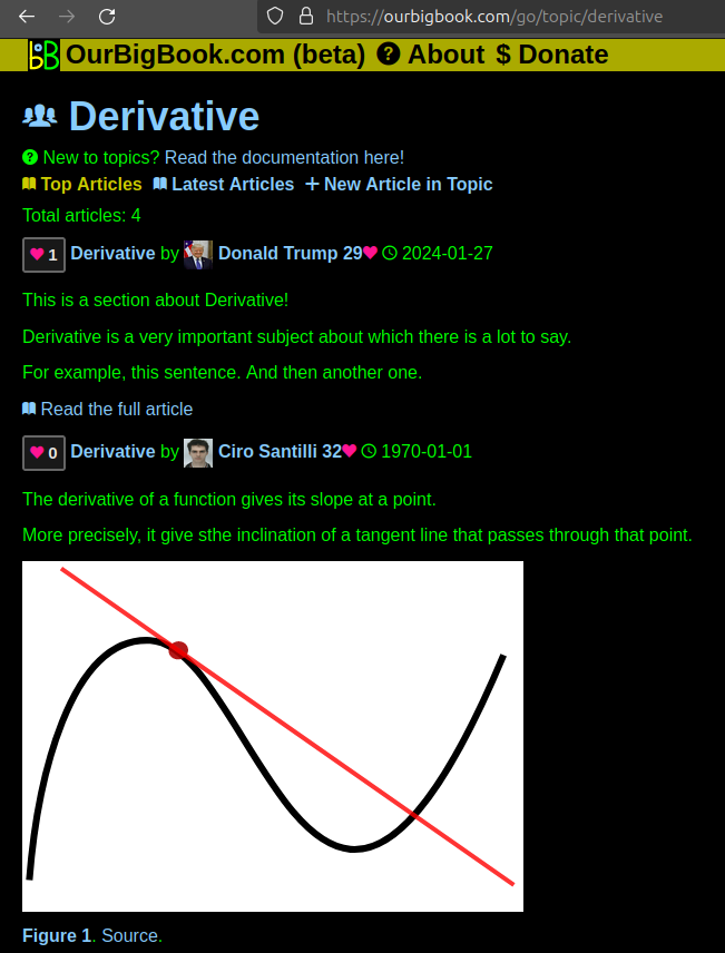
Figure 1. Screenshot of the "Derivative" topic page. View it live at: ourbigbook.com/go/topic/derivativeVideo 2. OurBigBook Web topics demo. Source. - local editing: you can store all your personal knowledge base content locally in a plaintext markup format that can be edited locally and published either:This way you can be sure that even if OurBigBook.com were to go down one day (which we have no plans to do as it is quite cheap to host!), your content will still be perfectly readable as a static site.
- to OurBigBook.com to get awesome multi-user features like topics and likes
- as HTML files to a static website, which you can host yourself for free on many external providers like GitHub Pages, and remain in full control

Figure 2. You can publish local OurBigBook lightweight markup files to either OurBigBook.com or as a static website.
Figure 3. Visual Studio Code extension installation.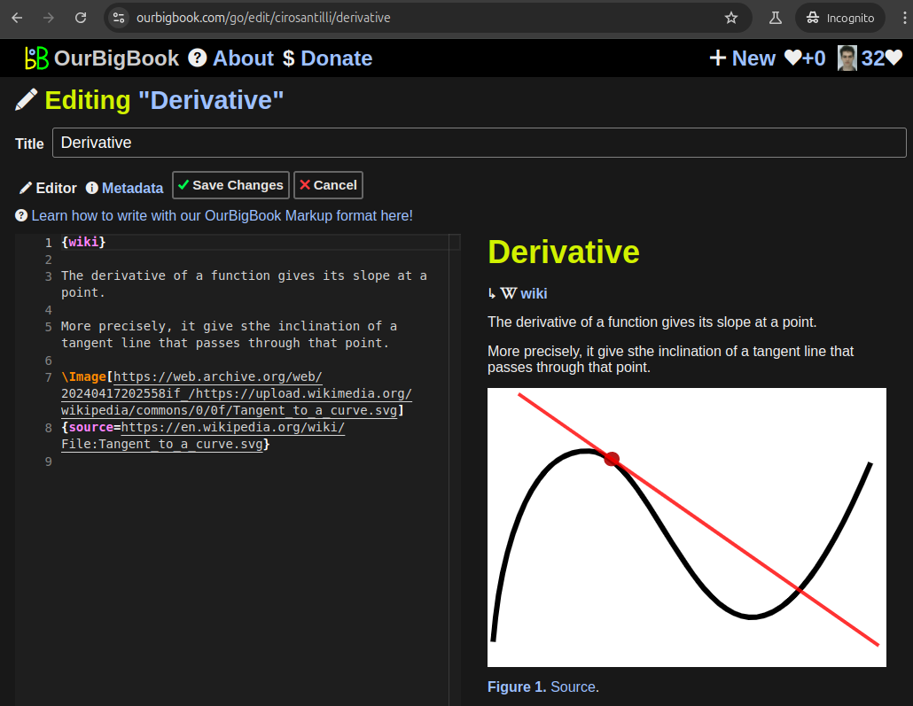
Figure 5. . You can also edit articles on the Web editor without installing anything locally. Video 3. Edit locally and publish demo. Source. This shows editing OurBigBook Markup and publishing it using the Visual Studio Code extension. 
- Infinitely deep tables of contents:
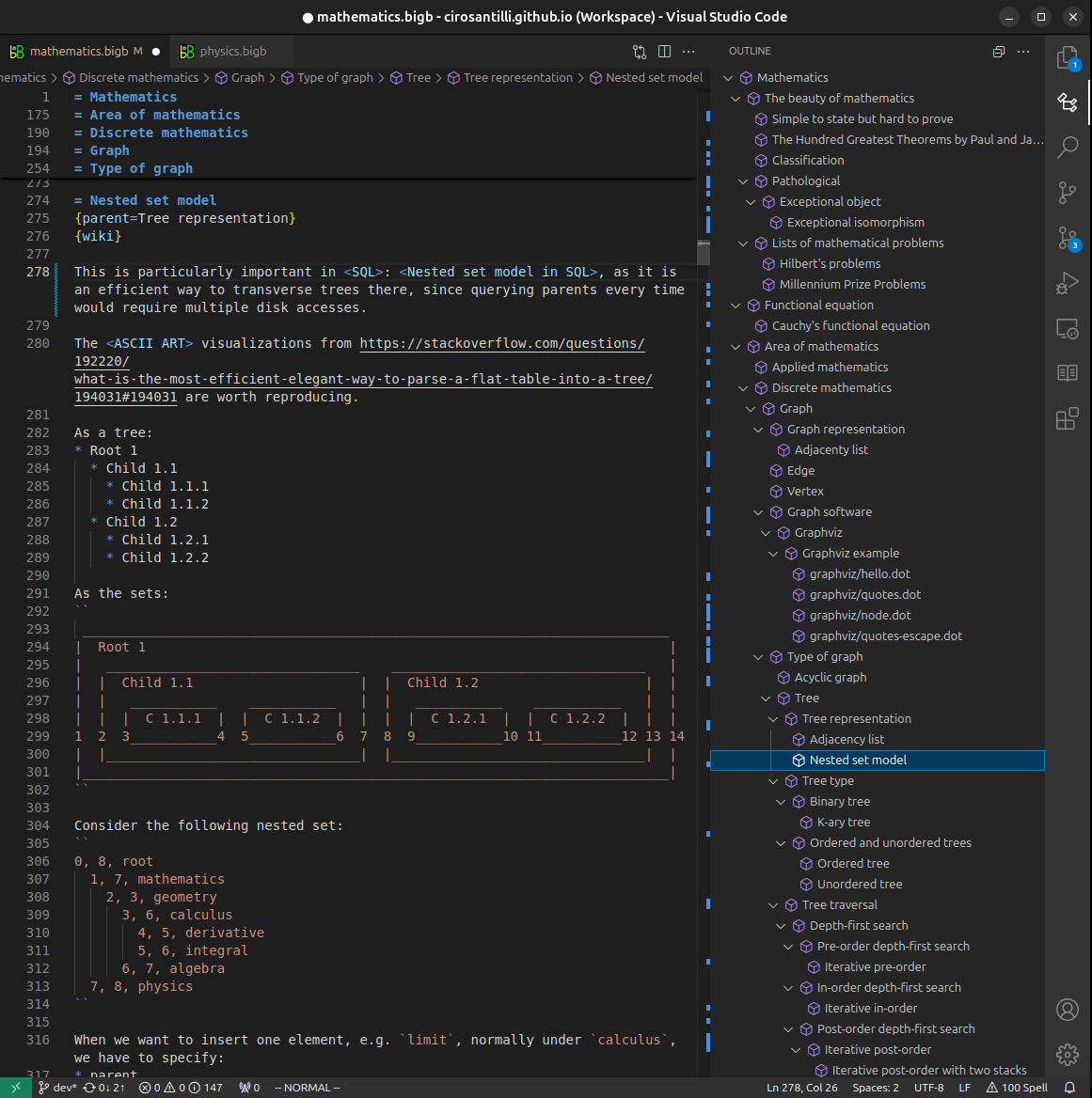

All our software is open source and hosted at: github.com/ourbigbook/ourbigbook
Further documentation can be found at: docs.ourbigbook.com
Feel free to reach our to us for any help or suggestions: docs.ourbigbook.com/#contact