Invasion is a crucial enabling step for the natural dissemination of most solid cancers, but it is neither synonymous with metastasis nor sufficient to complete it. A carcinoma must escape its original epithelial compartment and negotiate surrounding tissue before disseminated biological cells can form a distant lesion. An invasive primary tumour can nevertheless fail at circulation survival, distant tissue entry or colonization.
The metastatic cascade is a sequence of conditional transitions: local escape, access to blood or lymph, survival during transport, arrest and entry at a distant site, and successful outgrowth. Schematically,where is escape, transport, establishment of a surviving disseminated biological cell and sustained colonization. This is a chain of conditional probabilities, not an assumption that the stages are independent. An intervention can strongly affect one factor while leaving another limiting step dominant. Consequently, invasiveness and overall metastatic efficiency need not rank tumour populations identically.
Local invasion requires altered relationships with neighbouring biological cells and extracellular matrix. cancer biological cells may remodel matrix, traverse pre-existing paths or recruit stromal biological cells that facilitate movement. Adhesion changes, actomyosin forces, polarity and protease activity can contribute in different proportions depending on tissue geometry. Histological breach of a basement membrane is important evidence of invasive carcinoma, but a static section does not reveal whether the tumour biological cell itself degraded the barrier or exploited a route created by another biological cell. Intravital imaging, three-dimensional culture and perturbation of both tumour and stromal compartments help identify the mechanism.
Invasion does not require every biological cell to become an isolated mesenchymal migrant. biological cells can move collectively while maintaining junctions, with leader and follower biological cells performing different tasks. organoid and tumour experiments showed that basal-marker-positive leader biological cells can drive collective breast-cancer invasion, and functional perturbation of the programme impaired that behaviour.[ref]
Collective invasion in breast cancer requires a conserved basal epithelial program by Kevin J. Cheung and others. Collective strands, leader-cell programme and perturbation
Such evidence links a specific invasion mechanism to tissue behaviour, while leaving open which additional properties allow those invading biological cells to form distant colonies.An epithelial-to-mesenchymal transition can assist dissemination in some contexts, but epithelial-to-mesenchymal transition and invasion should not be treated as interchangeable variables. EMT-associated changes can alter motility, survival and therapy response; partial states can retain epithelial cohesion. In a breast-cancer lineage-tracing model, metastases occurred without passage through the reporter-defined epithelial-to-mesenchymal transition state, while that state was associated with chemoresistance.[ref]
Epithelial-to-mesenchymal transition is not required for lung metastasis but contributes to chemoresistance by Kari R. Fischer and others. Lineage tracing, lung metastasis and treatment response
The inference is limited by the state the reporter detects: failure to mark a biological cell is not proof that no transient or partial transition occurred. Other experimental work found that the timing of epithelial-to-mesenchymal transition activation and reversal affected dissemination and metastatic growth, emphasizing that a useful escape programme can differ from a useful colonization programme.[ref]Spatiotemporal regulation of epithelial-mesenchymal transition is essential for squamous cell carcinoma metastasis by Jeff H. Tsai and others. Temporal manipulation of EMT and metastatic progression
These findings challenge a universal epithelial-to-mesenchymal transition requirement, not the requirement to cross relevant anatomical barriers.A direct test of invasion's importance must preserve the stages being investigated. Start with an orthotopic primary tumour and inducibly perturb a proposed invasion factor after comparable tumours have formed. Quantify primary burden, local boundary crossing, circulating biological cells, early distant seeds and later colonies separately. A reduction in metastases accompanied by much smaller primaries could simply reflect fewer biological cells available to disseminate. Conversely, a perturbation that leaves growth unchanged but reduces escape and subsequent seeds supports an invasion-specific contribution. Rescue and independent perturbations help exclude nonspecific loss of viability or adhesion.
Intravenous injection is a complementary bypass experiment: it tests transport survival and later establishment while omitting much primary-site invasion and intravasation. If a mutant fails from a primary tumour but succeeds after injection, that supports an early-stage defect. If it fails in both assays, it may affect a later step or several processes. Injection is not a demonstration that invasion is dispensable for spontaneous metastasis. The route also changes the first capillary bed encountered and introduces an artificial bolus. Where feasible, temporally restricted perturbation, primary-tumour resection and tracking of individually labelled disseminated clones can further distinguish escape from dormancy and outgrowth.
The anatomical context matters. Blood cancers already occupy migratory compartments, so their dissemination cannot be forced into the same basement-membrane-escape model as an epithelial carcinoma. Within solid cancers, the rate-limiting step can also differ by organ and tumour state. Invasion provides access to the metastatic route in most solid tumours; distant colonization is an additional, demanding phenotype. Its importance is established by stage-specific causal measurements, not by equating an invasion assay, an epithelial-to-mesenchymal transition marker or a vascular injection result with the whole metastatic process.
Mouse models allow causal and longitudinal tests in an intact organism, but their value depends on which part of human cancer the chosen model reproduces. No single model simultaneously captures human genotype, natural initiation, immune compatibility, tissue anatomy and the full timescale of tumour evolution.
A major strength is experimental control. Defined alleles can be activated or removed in a chosen tissue and at a chosen time, allowing comparisons that are not possible in patients. Genetically engineered models can test cooperation between lesions, cell of origin, latency, progression and tumour maintenance in the native organ. Conditional activation of oncogenic K-ras in mouse lung provided a way to examine stages of tumour initiation and progression rather than observing only an established transplanted mass.[ref]
Analysis of lung tumor initiation and progression using conditional expression of oncogenic K-ras by Erica L. Jackson and others. Conditional activation and staged lung pathology
A reversible or inducible intervention after tumour formation can then distinguish an initiation requirement from a maintenance dependency. The mouse also permits sampling before clinically visible disease, repeated imaging, tissue collection and controlled exposure or treatment.The host environment is another advantage. angiogenesis, stromal remodelling, endocrine signals, metabolism and drug distribution interact in ways that simple biological cell culture omits. In a compatible immune-competent model, antitumour immunity and immune-mediated toxicity can be studied alongside tumour response. Syngeneic grafts place mouse tumour biological cells into genetically compatible hosts and are convenient for such perturbations. Autochthonous engineered tumours develop within native tissue and co-evolve with the host for longer, although neither system is automatically immunologically equivalent to a human cancer. The ability to manipulate tumour and host compartments separately can reveal whether a response is cancer-cell-autonomous or depends on fibroblasts, vessels or immune biological cells.
Human xenografts provide a different advantage. Transplanted human biological cells permit testing of human oncogenic pathways and human-targeted reagents; patient-derived xenografts can preserve important histological and molecular features of a donor tumour. Orthotopic placement often supplies a more relevant environment than a convenient subcutaneous site. Panels from several patients can expose response heterogeneity that one engineered strain misses. Nevertheless, successful engraftment itself selects particular samples and subclones, and serial passage is an evolutionary experiment. Ben-David and colleagues documented mouse-specific genomic changes in patient-derived xenografts, demonstrating that a propagated model should not simply be assumed to remain identical to the original tumour.[ref]
Patient-derived xenografts undergo mouse-specific tumor evolution by Uri Ben-David and others. Longitudinal copy-number changes during xenograft propagation
Compare early and late passages with the donor, and use the passage appropriate to the question.The greatest xenograft limitation for many questions is the host. Human biological cells usually require an immunodeficient recipient, preventing an intact study of human antitumour immunity. Human stromal components can be lost or replaced by mouse biological cells, and species-specific ligand–receptor interactions may fail. Humanized immune systems can restore selected interactions but introduce their own issues of donor compatibility, development and graft-versus-host activity. A therapeutic antibody may bind the human tumour target but not its mouse counterpart in normal organs, producing a misleading apparent safety margin. Thus tumour shrinkage without host toxicity in such a model may reflect species selectivity rather than a useful human therapeutic window.
Engineered and syngeneic models also have limitations. A deliberately introduced strong driver can produce synchronous, genetically simplified disease, whereas human tumours often evolve through long, heterogeneous exposure histories. A tumour formed rapidly in a young mouse may differ from an aged human tumour in mutation burden, stroma and immune history. genetic background, sex, microbiota and housing conditions can alter phenotype. Species differences in lifespan, organ structure, metabolism, telomere regulation and drug pharmacokinetics further limit direct extrapolation. The tumour spectrum after p53 loss, for example, is informative about tumour suppression but does not by itself reproduce the distribution of human TP53-mutant cancers.[ref]
The endpoint and intervention must fit the biological question. Subcutaneous growth measures a local mass under an artificial geometry; tail-vein injection measures selected post-entry metastatic steps. Neither establishes a treatment effect on natural invasion or initial dissemination. Drug tests should establish exposure and target engagement in both tumour and relevant normal tissue, use clinically motivated schedules where possible, and measure survival or durable disease control as well as short-term volume. Rapidly growing implants may make some treatments look more effective than they are against slow, heterogeneous human disease.
Good design uses random allocation after a defined eligibility point, blinded measurement, prespecified endpoints and justified numbers of independent animals. Multiple tumours within one mouse are not automatically independent treatment replicates. Match or record relevant background variables and validate the intended allelic change and molecular phenotype. Ethical constraints favour well-chosen questions and informative longitudinal measurements rather than duplicating a weak model at larger scale. Combine mouse results with human genomics, patient-derived cultures or organoids and, ultimately, human evidence. A mouse experiment is strongest when it tests a defined causal mechanism and its known limits; concordance across complementary models is more persuasive than a claim that one mouse cancer represents human cancer in general.
Direction arises from switching between mutually incompatible states, with feedback providing memory and regulated destruction resetting the system. Different transitions use related logic, but a steep response, a bistable switch and an irreversible molecular event are not identical. The cell cycle also requires checkpoints and once-per-cycle licensing so that the ordered biochemical states produce a correct sequence of physical events.
At the G1/S transition, growth signals promote cyclin-dependent kinase activity and relieve RB-mediated restraint on E2F. E2F-dependent expression of additional cell-cycle components can reinforce the transition. More generally, activation of a kinase together with removal of its inhibitor creates positive feedback. In yeast, multisite phosphorylation of Sic1 permits recognition by the Cdc4 ubiquitin-ligase system and degradation of an inhibitor of S-phase kinase. This supplies a threshold that prevents weak or premature signals from launching the next programme.[ref]
Multisite phosphorylation of a CDK inhibitor sets a threshold for the onset of DNA replication by Piers Nash and others. Phosphorylation-dependent Sic1 recognition and timing
The detailed regulators vary across organisms and biological cell types; the conserved point is a coordinated transition from an inhibited state to one that reinforces its own activation.Mitotic entry gives an experimentally accessible example of bistability. cyclin B binding increases the available CDK1 complex, but Wee1-dependent inhibitory phosphorylation can keep it inactive. Cdc25 removes the inhibitory phosphate. Active CDK1 promotes Cdc25 activity and suppresses Wee1, giving a positive-feedback loop and a double-negative loop. Over a suitable input range, both a low-activity and a high-activity state can be stable. If cyclin input rises from interphase, activation occurs at a higher threshold than the input required to maintain a pre-existing mitotic state. These distinct thresholds constitute hysteresis and prevent small fluctuations from repeatedly reversing a committed transition.
In egg extracts, nondegradable cyclin could be supplied while CDK activity was approached from different initial states. The same cyclin input then supported different persistent activities depending on history, directly distinguishing hysteresis from a single steep but history-independent response.[ref]
Building a cell cycle oscillator: hysteresis and bistability in the activation of Cdc2 by Joseph R. Pomerening and others. History-dependent activity at matched nondegradable cyclin input
These are extract measurements, not universal numerical thresholds for every intact biological cell. A useful conceptual representation iswhere represents effective activating input. hysteresis supplies memory within this interval; it does not mean that the switch can never be reset.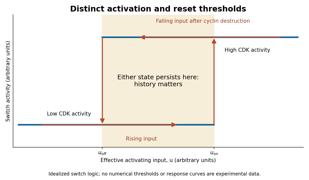
Hysteretic activation preserves state until a separate reset crosses the lower threshold
. Details. 
Reset is achieved by coupling activation to delayed negative feedback and proteolysis. Mitotic kinase activity helps prepare activation of the anaphase-promoting complex with Cdc20, while the spindle checkpoint prevents productive progression until attachment conditions are met. Once this restraint is satisfied, APC/C-dependent ubiquitination promotes destruction of securin and cyclin B. securin loss allows separase to cleave cohesin, making sister-chromatid separation possible. Experiments identifying cohesin cleavage connected regulated proteolysis to a physical transition that simple reversal of a phosphorylation state cannot undo.[ref]
Sister-chromatid separation at anaphase onset is promoted by cleavage of the cohesin subunit Scc1 by Frank Uhlmann and others. Cohesin cleavage and anaphase onset
Chromatids cannot be returned to a pre-anaphase chromosome merely by reducing separase activity.cyclin B destruction lowers CDK1 activity, allowing phosphatases to reverse mitotic phosphorylation and promoting an interphase state. Activation of anaphase-promoting complex with Cdh1 at low kinase activity can help keep cyclins low, while high kinase activity opposes that state. Thus low-CDK and high-CDK programmes are mutually antagonistic. Cyclin synthesis eventually provides a new activating input. Early experiments in extracts showed the importance of cyclin synthesis and destruction, including failure of normal exit with nondegradable cyclin.[ref]
The role of cyclin synthesis and degradation in the control of maturation promoting factor activity by Andrew W. Murray and others. Cyclin synthesis, degradation and nondegradable variants
A bistable activation module combined with an appropriate delayed reset can generate an ordered oscillation; a bistable module held at constant input need not oscillate on its own.replication licensing enforces another essential directionality. At low CDK activity, origin recognition complex with Cdc6 and Cdt1 loads MCM2-7 double hexamers, establishing competence to replicate. In S phase, activating kinases promote origin firing while high CDK activity and additional controls prevent renewed licensing. In animal biological cells, geminin inhibition of Cdt1 and regulated degradation help separate these states. Purified-protein reconstitution demonstrated loading of the replication-helicase precursor as a distinct biochemical event.[ref]
Concerted loading of Mcm2-7 double hexamers around DNA during DNA replication origin licensing by Dirk Remus and others. Origin licensing by double-hexamer loading
Separating licensing from firing allows many origins to be prepared but prevents fired regions from being copied repeatedly during the same cycle. Lowering mitotic kinase activity must eventually restore licensing competence for the next cycle, rather than permit uncontrolled rereplication during the current S phase.Checkpoint pathways modify or hold these switches when DNA is unreplicated, damaged or chromosomes are improperly attached; they do not supply the entire oscillator. Energy-consuming phosphorylation cycles, ubiquitination, proteolysis and resynthesis make the system a driven process, while feedback stabilizes distinct states. Thresholds provide commitment, hysteresis provides memory, destruction and cleavage enforce selected transitions, and licensing/checkpoints couple those transitions to one correct round of replication and segregation.
Replication fidelity uses successive discrimination and repair systems, because no single enzyme detects every error. The important distinctions are between an incorrect base pair, a slipped sequence register, the wrong sugar incorporated into DNA, a damaged template and an incompletely or incorrectly restored chromosome. Repair must also identify which information should be retained: recognizing a mismatch without strand discrimination could fix the wrong sequence.
A polymerase can select an incorrect deoxynucleoside triphosphate and insert the wrong base through imperfect discrimination, transient base configurations or an altered active site environment. If the mismatch is extended and survives into another replication round, it can become a stable base substitution. Many replicative polymerases possess a separate 3′→5′ exonuclease proofreading site. A mismatched primer end tends to slow extension and transfer toward this site; removal of the terminal nucleotide permits another attempt at correct synthesis. This is distinct from the polymerase's forward synthetic reaction and from proofreading in biological translation. In mice, selective disruption of DNA polymerase δ proofreading increased mutation and tumour susceptibility, demonstrating an organismal consequence of unrepaired copying errors.[ref]
High incidence of epithelial cancers in mice deficient for DNA polymerase δ proofreading by Robert E. Goldsby and others. Proofreading-domain mutation, mutator phenotype and tumour development
Post-replicative DNA mismatch repair corrects many errors that escape the polymerase. In eukaryotes, MutSα, formed by MSH2 and MSH6, recognizes base mismatches and many short insertion–deletion loops; MutSβ, containing MSH2 and MSH3, has an important role in larger loops. Recruitment of MutL complexes, strand incision, excision, resynthesis and ligation replaces a stretch of the newly synthesized strand. Nascent-strand discontinuities and replication-associated factors including PCNA help direct the process. Purified human activities reconstituted nick-directed repair from defined substrates, showing how a mismatch and a strand-break signal can support directional replacement.[ref]
Human mismatch repair: reconstitution of a nick-directed bidirectional reaction by Nicoleta Constantin and others. Reconstitution using defined nicked substrates and human factors
The classic Escherichia coli MutH system instead uses transiently undermethylated daughter DNA at suitable sites; that methyl-directed mechanism is not a universal description of eukaryotic DNA mismatch repair or even of every bacterium.replication slippage creates a different substrate. In repetitive sequence, transient misalignment can leave an extra loop in the nascent or template strand. A retained nascent-strand loop can produce an insertion; copying past a looped-out template segment can produce a deletion. Short repeats are therefore vulnerable to length changes, and coding-region insertions or deletions can cause a frameshift if their length is not a multiple of three. DNA mismatch repair recognizes many such loops, although substrate size and structure affect efficiency. Yeast mismatch-repair mutants destabilized simple repetitive tracts, linking repair defects with microsatellite instability.[ref]
Destabilization of tracts of simple repetitive DNA in yeast by mutations affecting DNA mismatch repair by Micheline Strand and others. Repeat-length instability in repair mutants
This is a DNA-sequence frameshift mechanism, distinct from a ribosome changing reading frame during biological translation.The layers can be represented by conditional escape probabilities,where the second and third factors refer to escape among errors reaching the corresponding stage. As a purely illustrative calculation, per copied base. These are not asserted universal measured rates: mismatch identity, sequence, polymerase and repair state all matter. The multiplicative logic explains why disabling a later layer can reveal errors that had previously escaped detection without eliminating DNA synthesis itself.
Polymerases can also incorporate a ribonucleotide instead of a deoxyribonucleotide. base pairing may be correct, so ordinary mismatch recognition is insufficient. RNase H2 can initiate ribonucleotide excision repair by incising near an embedded ribonucleotide, followed by DNA synthesis, removal of the displaced segment and ligation. Reconstitution established this dedicated pathway.[ref]
RNase H2-initiated ribonucleotide excision repair by Justin L. Sparks and others. RNase H2-initiated incision and downstream repair reactions
RNase H1 processing of an RNA–DNA hybrid should not be equated with removal of a single isolated ribonucleotide. Separately, normal RNA primers on Okazaki fragments must be processed and the remaining nicks sealed; persistent primers, flaps or unligated junctions threaten chromosome integrity even without an incorrect base.A chemically damaged template can miscode or stall a fork. Small base lesions can be addressed by base excision repair, whereas nucleotide excision repair removes a damaged oligonucleotide around many bulky, helix-distorting lesions. These pathways repair chemical damage, not every mismatch made on an undamaged template. Replication across an oxidized base can require coordinated recognition of the lesion and the incorrectly paired partner so that repair does not convert damage into a fixed mutation. translesion synthesis permits passage across some lesions with specialized polymerases; it is damage tolerance, not necessarily lesion removal or error-free copying. template switching can provide another route around a block.
Stalled or collapsed forks can leave gaps, broken DNA and structural rearrangements. homologous recombination can use a sister chromatid to restore information and restart appropriate substrates; inappropriate pairing or template switching can instead change sequence organization. non-homologous end joining can restore continuity after a break but need not preserve every base, particularly when end processing is required. Replication checkpoints help stabilize forks and delay progression, while under-replication or rereplication requires control of origin use as well as lesion processing. Numerical chromosome-segregation errors are a related genome-stability problem but are not corrected by simply excising one mismatched base. Base selectivity and proofreading act during synthesis, mismatch and ribonucleotide repair address distinct residual substrates, and damage/fork-repair pathways restore templates and continuity with different fidelity limits.
Yes: a microRNA can suppress tumour formation or progression by restraining an oncogenic programme, but that role must be demonstrated for a defined microRNA, tissue and genetic context. Reduced expression in a tumour or growth inhibition after massive microRNA mimic transfection is suggestive rather than sufficient evidence of an endogenous tumour-suppressor function.
microRNAs are short regulatory RNAs whose guide strands associate with Argonaute. Recognition through sequence complementarity, often involving a microRNA seed region and a target's 3′ untranslated region, can recruit translational repression and RNA decay machinery. Canonical precursor processing involves Drosha and Dicer, although not every microRNA follows every canonical step. A microRNA can affect many transcripts, and a transcript can receive input from several microRNAs. Therefore modest coordinated repression of several growth or survival factors can have a substantial phenotype even without complete elimination of one target. Suppression is a property of the regulatory network, not of an RNA being universally “anticancer”.
The let-7 family supplies a mechanistic example. Experiments connecting let-7-responsive sequences in Ras transcripts with regulation of Ras expression linked a developmental regulatory RNA to a conserved growth pathway.[ref]
RAS is regulated by the let-7 microRNA family by Steven M. Johnson and others. Target-site reporters, developmental genetics and human RAS regulation
Reduced let-7 in lung cancer is compatible with loss of this restraint, but the inverse expression relationship alone would not show causation. Functional experiments restoring let-7 in relevant lung-cancer systems suppressed tumour development, providing a stronger test of tumour-suppressive capacity.[ref]Suppression of non-small cell lung tumor development by the let-7 microRNA family by Manish S. Kumar and others. Functional let-7 manipulation in lung-tumour models
Expression should still be compared with a physiological range, and one must distinguish preventing tumour initiation from treating an established tumour.Genetic loss gives a complementary direction of evidence. The miR-15a and miR-16-1 cluster lies within a recurrently deleted region associated with chronic lymphocytic leukaemia. Deletion of the corresponding mouse regulatory region or the microRNA cluster increased B-cell proliferative abnormalities and produced clonal lymphoproliferative disease in a subset of animals.[ref]
DLEU2 microRNA cluster deletion study by Ulf Klein and others. Separate deletions of the broader region and the miRNA cluster; B-cell proliferation and disease
Comparing the smaller cluster deletion with a larger deletion matters because neighbouring genes can contribute to a human copy-number association. Disease after specific microRNA loss supports endogenous restraint more directly than an expression signature does; incomplete penetrance and latency imply cooperation and context rather than an automatic one-step conversion of every b cell.The p53–miR-34 connection illustrates both strength and limitation of a proposed pathway. p53 activation can induce miR-34-family transcription, and ectopic miR-34 family can restrain proliferation or promote death in suitable biological cells, connecting a transcriptional tumour suppressor with post-transcriptional repression.[ref]
A microRNA component of the p53 tumour suppressor network by Lin He and others. p53-dependent miR-34 induction and growth-related responses
Yet mice lacking all three miR-34 family members retained substantial p53-dependent arrest and apoptosis and did not show the predicted general increase in tumour susceptibility in the tested settings.[ref]Intact p53-dependent responses in miR-34-deficient mice by Carla P. Concepcion and others. Triple-family deletion and in vivo tumour-suppression tests
This does not prove that miR-34 family cannot suppress a tumour in any setting. It shows that a gain-of-function phenotype and placement downstream of p53 do not establish that the microRNA family is an indispensable general mediator of p53 tumour suppression.A convincing analysis combines several evidence types. Measure mature guide RNA in defined malignant and normal biological cell populations, distinguishing it from precursor abundance and changes in stromal composition. Determine whether deletion, promoter silencing or altered processing explains loss. Reduce or delete the endogenous microRNA and test initiation or progression in a suitable model, with restoration from a controlled expression system. Compare related family members to expose redundancy. Use seed-mutant controls and multiple reagents to distinguish sequence-specific regulation from delivery toxicity or innate immune activation; deleting an entire processing enzyme is not a specific test of one microRNA because it affects many regulatory RNAs.
To identify consequential targets, combine binding evidence, early RNA/protein responses and target-site tests. A 3′-region reporter whose seed-complementary site is mutated supports direct sequence-dependent regulation, but the decisive endogenous test is to edit the site or express a suitably controlled microRNA-resistant target and ask whether it rescues the phenotype. Failure of one target to rescue can reflect regulation of several cooperating targets rather than absence of specificity. Conversely, rescuing only bulk proliferation does not establish which target controls invasion or metastasis. Measure relevant tissue phenotypes and target protein levels, not merely a pathway enrichment score from predicted targets.
Tumour-suppressive activity also does not guarantee an effective therapy. Delivery must reach the relevant biological cells and release the RNA into the correct compartment without excessive exposure of normal tissues. A clinical miR-34a microRNA mimic study encountered serious immune-mediated toxicity, illustrating that an endogenous regulatory rationale does not establish safe systemic replacement.[ref]
Phase 1 study of MRX34, a liposomal miR-34a mimic, in patients with advanced solid tumours by David S. Hong and others. Clinical safety findings and limits of replacement therapy
Target abundance, isoform-specific 3′ regions and competing pathways can change the effect of the same microRNA between tumours. microRNAs can be genuine tumour suppressors; the strongest evidence is specific endogenous loss, controlled restoration and mechanistic target tests, interpreted alongside genetic redundancy and tissue context. Articles by others on the same topic
There are currently no matching articles.