
J. M. Martin 2026-10-13
I. Ito 2026-10-13
H. Sugiyama 2026-10-13
Geoffrey M. Cooper 2026-10-13
G. J. Augustine 2026-10-13
D. W. Tank 2026-10-13
D. Purves 2026-10-13
D. Fitzpatrick 2026-10-13
D. D. Hunter 2026-10-13
C. Hirono 2026-10-13
Bruce Alberts 2026-10-13
Bert Sakmann 2026-10-13
B. Hille 2026-10-13
Alexander Johnson 2026-10-13
Membrane potential 2026-10-13
The voltage difference across a cell membrane. Ion concentration gradients and selective membrane conductances determine it; changing conductance can change the voltage.
Red blood cell 2026-10-13
A red blood cell is a specialized biological cell that transports oxygen in blood. Its deformability lets it pass through vessels narrower than its unstressed size. A compliant-body lubrication model isolates the coupling between its deformation, the surrounding viscous film and a driving pressure difference; it is an idealization rather than a complete membrane or cytoplasm model.
Neutrophil 2026-10-13
A neutrophil is an immune biological cell. Its adhesive rolling on a surface involves contact mechanics, fluid stresses, and binding and unbinding of adhesive bonds.
Gliding bacterium 2026-10-13
A gliding bacterium moves along a surface without swimming through the bulk fluid. In a thin-fluid-film model, its relative motion dissipates energy through Couette flow, although the actuation mechanism can involve additional dissipation.
Ribosome 2026-10-13
A ribosome is the molecular machinery for biological translation: it reads messenger RNA and joins amino acids into a polypeptide that can become a protein. Both prokaryotes and eukaryotes have ribosomes. A ribosome consists of ribosomal RNA and proteins organized into small and large subunits, with no enclosing membrane. Ribosomal RNA contributes to both structure and the active site that forms peptide bonds; a ribosome is therefore not merely a collection of proteins. See Clark, Douglas and Choi, Biology 2e, 4.3: ribosome composition and Nissen et al., The structural basis of ribosome activity in peptide bond synthesis, Science 289, 920–930 (2000).
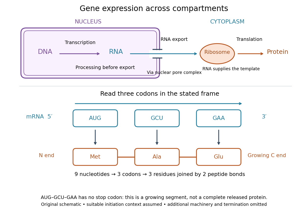

For a computed decoding example, choose the reading frame starting at the first displayed nucleotide of . Under the standard genetic code, AUG specifies methionine (Met), GCU specifies alanine (Ala), and GAA specifies a glutamic acid residue (Glu) in the growing polypeptide. Nine nucleotides divide into three codons, giving the ordered three-amino acid residue segment Met–Ala–Glu. The ribosome reads the messenger RNA , and the chain grows from its N-terminus toward its C-terminus. If synthesis begins at this AUG in an appropriate eukaryotic initiation context, these are the first three amino acid residues before any subsequent processing. See Clark, Douglas and Choi, Biology 2e, 15.1: standard codon assignments.
For this three-amino acid residue segment, joining the three delivered amino acids requires two peptide bonds, one between Met and Ala and another between Ala and Glu. There is no stop codon in the displayed nucleotide sequence, so this is a growing segment rather than a complete released protein. An isolated nine-nucleotide RNA is not claimed to supply all signals needed for initiation. This small calculation separates three jobs: the messenger RNA specifies order, transfer RNA delivers the amino acids, and the ribosome catalyzes their joining. Each new amino acid residue is added at the C-terminus. See Clark, Douglas and Choi, Biology 2e, 15.5: initiation and chain elongation.
The beauty of a ribosome is its reusable chemistry: the same machinery can assemble many different proteins by following different messenger RNA templates. The genetic code connects an alphabet of nucleotides to an alphabet of amino acids, while transfer RNA links recognition of the message to delivery of building material. Changing the message can therefore change the product without building a different ribosome for every protein.
In 1955 George E. Palade described small particles in the cytoplasm, both free and associated with membranes, in A small particulate component of the cytoplasm, Journal of Biophysical and Biochemical Cytology 1, 59–68. The paper documents the structural observations, rather than the full modern decoding mechanism. Complementary biochemical work by Hoagland, Stephenson, Scott, Hecht and Zamecnik described a soluble RNA intermediate in protein synthesis in 1958, helping establish the role now assigned to transfer RNA; see A soluble ribonucleic acid intermediate in protein synthesis, Journal of Biological Chemistry 231, 241–257. Together, structural observations and biochemical experiments connected visible cellular machinery with its working parts.
Cell nucleus 2026-10-13
The cell nucleus is a membrane-bounded compartment characteristic of eukaryotic biological cells, containing most of the cellular DNA in a typical nucleated biological cell. Its double nuclear envelope separates this DNA from the surrounding cytoplasm. Most does not mean all: mitochondria and chloroplasts retain their own DNA. Nor does every specialized eukaryotic biological cell retain a cell nucleus: mature mammalian red blood cells have lost theirs. Prokaryotes organize their DNA in a nucleoid without a nuclear envelope, and still have ribosomes. See NHGRI: nucleus, [ref]
https://openstax.org/books/biology-2e/pages/23-1-eukaryotic-origins. Clark, Douglas and Choi, Biology 2e, 23.1: organelle DNA
and Clark, Douglas and Choi, Biology 2e, 40.2: mature mammalian red blood cells.Follow one nuclear protein-coding gene: its DNA is used in transcription to make RNA; after the appropriate processing, messenger RNA is exported through a nuclear pore complex; a ribosome in the cytoplasm then uses it in biological translation to build an amino acid chain. The DNA remains in the cell nucleus. The RNA is a template, not the material converted into a protein. The ribosome diagram below follows this route; gene expression gives a complementary nucleotide sequence example. See Alberts et al.: selective RNA export and protein import.
This architecture makes gene expression interesting in a spatial way: nuclear transcription and cytoplasmic biological translation are separated by a selective boundary. A messenger RNA can be processed before export and use, while proteins needed for nuclear functions travel inward. Inside the cell nucleus, the cell nucleolus helps assemble ribosome subunits for export. Thus the compartment housing most DNA also helps build the machinery that interprets its messages elsewhere.
Robert Brown described and named the cell nucleus in his account of plant biological cells, read to the Linnean Society in 1831 and published in its Transactions in 1833. This historical observation identified a recurring structure, without establishing its later-understood role in DNA storage or gene expression. See Brown, On the Organs and Mode of Fecundation in Orchideæ and Asclepiadeæ, Transactions 16, 685–738 (1833). This is a documented contribution rather than a claim that no one had observed the structure earlier.
There are unlisted articles, also show them or only show them.