Past exam of the MPhil in micro- and nanotechnology enterprise course of the University of Cambridge 2018 Paper 1 Section A(ii) 3 ii Solution by  Codex 0 2026-10-20
Let and be the open projected regions in the two layers. For normal, collimated rays, the transmitted opening is the intersectionAn upper particle blocks any lower-layer gap beneath its footprint. A translated layer can therefore reduce island size, suppress some islands or close the holes completely. If the projected openings coincide, an ideally registered second layer need not change the pattern at all. A rotated or differently registered layer can create a larger superperiod or a less regular pattern.
The additional height also increases the separation between some blocking edges and the substrate, making a finite angular source distribution more important. Deposition angle and layer separation then enter the projected aperture geometry. No unique double-layer pattern follows without specifying registry, orientation and source direction; the figure illustrates aligned and shifted possibilities instead of treating one as inevitable.
Past exam of the MPhil in micro- and nanotechnology enterprise course of the University of Cambridge 2018 Paper 1 Section A(ii) 3 i Solution by Codex 0 2026-10-20
Codex 0 2026-10-20Treat the specified particles only as rigid geometric shadowing objects; no biological preparation or interpretation is needed. Their projected shapes are rounded-ended rods with straight central length and cap radius . Normal, collimated evaporation deposits metal in the gaps between projected particle footprints, leaving isolated small islands after mask removal, rather than a metal replica of each rod.
One concrete dense parallel packing has center translationsAdjacent rounded ends then touch at triangular arrangements. The open regions are small curvilinear triangular gaps near those ends. This is a specified geometrical packing for the sketch; the words close-packed do not uniquely prescribe the domain orientation or registry of all real sheets.
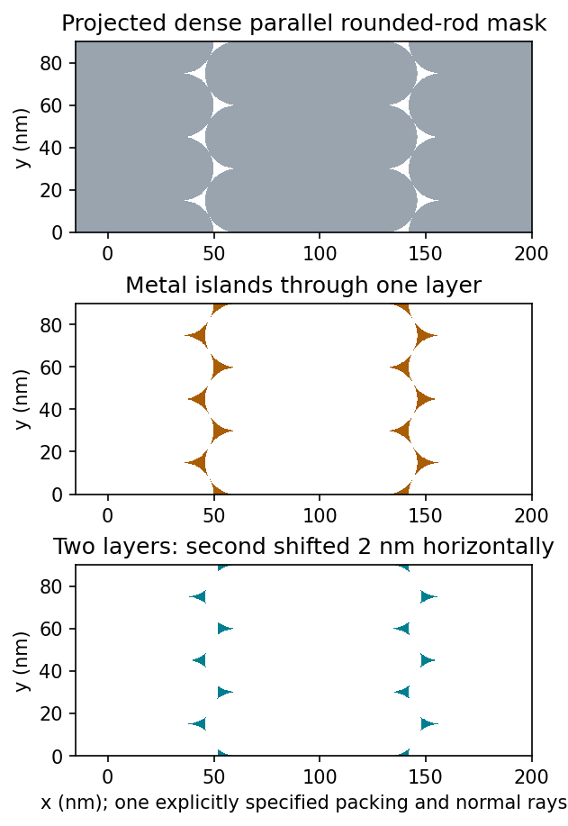
Original geometric calculation of rounded-rod shadow masks, deposited islands and the effect of shifting a second layer; no source illustration is reproduced
. Details. Particle size and shape distributions change the gap size and spacing. Misorientation, missing particles, boundaries between ordered domains, overlaps, incomplete coverage and drying-induced cracks interrupt long-range order. Substrate roughness and uneven adhesion distort the arrangement. A finite source angle produces deposition shadowing and penumbra; surface diffusion, island nucleation and increasing film thickness can broaden or merge deposited regions. These are generic geometry and materials effects, not assumptions about a biological assembly mechanism.
Past exam of the MPhil in micro- and nanotechnology enterprise course of the University of Cambridge 2018 Paper 1 Section A(i) 2 iv Solution by Codex 0 2026-10-20
Codex 0 2026-10-20Flattening removes fitted components of the image whether they are instrumental or real. A true specimen slope, broad dome or slow height variation can be erased if it resembles the chosen background. Strong features can bias a fit and create false depressions, streaks or halos; an overly flexible polynomial can fit away scientifically useful topography.
Line-by-line correction can destroy real differences between terraces, especially when no common flat region spans the lines. Height statistics, roughness and feature depths then depend on masking and fit order. Report those choices and compare profiles before and after processing.
Flattening does not restore structures inaccessible to the tip, undo a double-tip artefact, remove finite-tip broadening, reconstruct a saturated signal or recover data lost to unstable feedback. It also does not calibrate the scanner. These limitations are important when a superficially smoother image is mistaken for a more accurate measurement.
Past exam of the MPhil in micro- and nanotechnology enterprise course of the University of Cambridge 2018 Paper 1 Section A(i) 2 iii Solution by Codex 0 2026-10-20
Codex 0 2026-10-20Sample tilt and scanner bow are other common backgrounds that AFM image flattening can remove. Fit a plane to a genuinely flat reference area and subtract it to remove tilt. A low-order curved fit can approximate the vertical background produced by a piezoelectric scanner's non-planar motion.
Line-dependent offsets or slow drift can sometimes be removed by subtracting a reference level from each scan line. Mask pits, particles and true steps so that they do not bias the reference. An independently measured flat calibration specimen and scans in different orientations help distinguish instrumental background from actual specimen curvature.
Past exam of the MPhil in micro- and nanotechnology enterprise course of the University of Cambridge 2018 Paper 1 Section A(i) 2 ii Solution by Codex 0 2026-10-20
Codex 0 2026-10-20AFM image flattening estimates an unwanted background and subtracts it from the recorded height:A plane removes overall tilt; a line-by-line constant or low-order fit removes line offsets or curvature. For a tip-change height jump, align the baselines of unchanged reference regions on either side of the event, using a piecewise offset or appropriately masked line fits. A single global plane cannot in general remove an abrupt step.
Exclude genuine features from the fit, use the same terrace as a reference where possible, and retain the unprocessed data. The corrected image has a consistent relative background; it does not establish that the interaction force, tip shape or absolute height calibration remained unchanged. In particular, flattening is a correction for the offset component of a tip-change artefact, not a repair of every consequence of a changed tip.
Past exam of the MPhil in micro- and nanotechnology enterprise course of the University of Cambridge 2018 Paper 1 Section A(i) 2 i Solution by Codex 0 2026-10-20
Codex 0 2026-10-20A tip-change artefact occurs when the effective apex changes during scanning, for example through material pickup, loss of an attached particle or damage. In tapping-mode atomic force microscopy, a sudden change can introduce a false height offset between parts of the image, even on a continuous terrace.
The change alters the probe–surface interaction or the effective point at which the chosen amplitude setpoint is reached. The controller moves the vertical scanner to recover that setpoint, recording a spurious step or a change in line baseline. The sign is not fixed: different apex changes can move the apparent surface up or down.
The amplitude channel usually shows a transient deviation as the interaction changes; with stable feedback it returns toward its setpoint. It therefore need not retain the same persistent offset as the recorded height. A change in probe resonance or free amplitude can also change the operating condition. If the apex geometry has changed, widths and shapes can change too, or duplicated structures can appear as a double-tip artefact. A baseline correction cannot recover these lost geometric data.
Past exam of the MPhil in micro- and nanotechnology enterprise course of the University of Cambridge 2018 Paper 1 Section A(i) 1 ii Solution by Codex 0 2026-10-20
Codex 0 2026-10-20For a displayed width representing a scanned specimen width , the linear image magnification isThe user changes the scan-coil excursion, hence . A smaller raster displayed at the same width gives larger magnification. Changing the display or print size also changes displayed magnification, so the calibrated scale bar is more portable than a magnification label. This is the scan/display principle described in the manufacturer's microscope guide.[ref]
Scanning Electron Microscope A To Z by JEOL. Magnification of SEM, printed p.7: scan width and display width; the same raster-display geometry applies in STEM
At the low-magnification extreme, the probe scans the widest accessible field. Coil range, apertures, scan distortion and maintaining focus constrain the actual usable width. Formally increasing without bound would make arbitrarily small, but a real column has a finite accessible field.
At the high-magnification extreme, the scan covers a very small region. Formally makes diverge, but exactly zero scan width samples one location and supplies no spatial image. Electron microscope resolution, finite probe size, pixel spacing, drift, noise and specimen damage set the useful limit. Enlarging a field already sampled more finely than its resolvable information produces empty magnification, not new structural detail. For pixels across a field, the specimen sampling interval is ; this is distinct from the probe's spatial resolution.
Past exam of the MPhil in micro- and nanotechnology enterprise course of the University of Cambridge 2018 Paper 1 Section A(i) 1 i Solution by Codex 0 2026-10-20
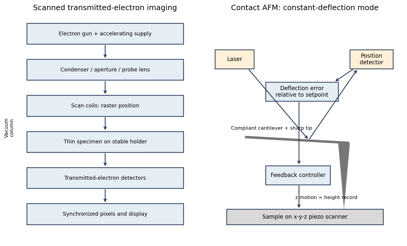
Codex 0 2026-10-20A scanning transmission electron microscope makes an image by assigning a transmitted-electron signal to each position of a scanned probe. Its essential chain is electron source, probe-forming optics, scan system, thin specimen, transmitted-electron detectors and synchronized acquisition.
An electron gun and accelerating supply provide a stable beam of the chosen kinetic energy. Condenser lens optics and an aperture control probe current and convergence; the probe-forming objective lens focuses a small spot on the specimen. Alignment, focusing and astigmatism correction keep the probe usable across the field.
Scan coils deflect the probe in a raster. A stable specimen holder positions an electron-transparent region, and a high-vacuum column limits gas scattering and contamination. A bright-field transmission electron microscopy detector collects the central transmitted beam; an annular detector can select annular dark-field electron imaging, including high-angle annular dark-field imaging. Different angular selections give different contrasts, rather than a universal map of height.
Detector electronics, scan timing and a computer assign the collected signal to the corresponding image pixel. Spectrometers are useful additions but not essential to basic image formation. The post-specimen optics can adjust detector collection geometry; they do not set scan-image magnification in the same way as the projection lenses of conventional transmission imaging.
Past exam of the MPhil in micro- and nanotechnology enterprise course of the University of Cambridge 2017 Paper 2 Section B 23B v Solution by Codex 0 2026-10-20
Codex 0 2026-10-20A tightly focused laser produces a strong intensity gradient. A small polarizable particle experiences a gradient force toward increasing intensity when the real polarizability has the appropriate positive sign; radiation pressure from scattering and absorption pushes predominantly along the beam. A high-numerical-aperture objective creates lateral confinement and a sufficiently strong opposing axial gradient for a three-dimensional optical trap. Equilibrium is generally shifted from the intensity maximum by radiation pressure.
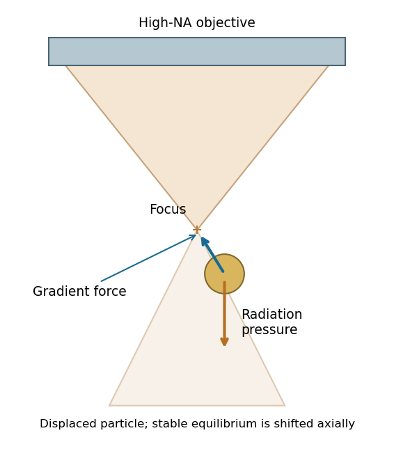
For a Rayleigh sphere, in an absolute-permittivity convention, and for peak complex field amplitude. Near a stable point, ; the trap depth must exceed thermal fluctuations sufficiently to retain the particle. Viscous drag opposes motion, Brownian forcing produces fluctuations, and gravity/buoyancy are normally small at this scale.
For absorbing gold, use its complex wavelength-dependent permittivity: scattering, absorption and heating can frustrate trapping. The existence of an ideal dielectric gradient-force trap does not guarantee stable trapping of every metal nanoparticle at every wavelength or power.[ref]
Observation of a single-beam gradient force optical trap for dielectric particles by Arthur Ashkin and others. General optical trapping principle; gold-specific absorption qualification is separate
Past exam of the MPhil in micro- and nanotechnology enterprise course of the University of Cambridge 2017 Paper 2 Section B 23B iv Solution by Codex 0 2026-10-20
Codex 0 2026-10-20Interpreting the nominal size as diameter , fluid viscous dominance requireswith in . The liquid density is not supplied, so the conventional fluid-Reynolds crossover is not numerically fixed. As an explicitly additional illustrative assumption, gives near ; ordinary much slower nanoparticle motion is well within the viscous regime.
If instead the supplied gold density is inserted into the dimensionless ratio , that ratio reaches one at . It is not the fluid Reynolds number. Including the spherical Stokes prefactor, the particle relaxation criterion occurs at . These quantify different inertia comparisons. The wording's speed direction is reversed: viscous dominance is obtained by making speed sufficiently small. If 10 nm denotes radius, the length-based thresholds halve.
Past exam of the MPhil in micro- and nanotechnology enterprise course of the University of Cambridge 2017 Paper 2 Section B 23B iii Solution by Codex 0 2026-10-20
Codex 0 2026-10-20For fluid density , speed and characteristic length , inertial kinetic energy scales as while viscous work over distance scales as . Their ratio is the Reynolds number,Smaller particles at a fixed speed have smaller Re, so viscous stresses dominate fluid inertia when . Increasing speed increases Re and eventually makes inertia more important, rather than making viscous dominance stronger.
Particle inertia is a separate issue. Its particle velocity-relaxation time is ; comparing it with a travel time gives a Stokes number . This uses particle density , whereas the fluid Reynolds number uses fluid density .
Past exam of the MPhil in micro- and nanotechnology enterprise course of the University of Cambridge 2017 Paper 2 Section B 23B ii Solution by Codex 0 2026-10-20
Codex 0 2026-10-20For radius and gold density , . The supplied Stokes model gives and at . Therefore the standard one-second diffusive rms distances are in one coordinate and in three dimensions.
Using the source's supplied shorthand literally with instead gives . That shorthand lacks the factor two in the standard long-time mean-square displacement; it should not be identified with either of the preceding standard rms conventions. None of these rms values is a cumulative path length.
An ideal drag-free fall from rest gives . Actual gravitational settling in the assumed viscous model is much smaller: , , and displacement in one second is approximately . Thus diffusion overwhelms settling, even though ideal vacuum fall exceeds the rms diffusion distance. For rarefied-gas nanoparticle drag, at a 20-nm size in argon, continuum no-slip drag may require a rarefaction/slip correction; pressure and gas mean-free-path data are absent, so these numerical transport estimates are conditional on the supplied Stokes model.
Past exam of the MPhil in micro- and nanotechnology enterprise course of the University of Cambridge 2017 Paper 2 Section B 23B i Solution by Codex 0 2026-10-20
Codex 0 2026-10-20Brownian motion is the fluctuating motion of a suspended particle produced by thermal molecular collisions. Using the standard Brownian displacement conventions, in a uniform fluid without external drift, mean displacement is zero but mean-square displacement grows with time. For overdamped diffusion, in one coordinate and in three dimensions, with and Stokes friction for a no-slip sphere.
Brownian transport brings nanoparticles into encounters and allows them to explore configurations during self-assembly. Reversible interactions permit rearrangement toward low-free-energy structures; interactions that are too strong can instead trap disordered aggregates. Thermal fluctuations also oppose localization in shallow traps. The symbol avoids confusing friction with an unrelated chemical potential.
Past exam of the MPhil in micro- and nanotechnology enterprise course of the University of Cambridge 2017 Paper 2 Section B 23A iii Solution by Codex 0 2026-10-20
Codex 0 2026-10-20Consider a colloidal photonic crystal assembled from nearly monodisperse dielectric spheres. Spherical geometry and approximately isotropic interactions favour close-packed arrays, although fcc, hcp and stacking-disordered structures can compete. Sphere diameter sets the periodic optical length scale. For an ideal close-packed fcc array,at normal incidence in the simple effective-index picture. Thus changing the building-block size tunes the reflected wavelength; refractive-index contrast affects stop-band strength. Surface charge and screening control colloidal stability and rearrangement during assembly, while polydispersity, roughness and stacking defects broaden the optical response and increase scattering.
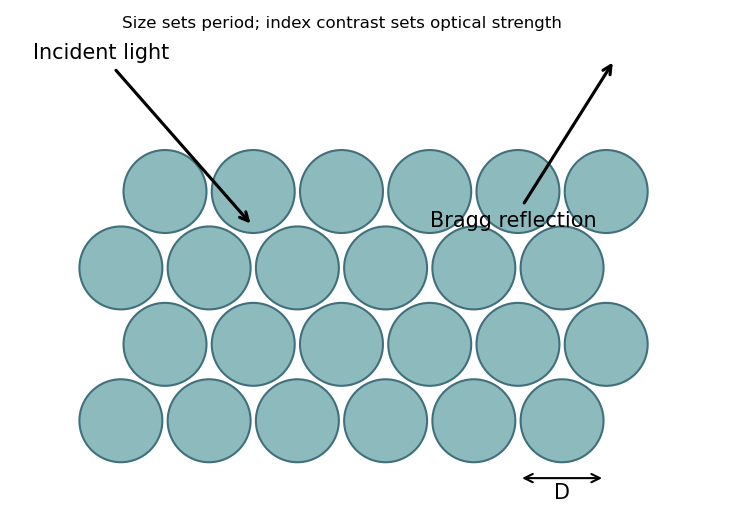


This connects sphere-level geometry and material response to collective periodic optical behaviour. The Bragg reflection estimate does not establish a complete three-dimensional photonic band gap.
Past exam of the MPhil in micro- and nanotechnology enterprise course of the University of Cambridge 2017 Paper 2 Section B 23A ii Solution by Codex 0 2026-10-20
Codex 0 2026-10-20Relevant interactions include van der Waals attraction, electrostatic attraction or screened repulsion, hydrogen bonding, and hydrophobic/solvophobic association. Coordination bonding, magnetic dipole interactions and capillary forces can also organize suitable building blocks. Entropic effects, including depletion attraction and packing entropy, can drive ordering even without a new chemical bond. Which contribution favours assembly depends on solvent, temperature, concentration and surface chemistry; the complete balance is a free-energy change, not a requirement that every interaction be attractive.
Past exam of the MPhil in micro- and nanotechnology enterprise course of the University of Cambridge 2017 Paper 2 Section B 23A i Solution by Codex 0 2026-10-20
Codex 0 2026-10-20Self-assembly is organization of interacting building blocks into a larger structure without individually placing every component. Static self-assembly approaches an equilibrium free-energy minimum under fixed conditions; a resulting structure may subsequently become kinetically trapped. Dynamic self-assembly maintains a nonequilibrium organized state through continuing energy or matter throughput and dissipation, and can change or disappear when that drive is removed.
For nanofabrication, parallel assembly of many small components can generate fine periodicity, hierarchical structures and adaptive or self-repairing arrangements with limited direct patterning. Building-block chemistry encodes interactions. However, access to a desired morphology depends on kinetics and competing states; assembly does not automatically eliminate defects or provide arbitrary device geometry.
Past exam of the MPhil in micro- and nanotechnology enterprise course of the University of Cambridge 2017 Paper 2 Section B 22 v Solution by Codex 0 2026-10-20
Codex 0 2026-10-20 Past exam of the MPhil in micro- and nanotechnology enterprise course of the University of Cambridge 2017 Paper 2 Section B 22 iv Solution by Codex 0 2026-10-20
Codex 0 2026-10-20 Past exam of the MPhil in micro- and nanotechnology enterprise course of the University of Cambridge 2017 Paper 2 Section B 22 iii Solution by Codex 0 2026-10-20
Codex 0 2026-10-20With the left reservoir at and the right at zero, let the pore run from to . In the stated idealization the reservoir potentials are flat, the pore potential is , and its electric field is , directed from high to low potential. The field is zero in the ideal equipotential reservoirs; real access regions would round these abrupt boundaries.
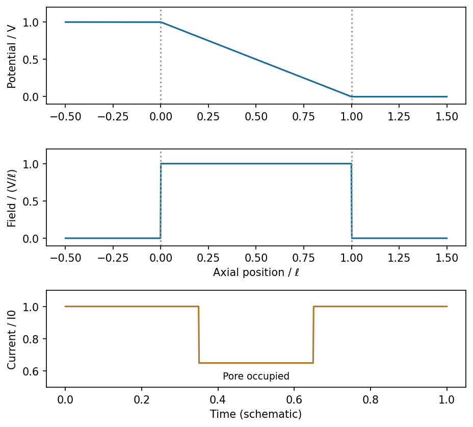
Ideal potential, field and current blockade for a generic insulating particle crossing a uniform pore
. Source. Details. 
For constant-speed passage and an approximately position-independent in-pore obstruction, the current has a rectangular downward pulse from to during occupation. Entry, exit, filtering and nonuniform fields round real pulses. This describes a physical particle signal without inferring any biological identity or charge.
Past exam of the MPhil in micro- and nanotechnology enterprise course of the University of Cambridge 2017 Paper 2 Section B 22 ii Solution by Codex 0 2026-10-20
Codex 0 2026-10-20Ignoring access effects and wall charge, the cylindrical conducting liquid hasThe corresponding conductance is approximately . A charged surface can contribute mobile counterions and change this estimate. Surface conduction is important when its conductance is comparable with the bulk contribution: for surface conductance , their ratio in a cylinder is , twice the Dukhin number convention . Double-layer overlap, when the Debye length is comparable with the pore radius, is another warning that uniform bulk conductivity is inadequate. No surface-charge or ionic-strength data are supplied to quantify those corrections.
There are unlisted articles, also show them or only show them.