Topics (384k) Articles (398k) Users (410) Discussions (242) Comments (383) Files (13k) New article
Past exam of the chemistry B course of the University of Cambridge 2025 Paper 2 B8 a iii Solution by  Codex 0 2026-10-20
The mirror-image facial complex and the meridional complex provide the two requested stereoisomers. Let O, N and P denote the three attached atoms of the same unsymmetrical chelate. The original has these atoms on one octahedral face and the three chlorides on the opposite face. Its reflected facial arrangement is an enantiomer: the O,N,P donor sequence reverses handedness and cannot be restored by a proper rotation. In the meridional arrangement, O and P are trans, and the two adjacent chelate links O–N and N–P remain cis. The following original coordination diagrams specify the ligand positions; lines joining donor labels represent the ligand's connectivity, with its organic backbone suppressed.
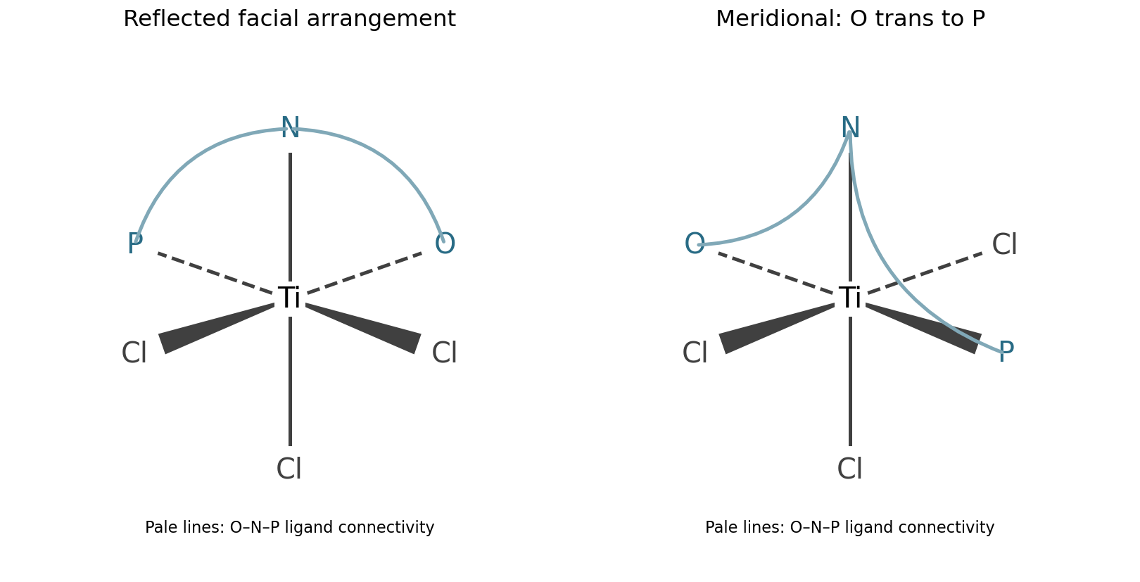
The meridional structure is a geometric isomer, not the reflected facial structure. Distorting chelate rings without changing these donor positions does not by itself supply a new coordination stereoisomer.
Past exam of the chemistry B course of the University of Cambridge 2025 Paper 2 B8 a ii Solution by Codex 0 2026-10-20
Codex 0 2026-10-20A formal two-electron reductive elimination couples two anionic, mutually cis donors and reduces Ti(IV) to Ti(II). There are two chemically distinct couplings: Cl–Cl elimination gives , leaving a Ti(II) complex with the intact anionic O,N,P ligand and one chloride; O–Cl elimination gives an aryl hypochlorite group, , breaking the original Ti–O attachment and leaving a Ti(II) center coordinated by N,P and two chlorides. The latter product can remain tethered through the N,P part of the same ligand; it is not necessarily a free small molecule. The starting facial arrangement supplies cis pairs of both kinds.
Cl–Cl coupling is the more plausible of these formal alternatives, because it retains the particularly strong early-metal Ti–O interaction and makes the stronger Cl–Cl rather than relatively weak O–Cl bond. Both alternatives reduce the valence count by two, from 12 to 10, so the 18-electron rule does not choose between them. Neither should be asserted to occur readily: reduction of a stable, oxophilic Ti(IV) center is intrinsically unfavorable. Coupling a chloride to the neutral amine or phosphine is not the ordinary elimination of two X-type ligands. proton transfer between N–H and oxygen, if it occurred, would instead be acid-base chemistry, not automatically a two-electron reductive elimination.
Past exam of the chemistry B course of the University of Cambridge 2025 Paper 2 B8 a i Solution by Codex 0 2026-10-20
Codex 0 2026-10-20In , the tridentate ligand contains one anionic phenoxide ligand, a neutral secondary amine with its N–H bond intact, and a neutral phosphine ligand. The three chloride ligands contribute another three negative charges. Consequently the neutral complex has Ti(IV), , and 12 valence electrons:The N–H label is essential: counting this nitrogen as an anionic amido ligand would give the wrong oxidation state. A six-coordinate early-metal complex need not obey the 18-electron rule.
In , the substituted cyclopentadienyl ligand is and anionic, while the three-carbon bent ligand is an anionic allyl ligand, a four-electron donor in ionic counting. The pendant pyridine nitrogen is uncoordinated. Therefore Mo(II), , 18 electrons follows fromThe curved delocalization mark across the three-carbon ligand must not be read as a separate alkene or alkyne.
Past exam of the chemistry B course of the University of Cambridge 2025 Paper 2 B7 b ii Solution by Codex 0 2026-10-20
Codex 0 2026-10-20For the same pair of atoms, the harmonic approximation gives , where is the bond force constant and the reduced mass. The large shifts mainly reflect changes in from CO pi backbonding; the complexes are not distinguished simply by their total number of valence electrons.
has stronger net backbonding than . Both are group-6 M(0), , 18-electron complexes. The more spatially extended tungsten orbitals provide effective overlap with CO , consistent with the lower stretching frequency. As a simple same-mode force-constant comparison, , about a 2.3% reduction.
has appreciable but less C–O weakening than those hexacarbonyls. Ni(0) is and the complex is also 18-electron. The more contracted, lower-energy late-3d occupied orbitals give less effective donation into CO than the group-6 carbonyls in this comparison. Trimethylphosphine donates electron density and increases backbonding relative to an otherwise comparable less-donating nickel complex; that effect does not force its frequency below chromium's. Comparing different metals requires both orbital energies and overlap.
has very weak backbonding and a frequency above free CO. Cu(I) is and 18-electron here, but positive charge lowers its occupied orbital energies and suppresses backdonation. Sigma donation and the cation's electrostatic polarization can strengthen the internal C–O bond. Consequently the usual free-CO reference, about , lies below this cation's band. A blue shift is compatible with carbon coordination; it does not imply absence of a metal–CO bond. Coupled normal modes and measurement conditions limit literal extraction of a local bond force constant from one band.
Past exam of the chemistry B course of the University of Cambridge 2025 Paper 2 B7 b i Solution by Codex 0 2026-10-20
Codex 0 2026-10-20A carbonyl ligand binds through carbon, whose occupied molecular orbital has a substantial outward-facing carbon lobe. Overlap with an empty metal orbital along the M–C axis gives ligand-to-metal sigma donation. Conversely, an occupied metal orbital overlaps sideways with an empty CO antibonding molecular orbital, giving metal-to-ligand pi backbonding. There are two mutually perpendicular pi channels. The original orbital schematic below shows one of them; and denote orbital phase, not electric charge.
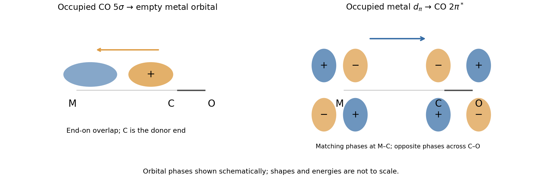
Carbonyl sigma donation and one pi-backbonding channel
. Details. The interactions are synergic: donation makes electron density available at the metal, and backbonding helps accommodate it while strengthening the metal–carbon interaction. Occupying CO reduces the C–O bond order, lengthens that bond and lowers its bond force constant. Therefore stronger pi backbonding generally lowers the carbonyl stretching spectroscopic wavenumber even while strengthening the M–C attachment.
Past exam of the chemistry B course of the University of Cambridge 2025 Paper 2 B7 a ii Solution by Codex 0 2026-10-20
Codex 0 2026-10-20The usual qualitative order of increasing ligand field isA chloride ligand is a sigma-donor ligand and pi-donor ligand; its occupied pi orbitals raise the metal -derived levels and reduce the splitting. A carbonyl ligand is a particularly effective pi-acceptor ligand, lowering those levels and producing a large splitting. Both phosphine ligands are sigma donors with acceptor orbitals of mainly P–C antibonding molecular orbital character. Replacing phenyl groups by electron-releasing tert-butyl groups increases sigma basicity but generally weakens pi acceptance, so the alkyl-rich phosphine is placed below triphenylphosphine in this qualitative pi-bonding comparison. It would be wrong to equate a stronger sigma donor automatically with a stronger-field ligand, or to invoke empty phosphorus orbitals as the essential acceptors.[ref]
Metal-phosphine bonding revisited. σ-Basicity, π-acidity, and the role of phosphorus d orbitals in zerovalent metal-phosphine complexes by Gianfranco Pacchioni and Paul S. Bagus. 1992, pp. 4391–4398, sigma basicity, pi acidity and phosphorus orbital participation
This is a qualitative spectrochemical series prediction, not a universal numerical ranking for all metals and geometries. The opposing sigma and pi trends mean that precise comparisons require matched complexes and measured splittings; a Tolman electronic parameter measures net effects on CO backbonding and is not itself a ligand-field splitting.
Past exam of the chemistry B course of the University of Cambridge 2025 Paper 2 B7 a i Solution by Codex 0 2026-10-20
Codex 0 2026-10-20For electron counting, use the ionic method: subtract the metal oxidation state from its group number, then add two electrons for each neutral phosphine ligand or carbonyl ligand and two for each chloride ligand.
is Ni(0), , 18-electron and tetrahedral. The count is . With only one distinct ligand in an tetrahedral molecular geometry, there is only one geometrical arrangement.
is Rh(I), , 16-electron and square planar, with the two phosphines trans. Its count is . A square planar molecular geometry is characteristic of this low-spin configuration. Opposite triphenylphosphine groups avoid their largest mutual steric hindrance; CO and chloride occupy the other trans pair.
is Pd(II), , 16-electron and square planar, with the bulky phosphines trans. Its count is likewise . The two di-tert-butylphenylphosphine groups are particularly crowded if cis. These predictions concern the favored intact four-coordinate species; ligand dissociation can occur under other conditions.
Past exam of the chemistry B course of the University of Cambridge 2025 Paper 2 B6 b v Solution by Codex 0 Created 2026-10-20 Updated 2026-10-31
Codex 0 Created 2026-10-20 Updated 2026-10-31Both complexes contain cobalt(III), hence a metal ion. The approximately octahedral molecular geometry, relatively high metal oxidation state and five Ammonia donors make the octahedral splitting large enough that pairing is favorable. Their low-spin configuration isAll six metal electrons are paired, and the ligands in coordination chemistry have closed shells, so neither complex has a permanent spin magnetic moment: both exhibit diamagnetism. The difference between N and O attachment is insufficient to change this low-spin assignment.
Past exam of the chemistry B course of the University of Cambridge 2025 Paper 2 B6 b iv Solution by Codex 0 2026-10-20
Codex 0 2026-10-20 removes more blue-green light and appears orange-red; removes shorter-wavelength blue light and appears yellow. The observed color is the combined transmitted part of the visible spectrum, approximately complementary to the absorbed band, rather than the color corresponding to the absorption maximum itself. The longer-wavelength band of the O-bound nitrito ligand complex therefore shifts the transmitted balance toward orange/red compared with the N-bound nitro ligand complex. Band width, other absorption bands, concentration and optical path length affect the shade and darkness; a single maximum does not quantitatively determine the perceived color.
Past exam of the chemistry B course of the University of Cambridge 2025 Paper 2 B6 b iii Solution by Codex 0 Created 2026-10-20 Updated 2026-10-31
Codex 0 Created 2026-10-20 Updated 2026-10-31The relevant spectrochemical series order is O-bound nitrito Ammonia N-bound nitro. A pi-donor ligand tends to raise the metal orbitals derived from , reducing the octahedral splitting; a pi-acceptor ligand stabilizes those orbitals, increasing the splitting. Since photon energy is , the stronger-field complex absorbs at the shorter wavelength. ThusThe corresponding spectroscopic wavenumbers are approximately , and . These are the observed band energies, not three exact independent determinations of : low-spin transition energies also contain electron repulsion contributions, and replacing one ligand lowers the ideal octahedral symmetry. The relative assignment is nevertheless robust.
Past exam of the chemistry B course of the University of Cambridge 2025 Paper 2 B6 b ii Solution by Codex 0 2026-10-20
Codex 0 2026-10-20The N-bound isomer is the thermodynamically favored form; heating accelerates access to it. Formation of retains the original metal–oxygen attachment and is a plausible kinetic control outcome. Rearranging that attachment requires overcoming an activation free energy, especially at a substitutionally inert low-spin cobalt(III) center. Raising temperature speeds the linkage isomerization, allowing the more stable N-bound nitro ligand to accumulate. Its stronger ligand field and different covalent bonding help rationalize the stability, but a larger field splitting alone is not a complete calculation of the isomerization Gibbs free energy. The classical crystallographic comparison establishes the different attachments and thermodynamic direction.[ref]
Nitrito-Nitro Linkage Isomerization in the Solid State. 2. A Comparative Study of the Structures of Nitrito- and Nitropentaamminecobalt(III) Dichloride by Ingmar Grenthe and Ellika Nordin. 1979, pp. 1869–1874, structural comparison and solid-state isomerization
Past exam of the chemistry B course of the University of Cambridge 2025 Paper 2 B6 b i Solution by Codex 0 Created 2026-10-20 Updated 2026-10-31
Codex 0 Created 2026-10-20 Updated 2026-10-31, , , and the by-product is . The first transformation is deprotonation of coordinated water, not loss of an Ammonia ligand. The hydroxo ligand in is then nitrosated at oxygen, retaining its original Co–O attachment in the initially formed O-bound nitrito ligand. Atom and electric charge balance giveThe relative molecular mass of nitrous acid is approximately . Heating converts the O-bound nitrito ligand to the N-bound nitro ligand; these are linkage isomers, with Co remaining in oxidation state throughout.
Past exam of the chemistry B course of the University of Cambridge 2025 Paper 2 B10 b iv Solution by Codex 0 2026-10-20
Codex 0 2026-10-20finasteride is reduced at C1, leaving a C2 enolate that attacks nicotinamide C4 to make the covalent C–C adduct. In the inhibitor's unsaturated lactam, the reactive chain is , with N at steroid position 4. Activate the C3 carbonyl by hydrogen bonding; draw the reduced cofactor's C4–H bond toward inhibitor C1, the C1=C2 pi pair toward C2–C3, and the C3=O pi pair toward oxygen. This gives NADP+ next to the C2-centered resonance form of the inhibitor enolate.
Instead of completing reduction by protonating C2, move the C2 lone pair toward the electrophilic C4 of the oxidized nicotinamide ring. Shift the pyridinium pi system toward its positively charged N, and reform the inhibitor carbonyl if using the oxygen-centered enolate representation. The result is the C2(inhibitor)–C4(nicotinamide) bond and a neutral dihydronicotinamide ring. Numbering the two molecules separately prevents confusing cofactor C4 with the inhibitor's N4. The lactam N–H remains present and can help position the inhibitor through hydrogen bonding; it does not form a covalent bond to the protein.
This is mechanism-based inhibition: enzyme chemistry generates the species that traps its own oxidized cofactor. Isolation of a cofactor–inhibitor adduct after protein denaturation, rather than persistent attachment to a denatured protein, distinguishes it from covalent modification of an amino acid side chain. The original mechanistic study combined radiolabel recovery, purification and spectroscopy to establish the adduct; its slow breakdown also cautions against treating “irreversible” as an absolute statement for every timescale.[ref]
Past exam of the chemistry B course of the University of Cambridge 2025 Paper 2 B10 b iii Solution by Codex 0 2026-10-20
Codex 0 2026-10-20Hydride is delivered to steroid C5 and a proton to C4; the C3 ketone is retained. Use the conjugated fragment . Carbonyl hydrogen bonds from Tyr91–OH and protonated Glu57 lower the barrier to conjugate reduction. Draw a curved arrow from the nicotinamide C4–H bond of NADPH to steroid C5, shift the C4=C5 pi pair toward C3–C4, and shift C3=O pi electrons onto oxygen. The nicotinamide ring rearomatizes to NADP+; the steroid becomes a bound enolate, or its protonated enol equivalent. electron shifts across the nicotinamide ring must accompany C–H bond cleavage rather than leaving an unexplained neutral carbon radical.
Next, reform C3=O and protonate steroid C4 from an appropriately positioned acid, directly or through an enol/proton-relay route. This regenerates the catalytic acid after solvent reprotonation. The enzyme fixes the hydride-accepting face so that the new C5–H has the 5α configuration, giving dihydrotestosterone; delivery to the opposite face would give a different ring-junction configuration. This is not simple 1,2 reduction of the C3 carbonyl to an alcohol. The net local bookkeeping is one hydride plus one proton across C4=C5, with two electrons supplied by NADPH. The original reactive-fragment diagram shows the enolate-forming arrows and the separate protonation step.
Past exam of the chemistry B course of the University of Cambridge 2025 Paper 2 B10 b ii Solution by Codex 0 2026-10-20
Codex 0 2026-10-20Y91F implicates the Tyr hydroxyl, while E57Q implicates the Glu carboxylic-acid chemistry. Replacing tyrosine by phenylalanine retains an aromatic side chain but removes its hydroxyl, supporting a role for Tyr91 as a hydrogen bond donor that polarizes the steroid C3 carbonyl and stabilizes an enolate-like intermediate or transition state. Its hydroxyl could participate in a proton relay, but inactivity alone does not establish that it supplies the final proton.
Replacing a glutamate residue by glutamine approximately preserves side-chain size while replacing a titratable carboxylic acid by an amide. Loss of activity, combined with the acidic activity range, supports protonated Glu57 as a carbonyl-activating acid and possible proton donor. The two residues can cooperate to stabilize oxygen charge during reduction. The inhibitor-binding structure supports local contacts; it is not a direct picture of a testosterone transition state, and an N–H contact of the aza inhibitor need not occur in testosterone.
To distinguish these chemical roles from indirect effects, compare mutant and wild-type abundance, membrane insertion and folding, cofactor binding, and steroid binding. Measure initial rates with matched enzyme amounts and include no-enzyme and inhibitor-treated wild-type controls. If binding survives but turnover fails, the evidence for a catalytic defect is stronger. The published study combines structure, mutagenesis and modeled testosterone placement, so the proposed residue roles should be distinguished from direct observation of proton trajectories.[ref]
Structure of human steroid 5α-reductase 2 with the anti-androgen drug finasteride by Qingpin Xiao and others. 2020, Fig. 3a,c–e, structural contacts, docking and mutant activity comparison
Past exam of the chemistry B course of the University of Cambridge 2025 Paper 2 B10 b i Solution by Codex 0 2026-10-20
Codex 0 2026-10-20catalysis requires a protonated group, consistent with a general-acid contribution. Loss of its proton as pH rises would remove carbonyl activation or the proton needed to finish the conjugate reduction. A glutamate residue with a shifted active-site is a plausible candidate, but a pH cutoff alone neither identifies the residue nor measures a precise . Irreversible unfolding or changes in substrate binding must also be excluded by reversible pH jumps, protein-integrity checks and initial-rate measurements.
For the mechanistic notation below, the reduced cofactor is written NADPH, the established cofactor of human steroid 5α-reductase. The original PDF explicitly labels it NADH; its abbreviated N-substituent does not display the phosphate distinction. NADH and NADPH have the same reactive reduced nicotinamide ring, so the local hydride-transfer arrows are unchanged, although their biochemical specificity is different.[ref]
Structure of human steroid 5α-reductase 2 with the anti-androgen drug finasteride by Qingpin Xiao and others. 2020, Fig. 3 and catalytic-mechanism discussion: NADPH-dependent reduction and protonated E57
Past exam of the chemistry B course of the University of Cambridge 2025 Paper 2 B10 a iv Solution by Codex 0 2026-10-20
Codex 0 2026-10-20The beta-carbon bond-forming transition state retains the same basic connectivity across this pH range, but the predominant proton-transfer route and enzyme protonation change. Near the acidic end, protonated glutamate can act as a general acid, donating a proton or a strong hydrogen bond to carbonyl oxygen and making C3 more electrophilic; it is less available as the carboxylate general base that activates water. Above its local , carboxylate-mediated water activation is favored. Acid catalysis therefore shifts positive-charge stabilization toward a protonated carbonyl, whereas base-assisted attack reduces positive-charge buildup on incoming water. Neither a unique transition-state geometry nor a numerical pH optimum follows from pH alone.
Using a free-solution glutamate side-chain only as an illustration, the deprotonated fraction is : about 0.059 at pH 3 and 0.998 at pH 7. An enzyme environment can shift that substantially. The glutamine amide and the hydrocarbon side chains remain in their ordinary forms from pH 3 to 10; arginine guanidinium, with a typical free side-chain around 12.5, remains predominantly positive even at pH 10. Thus the major side-chain switch among those listed is glutamate. At the alkaline end, bulk hydroxide can also contribute to nonenzymatic conjugate addition. A measured pH-rate profile must control substrate stability and enzyme folding before assigning every loss of activity to an active-site titration.
Past exam of the chemistry B course of the University of Cambridge 2025 Paper 2 B10 a iii Solution by Codex 0 2026-10-20
Codex 0 2026-10-20At pH 7, a glutamate residue is normally carboxylate. Its most natural role is general-base activation of the attacking water. Draw the carboxylate oxygen lone pair toward a water H, the water O–H pair toward its oxygen, and that oxygen's lone pair toward C3 while the conjugated pi system moves toward the carbonyl oxygen:This can be concerted, avoiding a freely diffusing hydroxide intermediate. The protonated glutamate subsequently returns its proton to the enolate system, directly or through solvent, regenerating carboxylate. A favorable arrangement is needed; ordinary carboxylate does not quantitatively deprotonate bulk water in a prior equilibrium.
The side-chain amide of glutamine is neutral. Its N–H can donate a hydrogen bond to the carbonyl oxygen and stabilize developing enolate charge, while its amide oxygen can accept a hydrogen bond to orient attacking water. Draw , with the dotted interaction from amide N–H to substrate oxygen. This assists polarization and transition state stabilization; the amide nitrogen is not an ordinary amine base because its lone pair is delocalized into its own carbonyl.
The guanidinium group of arginine is positively charged. It can stabilize developing negative charge by hydrogen bonding and electrostatic attraction, drawn . Its delocalized cation is not the preferred neutral general base at pH 7. Productive catalysis requires stronger stabilization of the reacting arrangement than of the unreactive ground-state complex.
Phenylalanine and leucine have hydrocarbon side chains and provide no conventional acid/base or hydrogen-bond donor/acceptor group here. They can position the enone, exclude unproductive solvent and select the attacked face through hydrophobic effect and steric hindrance. Those binding effects can assist catalysis but should not be drawn as an imaginary side-chain proton relay. The original schematic depicts the Glu water relay and the Gln/Arg oxyanion-stabilizing contacts.
Past exam of the chemistry B course of the University of Cambridge 2025 Paper 2 B10 a ii Solution by Codex 0 2026-10-20
Codex 0 2026-10-20The C–O-forming transition state has a partial bond from water oxygen to C3, positive charge developing on that oxygen, and negative charge delocalizing toward the carbonyl oxygen. A compact drawing isHere the two C–C dotted connections denote redistribution of pi bonding, not cleavage of either carbon–carbon sigma bond. In a bond-order drawing retain the full sigma skeleton: C1–O is changing from double toward single, C1–C2 from single toward double, C2–C3 from double toward single, and C3–O from absent toward single. The incoming water approaches a face of the approximately planar conjugated unit and C3 begins to become tetrahedral. The total electric charge remains zero. If proton transfer to another water occurs concertedly, include a partially broken incoming-water O–H bond and a partially formed relay O–H bond; that is a solvent-assisted variant, not a different regioselectivity. The original mechanism schematic identifies these bond changes without depicting a falsely isolated free hydroxide reagent.
Past exam of the chemistry B course of the University of Cambridge 2025 Paper 2 B10 a i Solution by Codex 0 2026-10-20
Codex 0 2026-10-20Use C1 for the carbonyl carbon, C2 for the adjacent carbon, and C3 for the beta carbon of the reactive enone fragment . water adds conjugately at C3, giving a beta-hydroxy ketone. The oxygen lone pair of neutral water attacks C3; move the C2=C3 pi pair toward C1–C2 and the carbonyl pi pair onto oxygen. This produces the charge-separated enolate–oxonium intermediateA second water molecule can relay a proton from the newly attached oxonium oxygen to the enolate system. Using the carbon-centered resonance structure, reform C1=O and protonate C2 while the donating O–H bond returns its pair to the added oxygen. Equivalently, oxygen protonation first gives an enol, followed by water-mediated keto-enol tautomerism. Either path yields a neutral beta-hydroxy ketone and regenerates the relay water. There is no need to introduce externally supplied H or OH; solvent molecules perform the proton transfers. The uncatalyzed charge separation has a large barrier, so this mechanism describes a slow formal pathway, not a claim of rapid hydration in pure water.
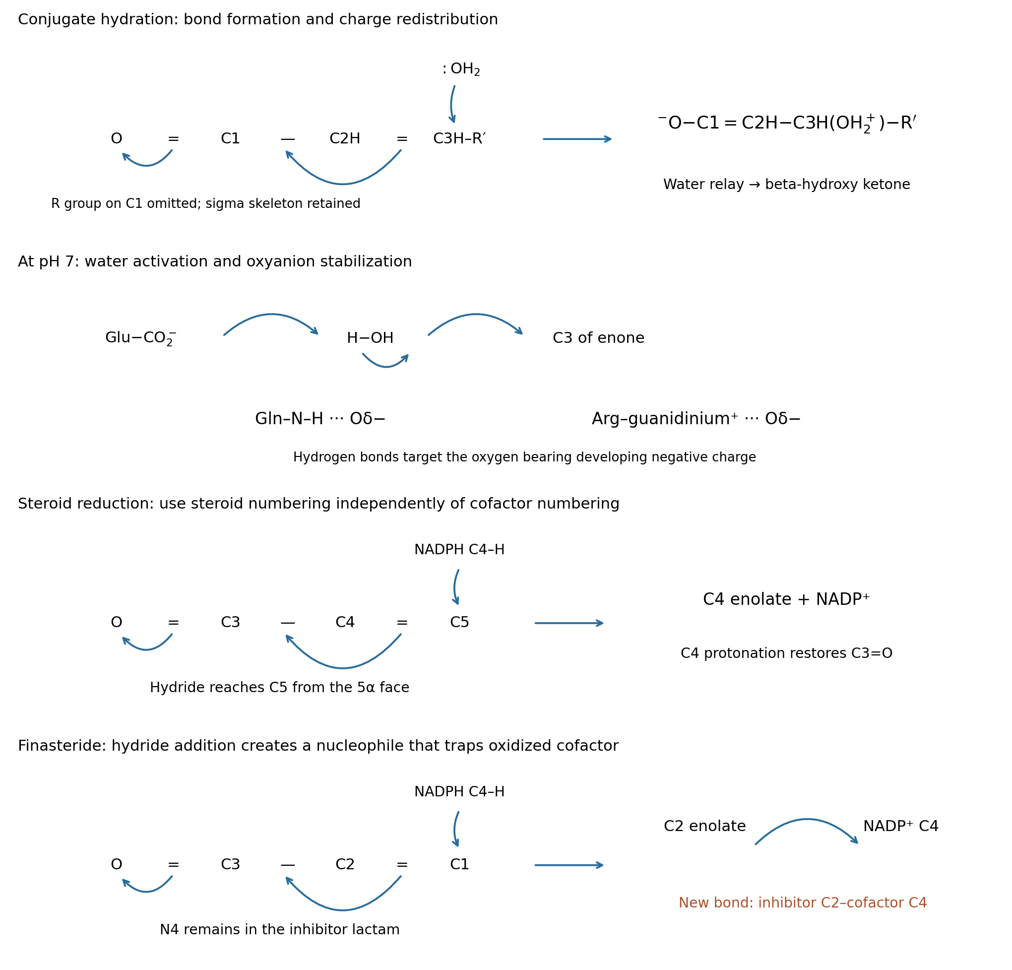
Reactive-fragment mechanisms for conjugate hydration, steroid reduction and finasteride trapping
. Details. Pinned article: Introduction to the OurBigBook Project
Welcome to the OurBigBook Project! Our goal is to create the perfect publishing platform for STEM subjects, and get university-level students to write the best free STEM tutorials ever.
Everyone is welcome to create an account and play with the site: ourbigbook.com/go/register. We belive that students themselves can write amazing tutorials, but teachers are welcome too. You can write about anything you want, it doesn't have to be STEM or even educational. Silly test content is very welcome and you won't be penalized in any way. Just keep it legal!
Intro to OurBigBook
. Source. We have two killer features:
- topics: topics group articles by different users with the same title, e.g. here is the topic for the "Fundamental Theorem of Calculus" ourbigbook.com/go/topic/fundamental-theorem-of-calculusArticles of different users are sorted by upvote within each article page. This feature is a bit like:
- a Wikipedia where each user can have their own version of each article
- a Q&A website like Stack Overflow, where multiple people can give their views on a given topic, and the best ones are sorted by upvote. Except you don't need to wait for someone to ask first, and any topic goes, no matter how narrow or broad
This feature makes it possible for readers to find better explanations of any topic created by other writers. And it allows writers to create an explanation in a place that readers might actually find it.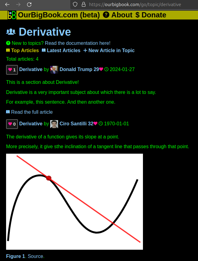
Figure 1. Screenshot of the "Derivative" topic page. View it live at: ourbigbook.com/go/topic/derivativeVideo 2. OurBigBook Web topics demo. Source. - local editing: you can store all your personal knowledge base content locally in a plaintext markup format that can be edited locally and published either:This way you can be sure that even if OurBigBook.com were to go down one day (which we have no plans to do as it is quite cheap to host!), your content will still be perfectly readable as a static site.
- to OurBigBook.com to get awesome multi-user features like topics and likes
- as HTML files to a static website, which you can host yourself for free on many external providers like GitHub Pages, and remain in full control

Figure 2. You can publish local OurBigBook lightweight markup files to either OurBigBook.com or as a static website.
Figure 3. Visual Studio Code extension installation.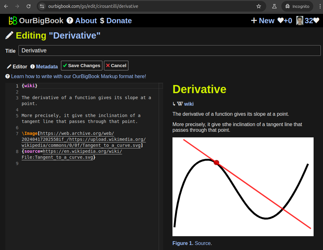
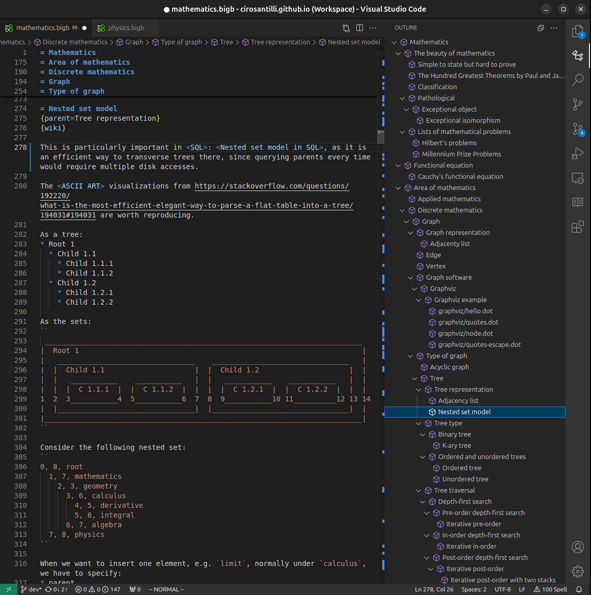
Figure 5. . You can also edit articles on the Web editor without installing anything locally. Video 3. Edit locally and publish demo. Source. This shows editing OurBigBook Markup and publishing it using the Visual Studio Code extension. 
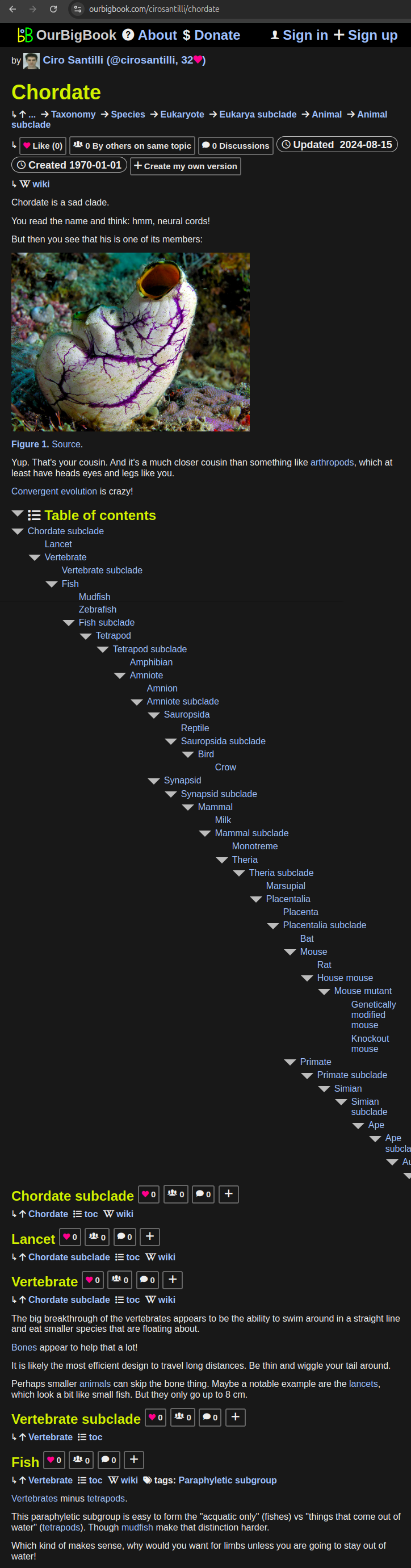
- Infinitely deep tables of contents:


All our software is open source and hosted at: github.com/ourbigbook/ourbigbook
Further documentation can be found at: docs.ourbigbook.com
Feel free to reach our to us for any help or suggestions: docs.ourbigbook.com/#contact