
Western blot response saturation means that increasing target amount no longer increases the recorded signal proportionally. It can arise before imaging, through limited binding or reagent availability, as well as through detector limits. Shortening exposure can address a detector limit without repairing an already unresponsive assay. Test the target and normalization readouts with a dilution series under the actual assay conditions; merely seeing a gray band does not establish a proportional response.
In a 2013 methods experiment, a biological cell extract containing added purified yeast protein was separated on gels, transferred with a Trans-Blot Turbo to polymer membranes, and read with an enzyme-linked secondary antibody. A ChemiDoc MP imager and film recorded the same dilution-series blots. The target and an abundant reference protein had different usable response ranges; transferred total-protein imaging and the reference bands did not track loading equally well. This demonstrates the need for assay-specific response checks, rather than a universal conversion from darkness to amount. [ref]
An illustrative response model separates background , target amount , retained fraction , response scale and characteristic amount :This is an assumed model, not a fitted Western blot calibration. When , . When , approaches . For two otherwise identical lanes, doubling gives the background-corrected ratioAt the ratio is , despite a true twofold amount change. Thus Western blot normalization cannot recover a lost amount difference merely by dividing two saturated readouts.
Total-protein Western blot normalization compares a target-band signal with a signal representing many transferred proteins in that lane. It is distinct from measuring one reference protein, and from measuring the target itself. Its denominator is a loading proxy: normalization estimates target abundance relative to detected total protein, rather than target amount per biological cell. Different sample compositions, selective Western blot transfer losses or unequal stain responses can still matter.
For an original calculation, suppose background-corrected target signals , and total-protein signals , are in their respective proportional response ranges. ThenThe invented signals double together, so this calculation supplies no evidence for a higher target fraction. With but , the same calculation gives . These examples assume a common target response and a common total-protein response between lanes, negligible remaining background, and comparable target recovery; they do not equate total-protein staining with target abundance. Multiplying both signals in a lane by the same loading factor cancels in their ratio; a selective target loss does not cancel.
Western blot loading controls ask how much sample entered each lane; Western blot transfer controls ask what reached the receiving sheet; Western blot antibody specificity controls ask what the antibodies recognized. Each closes a different gap in Western blot band interpretation.
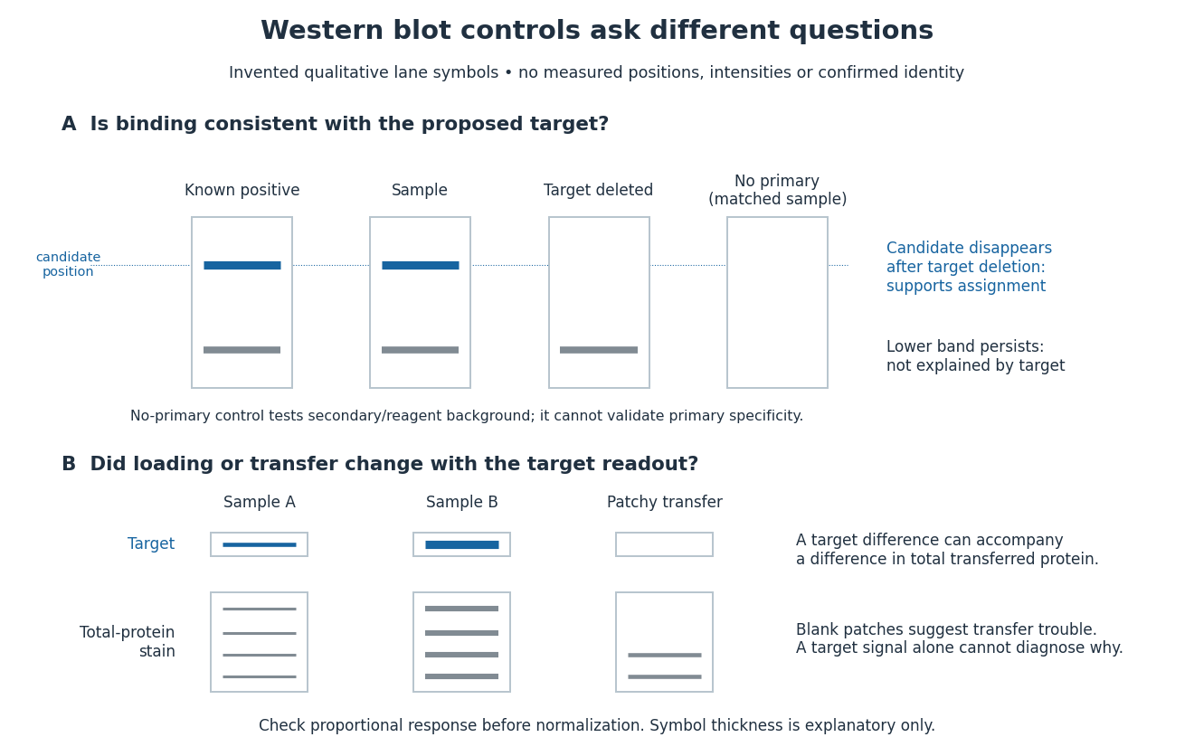
Conceptual lanes for Western blot controls
. Original schematic, with invented qualitative marks only. Candidate bands disappear in a target-deleted sample while a lower band persists; a separate panel shows how a loading or transfer difference can accompany a target-band difference. No lane, darkness, band position or relative amount represents a measurement, a specific real protein or confirmed identity. The no-primary comparison denotes a matched strip processed without primary antibody. Details.
Band position gives an apparent mass, not unique protein identity. Intensity depends on retention, epitope recognition and detection response; it does not establish folded state, enzyme activity or successful protein targeting. Missing bands can reflect missing targets or failed Western blot transfer or Western blot antibody detection. [ref]
Considerations when quantitating protein abundance by immunoblot by Alicia A. McDonough and others. McDonough and others, “Consider the Specificity of the Antibody for the Target” and “Consider the Linearity of the Detection System”
The yeast cross-species complementation experiment consequently combines two logically different readouts: Western blot evidence about production of an antibody-recognized protein, and growth evidence about functional complementation. A detectable protein need not perform the biological task required for growth.
Blocking occupies unused binding sites on the Western blot membrane. The primary antibody is applied, unbound material is washed away, and a labelled secondary antibody detects retained primary antibody. A fluorescence label emits detectable light after excitation; an enzyme-linked label can instead generate a colored product or light. Washing reduces background but does not prove that remaining binding is selective.
In the original experiment, blocking used excess protein. An anti-ribosome-protein serum followed by labelled secondary antibody produced colored or fluorescence bands; preimmune serum supplied comparison strips. The color readout was an enzyme reaction product, rather than a measurement of target enzyme activity. [ref]
Electrophoretic transfer of proteins from polyacrylamide gels to nitrocellulose sheets: procedure and some applications by Harry Towbin and others. Towbin, Staehelin and Gordon, pp. 4351–4353: Immunological Detection of Proteins on Nitrocellulose and Figure 4, including preimmune-serum controls
Western blot transfer moves separated proteins out of the gel onto a Western blot membrane, using an electric field across the gel thickness. Maintaining close contact preserves the lane pattern. Contact gaps, incomplete removal from the gel and passage through the receiving sheet can change how much protein is retained.
The original 1979 experiment pressed a wetted, -pore nitrocellulose Western blot membrane against the gel between scouring pads and plastic pipette-tray grids, held by elastic bands. The urea-gel assembly faced the cathode in acidic solution, with applied for one hour; the SDS-PAGE variant reversed polarity and used a glycine/methanol buffer solution. Stained gel/blot pairs checked pattern preservation. Corresponding labelled ribosome protein spots from two gels were compared through radioactive decay readouts after combustion. The stained gel remaining after transfer supplied a removal check; a second receiving sheet revealed breakthrough on overloading. These tests supported recovery for the examined urea-gel proteins, within experimental variation, while the SDS-PAGE results showed unequal staining recovery. Preserved band positions therefore did not establish universal quantitative transfer. [ref]
Electrophoretic transfer of proteins from polyacrylamide gels to nitrocellulose sheets: procedure and some applications by Harry Towbin and others. Towbin, Staehelin and Gordon, pp. 4350–4352: Electrophoretic Blotting Procedures, Figure 1, Figures 2–3 and Table 1
In the common SDS-PAGE version, protein denaturation and binding of an anionic surfactant reduce differences due to folded shape and intrinsic electric charge. An electric field drives the resulting protein complexes through the gel; smaller chains generally migrate farther. A lane of reference proteins supplies positions for estimating apparent mass. This estimate depends on the separation conditions: it is not a direct weighing of a molecule, and different proteins can migrate together.
For a scalar complex wave amplitude , the squared-amplitude scalar wave intensity convention is . It is nonnegative and unchanged by multiplying by a unit-modulus wave phase factor. This convention is useful in paraxial propagation and phase screen models. Conversion to physical energy flux also depends on the field variable and medium; an arbitrary superposition need not have its full flux determined by this scalar alone. For a random field, is the mean wave intensity, whereas is the squared coherent field; these generally differ.
There are unlisted articles, also show them or only show them.