Topics (384k) Articles (398k) Users (410) Discussions (242) Comments (383) Files (13k) New article

Pyridoxine is the alcohol form of Vitamin B6, with molecular formula . Its pyridine ring has a methyl group at position 2, a hydroxyl group at position 3 and hydroxymethyl groups at positions 4 and 5.[ref]
At position 4, pyridoxine differs from the aldehyde pyridoxal and the amine pyridoxamine. Replacing by changes the formula by two hydrogen atoms; replacing it by changes both the oxygen and nitrogen counts. This provides a concrete structural meaning for the three names, beyond their membership of the same vitamin family.
The distinction also matters for biochemistry. Pyridoxal phosphate and pyridoxamine phosphate are active coenzyme forms; free pyridoxine is not simply another name for either phosphorylated molecule.[ref]
Vitamin B6 Fact Sheet for Health Professionals by the Office of Dietary Supplements. Introduction, chemical forms and active coenzymes
For example, an experiment comparing equal molar amounts of two B6 forms holds the number of molecules constant, while their functional groups and ability to enter a particular reaction can still differ. Equal mass would additionally fail to hold molecular amount constant because the forms have different molar mass values.Pyridoxamine is an amine form of Vitamin B6. Its substituted pyridine ring carries a methyl group at position 2, a hydroxyl group at position 3, an aminomethyl group at position 4 and a hydroxymethyl group at position 5. Its molecular formula is .[ref]
The position-4 group makes the B6 family a useful example of how a small structural change distinguishes related molecules. Pyridoxine has there, pyridoxal has , and pyridoxamine has . These are respectively alcohol, aldehyde and amine forms, rather than three interchangeable names for one structure.[ref]
Vitamin B6 Fact Sheet for Health Professionals by the Office of Dietary Supplements. Introduction, six B6 vitamers
Adding a phosphate at the position-5 hydroxymethyl group produces pyridoxamine phosphate, an active coenzyme form. Pyridoxal phosphate is the other principal active B6 coenzyme form. The free vitamin and its phosphorylated derivative must therefore be distinguished when describing an enzyme assay: the name of the vitamin family alone does not identify the molecular species supplied to the enzyme.[ref]
Vitamin B6 Fact Sheet for Health Professionals by the Office of Dietary Supplements. Introduction, PLP and PMP
Pellagra is a disease caused by severe vitamin B3 deficiency. Its manifestations can include sun-sensitive dermatitis, diarrhea and neurological changes, including dementia. These familiar “three Ds” summarize possible effects; they are not a requirement that every affected person show all three. Vitamin B3 supplies precursors of nicotinamide adenine dinucleotide and nicotinamide adenine dinucleotide phosphate, so adequate dietary energy alone does not guarantee adequate support for metabolism.[ref]
Niacin Fact Sheet for Health Professionals by the Office of Dietary Supplements. Introduction and Niacin Deficiency
In 1915, Joseph Goldberger and G. A. Wheeler tested a restricted, cereal-heavy diet at a Mississippi prison farm. At least six of eleven remaining volunteers developed signs judged to be pellagra within five months; none were observed among the comparison prisoners. The reported menu supplied about 2,952 calories per man per day, making the contrast between calories and dietary composition especially revealing. Prisoners were offered pardons for participation; allocation was not described as randomized.[ref]
Experimental Pellagra in the Human Subject Brought About by a Restricted Diet by Joseph Goldberger and G. A. Wheeler. pp.3336–3339, Experiment, Controls, Results and Conclusion
Dark adaptation is recovery of visual sensitivity after exposure changes from brighter surroundings to darkness or much dimmer conditions. Operationally, a fixed test stimulus becomes detectable at a lower intensity as adaptation proceeds. This is more than the increase in light capture caused by dilation of the pupil: changes in retinal sensitivity and visual cycle chemistry also matter.
The direction of a threshold plot can be misleading. In an invented example, detection threshold falls from 1,000 to 1 arbitrary intensity unit under otherwise matched testing conditions. Sensitivity defined as reciprocal threshold has increased 1,000-fold; the threshold has fallen by three logarithmic units. A descending curve therefore represents improving sensitivity.
Selig Hecht and collaborators measured how preceding light exposure changed subsequent human dark adaptation. Stronger pre-exposure produced an initial cone-dominated phase followed by a delayed rod phase in their experiments, while lower pre-exposures did not show the same two-phase pattern. The exposure history is thus part of the measurement, not an irrelevant preparation detail.[ref]
The influence of light adaptation on subsequent dark adaptation of the eye by Selig Hecht and others. 1937 abstract: preadaptation intensity, conditional cone/rod phases and delayed rod recovery.
The visual cycle provides a biochemical connection. RPE65-deficient mice lacked normal rod pigment and rod responses despite retained opsin, showing that producing the receptor chain alone cannot replace the chemical supply needed for functional rhodopsin.[ref]
Rpe65 is necessary for production of 11-cis-vitamin A in the retinal visual cycle by T. M. Redmond and others. Abstract: retinoid composition, opsin and rod electroretinographic phenotype.
The knockout is not a normal human recovery curve. Psychophysical thresholds, electrical electroretinography responses and pigment chemistry measure different levels of the system. Comparisons of adaptation curves should specify stimulus properties and preceding exposure rather than assuming a universal recovery time.
A virtual displacement is an infinitesimal change of configuration compatible with the instantaneous mechanical constraints, evaluated at fixed time. It is a variation among admissible configurations, rather than necessarily the displacement made during a time step. In generalized coordinates, writeSubstitution into the applied virtual work identifies the generalized forces conjugate to the coordinates.[ref]
Newtonian Dynamics by Richard Fitzpatrick. Lagrangian Dynamics, Generalized Coordinates, equations592–593; Generalized Forces, equations594–597
An original moving-constraint example makes the fixed-time condition visible. Consider a particle constrained to the circleFor a virtual displacement, freeze and therefore ; first-order compatibility gives . For an actual motion, differentiation with respect to time instead givesAt with , every admissible virtual displacement has , while an actual velocity must have . If the circle is expanding at , the actual radial velocity is nonzero even though every virtual displacement is tangent to the instantaneous circle.
Consequently, an ideal radial constraint force can have zero virtual work but nonzero instantaneous power during this driven motion. Zero work on admissible variations is not the statement that a moving support never transfers energy. The example is a time-dependent holonomic constraint; the variations satisfy its instantaneous linearized condition. In a beam, the same distinction lets one impose zero endpoint virtual displacement while leaving the virtual endpoint slope free, as in the moment-free natural boundary condition for a beam.
A virial coefficient quantifies an interaction correction to the dilute-gas equation of state. In the number-density convention, , where . Thus has units of volume; a molar-density expansion uses differently scaled coefficients.
For classical particles with a pair potential , . Hard spheres of diameter exclude separations below , giving . Attractive interactions can instead contribute negatively. The expansion is a low-density organization of correlations, not a guarantee of convergence near a phase transition.
A merge conflict occurs when a version-control merging operation cannot automatically reconcile changes under its merge rules. For a typical three-way merge, the tool compares two revisions with their common base. Incompatible edits to the same region, or deletion on one side and modification on the other, can require a human decision.[ref]
git-merge in the Git documentation. True merge, How conflicts are presented and How to resolve conflicts
For example, the base contains
threshold=5; one side changes it to 7 and the other to 9. Choosing either value or a new value requires understanding the intended behavior. After editing the combined result, the resolver records that decision in version control. A textually conflict-free merge can still break a function's contract, so successful automatic combination supplies weaker evidence than software testing of the integrated program.A commit graph represents version-control commits as vertices and their parent relationships as directed edges. In Git, taking an edge from a child to each parent gives a directed acyclic graph: an existing version-control commit has fixed parents, and a newly created child cannot become its own ancestor.[ref]
For example, if and both descend directly from , and merge records parents , its edges are , , and . The merge retains both lines of ancestry. A version-control branch labels a selected tip; deleting that label does not by itself remove every reachable vertex. This graph supports ancestry tests and finding common bases for version-control merging.
A version-control snapshot records the logical state of tracked files at a revision. In Git, tree objects describe directory entries and refer to content objects; a version-control commit identifies the root tree alongside history metadata. Unchanged content can be shared among many version-control snapshots.[ref]
Pro Git by Scott Chacon and Ben Straub. §10.2, tree objects and commit objects
If a repository contains
a.txt and b.txt, changing only a.txt creates a new logical version-control snapshot containing the new a.txt and unchanged b.txt. It does not require a second physical copy of every byte. Two version-control commits can identify the same version-control snapshot but have different parents or messages. The recorded files and the ancestry explaining how they arose are therefore distinct information.A Vernier zero correction removes an indicated offset observed at a known zero setting of a Vernier scale. If the raw indication is and the indication at true zero is , the additive model givesThe zero error is ; the correction applied to a later reading is its negative. This model addresses an offset, not every possible scale or contact error.
For an original signed example, a raw reading with closed-jaw indication gives . If the zero indication is instead , the corrected reading is . A negative zero indication therefore requires adding its magnitude. It does not imply a negative object length.
The reference setting must represent the intended zero: clean, properly contacting outside faces on a caliper are one example. Record the actual signed indication and the scale convention, rather than relying on a memorized rule about which engraved zero lies to the left. The manufacturer guide explains the coincidence reading and identifies jaw tilt, force and viewing geometry as separate error sources.[ref]
Quick Guide to Precision Measuring Instruments: Calipers by Mitutoyo. printed pp. 16–17, scale reading and Sources of Error
For standard uncertainties, the additive model givesWith stipulated independent and , the combined value is approximately . Subtracting a measured offset does not make that offset known exactly. Correlation can matter when both observations share an uncertain reference or drift contribution.[ref]
Guide to the expression of uncertainty in measurement by JCGM. §5.2, correlated input estimates
A second reference checks the limitation. Suppose true zero reads but a known separation reads . Removing the zero offset leaves , still high. A model would have here. Measurement calibration across the working interval is needed to distinguish scale error from a zero correction, with the relevant measurement uncertainty retained.
The third ventricle is the narrow midline cavity of the diencephalon, between the paired thalami and adjoining the hypothalamus. It contains cerebrospinal fluid and forms part of the ventricular system. The lateral ventricles communicate with it through the interventricular foramina; the cerebral aqueduct connects it to the fourth ventricle.[ref]
This cavity is more than a passive container. A 2016 study of mouse third ventricles found organized groups of cilia producing a network of cerebrospinal fluid flows, including a periodically changing pattern. Related complex flows were observed in rat and pig third ventricles.[ref]
Cilia-based flow network in the brain ventricles by Regina Faubel and others. Abstract; ciliary modules and time-dependent flow
An opening specifies a possible route between cavities; local cilia-driven transport specifies how material moves within a cavity. A single well-mixed compartment model can therefore conceal spatial differences in the delivery of signaling molecules.
A lateral ventricle is one of the two large cerebrospinal fluid-filled cavities within the cerebral hemispheres. Each connects with the third ventricle through an interventricular foramen. Its location inside a hemisphere makes it a useful landmark when interpreting sections or images of the brain.[ref]
Lateral Ventricles in MeSH. Definition, paired cerebral cavities and communication with the third ventricle
The choroid plexus projects into ventricular cavities and contributes to cerebrospinal fluid production; the ependyma lines the ventricular wall. The resulting environment joins a secretory interface, a fluid compartment and neighbouring neural tissue. These components have different roles even though they appear close together in an anatomical section.[ref]
Anatomy and Physiology 2e by J. Gordon Betts and others. §12.2, ependymal cells; §13.3, cerebrospinal fluid and choroid plexuses
The connections below provide a simplified route through the ventricular system. Actual transport also has a time dependence: phase-contrast magnetic resonance imaging has resolved cerebrospinal fluid movement over the cardiac cycle, while work on the third ventricle has revealed structured local flows driven by motile cilia.[ref]
Normal flow patterns of intracranial and spinal cerebrospinal fluid defined with phase-contrast cine MR imaging by D. R. Enzmann and others. abstract, cardiac-cycle-dependent flow encoding
[ref]Cilia-based flow network in the brain ventricles by Regina Faubel and others. abstract, ciliary modules and ventricular flow networks
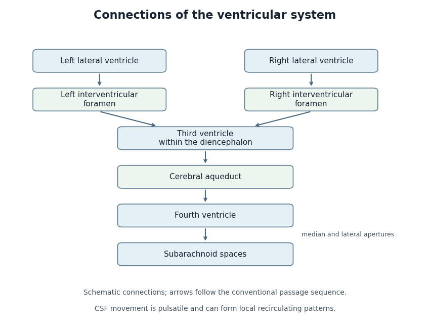
Anatomical connections of the ventricular system
. Original schematic showing paired lateral ventricles, the interventricular foramina, third ventricle, cerebral aqueduct, fourth ventricle and subarachnoid spaces. Arrows follow the conventional passage sequence; cerebrospinal fluid also undergoes pulsatile movement and local recirculation.[ref]Neuroscience, 2nd edition edited by D. Purves and others. The Ventricular System, Figure 1.17, anatomical connections
Details.The interventricular foramina are the paired openings through which the lateral ventricles communicate with the third ventricle. Each opening connects its own side's lateral ventricle to the shared midline cavity. This arrangement provides a route for cerebrospinal fluid to reach the rest of the ventricular system.[ref]
Lateral Ventricles in MeSH. Definition, each lateral cavity communicating with the third ventricle
Both sides reach the shared third ventricle, which joins the fourth ventricle through the cerebral aqueduct.
Measurements add a temporal picture to this connection map. In a 1991 study, phase-contrast magnetic resonance imaging encoded the direction of cerebrospinal fluid movement across the cardiac cycle. The ventricular measurements contained systolic and diastolic flow components, with essentially simultaneous timing at the interventricular opening and cerebral aqueduct. Anatomical continuity and measured pulsatile transport describe complementary features of the same pathway.[ref]
Normal flow patterns of intracranial and spinal cerebrospinal fluid defined with phase-contrast cine MR imaging by D. R. Enzmann and others. abstract, flow encoding and timing at ventricular passages
A venule is a small blood vessel that collects blood leaving capillaries and conveys it toward larger veins. It lies on the returning side of the microcirculation: arterioles supply the capillary network, while venules drain it. The smallest postcapillary venules have thin walls lined by endothelium.[ref]
A venule is also an important interface between flowing blood and surrounding tissue. In an experiment using time-lapse intravital microscopy, investigators followed neutrophils within inflamed mouse postcapillary venules. After adhering, the neutrophils crawled along the inner surface toward sites where they could leave the vessel. Observing movement over time distinguished this searching step from the initial arrest.[ref]
Intraluminal crawling of neutrophils to emigration sites: a molecularly distinct process from adhesion in the recruitment cascade by Mia Phillipson and others. Abstract and Results: intraluminal crawling after adhesion
Pinned article: Introduction to the OurBigBook Project
Welcome to the OurBigBook Project! Our goal is to create the perfect publishing platform for STEM subjects, and get university-level students to write the best free STEM tutorials ever.
Everyone is welcome to create an account and play with the site: ourbigbook.com/go/register. We belive that students themselves can write amazing tutorials, but teachers are welcome too. You can write about anything you want, it doesn't have to be STEM or even educational. Silly test content is very welcome and you won't be penalized in any way. Just keep it legal!
Intro to OurBigBook
. Source. We have two killer features:
- topics: topics group articles by different users with the same title, e.g. here is the topic for the "Fundamental Theorem of Calculus" ourbigbook.com/go/topic/fundamental-theorem-of-calculusArticles of different users are sorted by upvote within each article page. This feature is a bit like:
- a Wikipedia where each user can have their own version of each article
- a Q&A website like Stack Overflow, where multiple people can give their views on a given topic, and the best ones are sorted by upvote. Except you don't need to wait for someone to ask first, and any topic goes, no matter how narrow or broad
This feature makes it possible for readers to find better explanations of any topic created by other writers. And it allows writers to create an explanation in a place that readers might actually find it.
Figure 1. Screenshot of the "Derivative" topic page. View it live at: ourbigbook.com/go/topic/derivativeVideo 2. OurBigBook Web topics demo. Source. - local editing: you can store all your personal knowledge base content locally in a plaintext markup format that can be edited locally and published either:This way you can be sure that even if OurBigBook.com were to go down one day (which we have no plans to do as it is quite cheap to host!), your content will still be perfectly readable as a static site.
- to OurBigBook.com to get awesome multi-user features like topics and likes
- as HTML files to a static website, which you can host yourself for free on many external providers like GitHub Pages, and remain in full control

Figure 2. You can publish local OurBigBook lightweight markup files to either OurBigBook.com or as a static website.
Figure 3. Visual Studio Code extension installation.
Figure 5. . You can also edit articles on the Web editor without installing anything locally. Video 3. Edit locally and publish demo. Source. This shows editing OurBigBook Markup and publishing it using the Visual Studio Code extension. 
- Infinitely deep tables of contents:



All our software is open source and hosted at: github.com/ourbigbook/ourbigbook
Further documentation can be found at: docs.ourbigbook.com
Feel free to reach our to us for any help or suggestions: docs.ourbigbook.com/#contact