Topics (384k) Articles (398k) Users (410) Discussions (242) Comments (383) Files (13k) New article

A cardiac vein carries blood away from the heart's own tissue after passage through its supplying vessels. This is the return side of the coronary circulation, distinct from the large volume of blood passing through the ventricular cavities. Much of this return reaches the right atrium through the coronary sinus; anterior cardiac veins can return directly to the right atrium.[ref]
The great cardiac vein, middle cardiac vein and small cardiac vein provide named routes through this network. An illuminating anatomical exercise pairs each route with its connections rather than simply matching it to the nearest coronary artery. The artery supplies tissue and the vein drains tissue, while their branching and crossings need not be identical.
Primary evidence for that qualification comes from a study dissecting 37 fixed human hearts. It documented varying relationships among the coronary sinus, its tributaries and neighboring arteries.[ref]
The anatomy of the coronary sinus and its tributaries by J. R. Ortale and others. Abstract, methods and vessel relationships
Such a study observes actual connected vessels in specimens; a schematic drawing deliberately compresses the variation into a readable route map.For a transport measurement, identifying the correct cardiac vein matters because a regional sample has a regional venous oxygen content. Substituting it for mixed venous oxygen content in a whole-body Fick principle calculation would change the system being sampled. The anatomical return route and the corresponding blood flow must match the oxygen-balance question.
A pulmonary vein returns blood from a lung to the left atrium. The pulmonary veins complete the return side of the pulmonary circulation, after blood has received oxygen in the lungs. They are veins because they carry blood toward the heart, even though their oxygen content is higher than that of the blood arriving through the pulmonary artery.[ref]
Following a parcel of blood makes the sequence clear: right ventricle, pulmonary trunk, vessels of the lungs, pulmonary vein, left atrium, then left ventricle. The pulmonary vein connects the lung circuit to the receiving chamber of the left heart; it does not drain directly into the right atrium.
A simple oxygen balance illustrates why both content and flow matter. Suppose two returning streams have blood flow rates and oxygen contents . If they mix without further oxygen exchange, the left atrium's inflow content isFor constructed values L/min, L/min, and mL/mL, this gives mL/mL. An unweighted average would give mL/mL. A specimen's vessel connections establish where the streams meet; conservation of mass establishes how their transported oxygen combines.
A brachiocephalic vein is one of the paired vessels that unite into the superior vena cava. On each side, the internal jugular vein and subclavian vein contribute major head/neck and upper-limb return to this route.[ref]
Anatomy and Physiology 2e by J. Gordon Betts and others. §20.5, The Superior Vena Cava, Table 20.11 and Venous Drainage of the Head and Neck
The paired venous arrangement differs from the arterial naming pattern: a brachiocephalic artery is not a left/right counterpart for each of these veins.The connection explains why a regional venous sample is not automatically a fully mixed whole-body sample. The two brachiocephalic veins bring together different drainage territories, and additional return enters through other routes, including the azygos vein into the superior vena cava.
An original tracer-mixing example gives the left and right streams flows 1.5 and 1.0 L/min. If an illustrative marker concentration is 10 mg/L on the left and zero on the right, complete mixing of those two streams givesAdding a further unmarked return of 0.5 L/min reduces the mixed concentration to mg/L while preserving the same transported marker amount, 15 mg/min. Dilution can change concentration without destruction of the marker.
These are hypothetical well-mixed steady streams, not an injection protocol or measured human flow distribution. A sample taken before complete mixing can differ from the calculated average. The example connects vessel junctions with indicator dilution: the amount balance must include every stream contributing at the actual detector location.
A common iliac vein is a paired pelvic return vessel formed by the union of an external iliac vein and an internal iliac vein. The right and left common iliac veins join to form the inferior vena cava.[ref]
Anatomy and Physiology 2e by J. Gordon Betts and others. §20.5, Veins Draining the Lower Limbs; Table 20.16
The external route collects lower-limb return, while the internal route collects pelvic territories.The word common records a junction in the drainage tree. It does not imply identical left and right flows or a fixed ratio between the two tributaries. Tracing a parcel of returning blood therefore requires both its originating territory and the vessel connections.
An original incompressible-flow example takes external and internal inflows of 1.2 and 0.8 litres per minute. With no local storage change, common-vessel flow is L/min. If its lumen area is a stipulated , mean axial velocity isThe two input flow rates add; their velocities do not. A different lumen area changes velocity at the same volumetric flow. These values illustrate continuity and are not normal anatomical dimensions.
If an observation instead records changing vessel volume, the local balance is . Temporary storage can therefore distinguish simultaneous inflow and outflow without violating conservation of mass. Knowing a vessel name or measuring a local velocity is not enough to reconstruct every regional blood flow without geometry, timing and the appropriate network balance.
A hepatic vein carries blood out of the liver toward the inferior vena cava. It is an outflow vessel, whereas the portal vein and hepatic artery supply the liver through distinct routes.[ref]
Anatomy and Physiology 2e by J. Gordon Betts and others. §20.5, The Inferior Vena Cava and Hepatic Portal System
The shared word hepatic locates the organ; it does not make these vessels interchangeable.The two-input arrangement matters to a substance balance. In an original steady-flow model, let portal and arterial inflows be , with concentrations , and let hepatic outflow concentration be . With negligible fluid accumulation, . Net production of the substance by the liver iswhere positive values denote net release and negative values net removal. The calculation assumes matched sampling, a common concentration basis and negligible storage change over the measurement interval.
For stipulated L/min, L/min, mmol/L, mmol/L and mmol/L,The liver removes the modeled substance overall even though the hepatic-vein concentration exceeds the arterial concentration. Comparing the outflow with only one of two inputs would give a misleading interpretation.
A hepatic-vein measurement thus describes an organ-specific outflow. It need not equal mixed venous oxygen content or the composition of every region of the liver. Identifying the drainage territory and allowing for unequal flow among regions are part of interpreting a sample. Anatomy defines the paths; conservation of mass defines how the transported quantities must combine.
An external iliac vein continues the femoral vein proximally as lower-limb venous return passes beneath the inguinal ligament. It joins the internal iliac vein to form the common iliac vein, which connects the route with the inferior vena cava.[ref]
Anatomy and Physiology 2e by J. Gordon Betts and others. §20.5, Veins Draining the Lower Limbs, external/internal/common iliac sequence
External distinguishes this branch from the pelvic internal route; it does not mean that the vessel lies outside the body.Following the pathway separates a route name from a measured quantity. The external iliac vein can transport a changing blood flow, and its local velocity also depends on lumen area. A velocity observation is therefore not by itself a volumetric-flow measurement.
In an original incompressible-flow example, let litres per minute and lumen area . Mean axial velocity isIf the area increases by 25 percent while flow remains fixed, mean velocity falls to cm/s. The vessel can carry the same volume per minute with a different mean speed. The dimensions and flows are illustrative rather than measured anatomical norms.
This distinction also matters when comparing measurements taken at different locations. At a junction, flow changes by the added tributary contributions, while vessel geometry may change at the same time. Defining the observation site and geometric model keeps an anatomical statement about lower-limb return separate from a quantitative estimate of that return.
An internal iliac vein collects venous return from pelvic territories and joins the external iliac vein to form a common iliac vein. The paired internal routes are part of the return network leading toward the inferior vena cava and right atrium.[ref]
Anatomy and Physiology 2e by J. Gordon Betts and others. §20.5, Veins Draining the Lower Limbs and Table 20.16
Internal identifies a regional branch, not a general category containing every vein inside the body.The branching distinction matters to sampling. A pelvic return sample and a lower-limb return sample can have different concentrations because their tissues exchange different amounts of a substance. Their common destination does not make their compositions equal.
For an original two-tributary example, suppose an internal iliac vein receives flows 300 and 200 mL/min with concentrations 4 and 1 mmol/L. Complete mixing without local exchange givesThe unweighted mean 2.5 would ignore how much blood each branch contributes. Multiplying the mixture concentration by total flow recovers the incoming substance rate, mmol/min.
These stipulated values do not represent reference human flows or a complete map of pelvic drainage. A specimen taken before mixing can differ from the calculated average, and collateral routes can change which flows enter a selected vessel. Anatomical continuity defines the regional question; blood flow and conservation of mass determine how its transported substances combine.
The femoral vein is a deep vein carrying blood from the lower limb towards the external iliac vein. It continues proximally beneath the inguinal ligament as the external iliac vein, which later meets the internal iliac vein. The femoral vein therefore names a connected drainage route, rather than a fixed amount of blood flow.[ref]
Anatomy and Physiology 2e by J. Gordon Betts and others. §20.5, Veins Draining the Lower Limbs and Table 20.16
A flow measurement must distinguish the velocity sampled by an instrument from the average velocity over the vessel's cross-sectional area. For an original circular-lumen model with radius and an illustrative axial velocity profile ,At stipulated mm and cm/s, the blood flow is approximately . Multiplying the central speed by the whole area would give twice that value. These are chosen model values, not normal human measurements or a claim that every venous velocity profile is parabolic.
In Doppler ultrasound, the measured frequency shift depends on the velocity component along the acoustic beam. Imaging the femoral vein and measuring that component address different parts of the inference: geometry identifies the vessel and its lumen, while the velocity measurement needs a direction and spatial-sampling model. The professional ultrasound description identifies sound-wave imaging and Doppler ultrasound as tools for evaluating venous blood flow.[ref]
Venous Ultrasound by RadiologyInfo.org. How does the procedure work?, conventional imaging and Doppler information
A useful measurement comparison holds the anatomical segment fixed and records lumen area and the velocity distribution over the same interval. Changes in area or the profile can alter the relation between a central reading and total blood flow. The resulting quantity belongs to that region's return; it is not automatically whole-body cardiac output.
A plant cutting is a detached piece of a plant, such as part of a plant stem, leaf or root system, used to produce another plant by vegetative reproduction. A stem cutting must maintain a living shoot while forming new adventitious roots. Because this bypasses meiosis and fertilization, it can preserve the parental genotype, apart from mutations and pre-existing variation within the parent.
A practical example is a soft young shoot cut below a node, with lower leaves removed and its base placed in a moist, aerated medium. High humidity reduces water loss before the new root system can supply it; aeration avoids treating a rootless cutting as if it were an established waterlogged plant. The balance illustrates a physiological bottleneck: living leaves need water immediately, whereas a functional root system takes time to develop.[ref]
Cuttings: softwood by the RHS Advice Team. Preparing nodal softwood cuttings and aftercare
A plant clone is a lineage of plants descended from one founder by asexual reproduction. Vegetative propagation, for example rooting several plant cuttings from one stock plant, can multiply a selected genotype while avoiding the reshuffling associated with ordinary sexual reproduction. This is why such methods are useful for maintaining a named cultivar that does not reproduce its characteristics reliably from seeds.[ref]
A plant clone is defined by descent, not by identical appearance. Its members can experience different light, water or nutrient conditions, and somatic mutations can arise during growth. Shared ancestry consequently does not imply that every phenotype or every later DNA sequence must remain identical.
For an illustrative experiment, divide rooted plant cuttings from the same founder between brighter and dimmer conditions while keeping other conditions comparable. Differences in leaf size need not establish a difference in inherited genotype: the design deliberately holds ancestry relatively constant while changing the environment. Repeating this across several distinct plant clones can test whether different genotypes respond differently, an example of genotype-environment interaction.
Endovascular surgery comprises interventions performed from within a blood vessel, commonly using catheters and imaging to reach and treat a vascular region. It differs in access and apparatus from an operation that directly exposes the entire treated segment. The field includes methods such as balloon angioplasty and placement of a vascular stent; the appropriate intervention depends on the clinical problem.[ref]
What is a Vascular Surgeon? by the Society for Vascular Surgery. Open and endovascular procedure scope
An angiographic system combines vascular access with images of a contrast-filled vessel. The Society for Vascular Surgery describes an angiogram as potentially diagnostic or therapeutic, with balloon angioplasty and stent placement among the possible accompanying interventions.[ref]
Angiogram by the Society for Vascular Surgery. Procedure overview and possible therapies
The image supplies information about the lumen and device position, but an image of an apparently open region is not by itself a complete measurement of downstream tissue function.A simple imaging example shows why geometry matters. In a bench phantom, suppose a marker of known length spans 25 image pixels. At that marker’s plane, the scale is per pixel, so an 18-pixel feature would represent under the same projection conditions. Applying that scale to an object at a different depth can be wrong because magnification changes. These chosen values illustrate measurement calibration, not device sizing for a patient.
Fluoroscopy provides time-dependent projection images that help relate device motion to anatomy. Projection, motion, overlapping structures and image resolution limit what can be inferred from one view. A sound technical account therefore distinguishes the device’s mechanical action, the image measurement and the biological outcome.
The scientific questions extend beyond immediate access: how the intervention changes the flow pathway, how a device interacts with the vessel wall and whether the intended function persists. Those questions connect endovascular surgery to fluid dynamics, biomaterials, imaging and the measurement of clinical outcomes.
A bypass graft is a conduit that creates an alternative route for blood flow around a diseased or obstructed vessel segment. It can use biological vessel tissue or a synthetic material. Creating the route does not itself remove the original obstruction or eliminate its underlying disease.[ref]
Surgical Bypass by the Society for Vascular Surgery. What is Surgical Bypass?
A bypass connects an inflow region to an outflow region through a surgical anastomosis at each end. The graft’s geometry, material response, connections and interaction with blood all affect performance. Graft patency describes whether the conduit remains open; an open lumen, sufficient flow and useful downstream tissue function are related but distinct observations.
An idealized resistance model clarifies the new flow path. If a remaining native segment has hydraulic resistance and a graft connects the same two junctions with resistance , the two paths are in parallel:For illustrative and in consistent resistance units, . The local flow would increase sixfold at fixed pressure difference across those junctions. But if other vessels contribute a series resistance , the total resistance changes from to , giving a flow ratio at fixed overall pressure difference. The example shows why a local geometric improvement does not determine the whole circulation’s response.
The model assumes steady linear resistance and fixed driving conditions. Real grafts and vessels are compliant, branching biological structures with pulsatile flow. Bench measurements of pressure and flow, mechanical tests of the conduit and its connections, and imaging follow-up address different parts of the problem. The Society’s primary description includes ultrasound follow-up for graft monitoring.[ref]
Surgical Bypass by the Society for Vascular Surgery. Follow-up discussion
A bypass graft is therefore both a fluid pathway and a material–tissue interface. Its scientific evaluation connects vascular surgery, biomechanics, biocompatibility and, for engineered substitutes, tissue engineering.
Vascular stenosis is narrowing of the lumen of a blood vessel. The location, length, shape and cause of the narrowing matter, as does the reference used to express its severity. Narrowing can leave an open lumen; it is not synonymous with complete occlusion. A vascular stent supports an existing route, while a bypass graft creates an alternative route around a diseased region.
Diameter versus area stenosis illustrates an important measurement distinction. If is the narrowest measured diameter and is the stated reference diameter, define the illustrative diameter reductionFor circular lumens the associated area reduction isA narrowing from to is therefore by diameter and by area. These are geometric calculations using a specified reference, not interchangeable clinical grading conventions. Different reference segments or reconstruction methods can give different reported percentages.
An original ideal-flow example connects geometry to an observation. For incompressible flow with the same instantaneous volume flow through both sections, conservation of mass gives . Reducing circular diameter by half reduces area to one quarter and raises mean velocity fourfold at that maintained . The real circulation can change , so this ratio is conditional, not a universal diagnostic cutoff or prediction of tissue perfusion.
Ultrasound can examine both vessel appearance and moving blood. The Society for Vascular Surgery’s carotid-duplex description identifies a hand-held sound-wave probe and coupling gel, used to look for narrowing and examine flow. Such an apparatus makes anatomy and flow-related observations available together; a velocity estimate is not itself a direct measurement of every lumen dimension.[ref]
Characterizing vascular stenosis therefore combines the geometric convention, imaging method and physiological question. A percentage alone does not establish the pressure loss, the cause of symptoms or the best intervention. Persistent patency and clinical consequences require separate follow-up observations.
A vascular graft is a conduit used to replace or reconstruct a blood vessel or to create a new flow route. A bypass graft is one application: the conduit directs blood flow around a diseased segment. The graft can use biological tissue or a manufactured material; a tissue engineering approach attempts to build a vessel substitute with living cells and supporting matrix.
The engineering requirements are simultaneous. The lumen must permit flow, the wall must withstand loading, the connections must remain intact, and the blood-contacting surface must interact appropriately with circulating blood. Graft patency records whether the route remains open; it is a different endpoint from the wall's strength or its appearance under microscopy.
A simplified thin-walled circular tube illustrates one mechanical demand. Balancing the pressure force on a lengthwise half-cylinder against circumferential wall tension gives hoop stresswhere is lumen radius, wall thickness and the pressure difference across the wall. For stipulated , and , this gives approximately . The approximation assumes a thin wall and simple geometry; it does not predict the strength of a suture connection or a real multilayer graft.
A primary engineered-vessel study by Laura E. Niklason and colleagues measured static and dynamic compliance on a bench using controlled pressures and video-based tracking of vessel diameter. Separate tests assessed rupture strength. These apparatus measurements distinguished resistance to expansion, response during pulsatile loading and failure pressure.[ref]
Morphologic and mechanical characteristics of engineered bovine arteries by Laura E. Niklason and others. Compliance measurements, printed pp.631–632; Figure9, p.636
That distinction motivates vascular graft compliance mismatch and vascular graft burst pressure as separate assessments. A conduit can be strong at rupture yet differ mechanically from the vessel it joins. Conversely, surviving an initial pressure test does not establish long-term graft patency or biological remodeling. The measurement, tested material, loading history and follow-up interval must accompany claims about graft function.
Ignition is the register-based interpreter used within V8. It executes an internal representation of JavaScript computations rather than being a separate programming language with different source-level results.[ref]
Ignition in the V8 documentation. Opening description, low-level register-based interpreter
A small abstract bytecode example explains the role of such a representation. To compute a function returning
x + 2, imagine instructions load_arg r0; const r1, 2; add r2, r0, r1; return r2. For argument 5, the represented values are r0 = 5, r1 = 2 and r2 = 7, so the result is 7. An interpreter selects and performs the operation named by each instruction.This instruction spelling is invented for explanation; it is not an Ignition disassembly or a promise about a particular V8 release. The example separates source syntax, an intermediate instruction representation and execution. A register machine supplies a related abstract way to reason about stored intermediate values and operations, without identifying its formal instruction set with a production engine's bytecode.
Actual JavaScript operations require more than an arithmetic mnemonic. The expression
x + 2 can produce numerical addition or string concatenation depending on the value of x. Supplying "5" yields "52" rather than 7. Whatever internal instructions implement it must preserve the applicable ECMAScript behavior.[ref]ECMAScript Language Specification by Ecma International. Addition Operator, string concatenation and numeric addition
Understanding Ignition is therefore useful for studying execution and implementation costs, while the language specification remains the reference for observable program meaning.
d8 is V8's developer shell for running JavaScript programs and investigating engine behavior. It supplies a small host environment around the engine, with facilities for output, loading files and passing arguments.[ref]
Using d8 in the V8 documentation. Opening description; Print to the command line; Pass flags into JavaScript
For example, a file named
sum.js can contain console.log([1, 2, 3].reduce((a, b) => a + b, 0));. Running d8 sum.js prints 6. The array reduction and addition follow ECMAScript semantics, while console.log is an output facility supplied by the environment.The distinction also appears in arguments. The documentation places arguments after
-- and exposes them through a top-level arguments array. A script that prints arguments[0] can therefore inspect a supplied value. This is a shell convention, not a rule that every JavaScript host passes arguments through that same global object.[ref]Using d8 in the V8 documentation. Pass flags into JavaScript, argument separator and arguments object
d8 also documents
read for reading file content and load for executing another script in the current context. Those host operations are different from merely evaluating an arithmetic expression, even though both are invoked with JavaScript syntax.[ref]Using d8 in the V8 documentation. Read input; Load external scripts
A developer shell is useful for separating an engine computation from a larger application. A web browser additionally supplies document and interaction machinery; the presence of V8 alone does not imply a Document Object Model. When using shell observations as performance evidence, record the engine version, flags, workload and measurement method rather than treating one printed result as a benchmark.
The V4L2 plane data offset is the byte distance from the beginning of an image memory plane to its image data. It is the
data_offset member of a V4L2 plane descriptor. In this interface, bytesused includes that prefix, so the image-data extent is bytesused - data_offset.[ref]Write for
data_offset, for bytesused and for the plane's length, all in bytes. A consistent nonnegative extent satisfiesIf is the mapped plane-base address, the image begins at and ends immediately before . This is an offset within a plane, distinct from the descriptor's mem_offset used when arranging a memory mapping.For an original bounds example, stipulate , and . The image occupies 5888 bytes beginning at byte offset 256. Its last byte is at offset 6143. Taking a length of 6144 from the shifted starting address would instead read through offset 6399, including 256 bytes beyond the declared extent. Conversely, discarding the prefix twice would lose 256 valid bytes.
The offset does not specify framebuffer stride or component order. Once the correct image start is found, the negotiated pixel format still determines how rows and color components are read. A correct extent prevents a boundary mistake; it does not by itself establish that the samples represent the intended image.
Pinned article: Introduction to the OurBigBook Project
Welcome to the OurBigBook Project! Our goal is to create the perfect publishing platform for STEM subjects, and get university-level students to write the best free STEM tutorials ever.
Everyone is welcome to create an account and play with the site: ourbigbook.com/go/register. We belive that students themselves can write amazing tutorials, but teachers are welcome too. You can write about anything you want, it doesn't have to be STEM or even educational. Silly test content is very welcome and you won't be penalized in any way. Just keep it legal!
Intro to OurBigBook
. Source. We have two killer features:
- topics: topics group articles by different users with the same title, e.g. here is the topic for the "Fundamental Theorem of Calculus" ourbigbook.com/go/topic/fundamental-theorem-of-calculusArticles of different users are sorted by upvote within each article page. This feature is a bit like:
- a Wikipedia where each user can have their own version of each article
- a Q&A website like Stack Overflow, where multiple people can give their views on a given topic, and the best ones are sorted by upvote. Except you don't need to wait for someone to ask first, and any topic goes, no matter how narrow or broad
This feature makes it possible for readers to find better explanations of any topic created by other writers. And it allows writers to create an explanation in a place that readers might actually find it.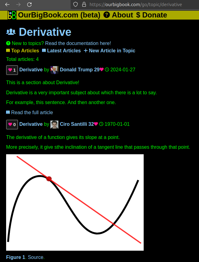
Figure 1. Screenshot of the "Derivative" topic page. View it live at: ourbigbook.com/go/topic/derivativeVideo 2. OurBigBook Web topics demo. Source. - local editing: you can store all your personal knowledge base content locally in a plaintext markup format that can be edited locally and published either:This way you can be sure that even if OurBigBook.com were to go down one day (which we have no plans to do as it is quite cheap to host!), your content will still be perfectly readable as a static site.
- to OurBigBook.com to get awesome multi-user features like topics and likes
- as HTML files to a static website, which you can host yourself for free on many external providers like GitHub Pages, and remain in full control

Figure 2. You can publish local OurBigBook lightweight markup files to either OurBigBook.com or as a static website.
Figure 3. Visual Studio Code extension installation.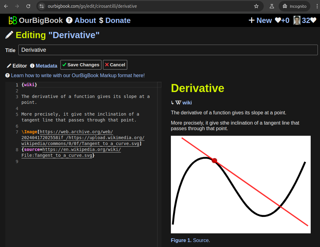
Figure 5. . You can also edit articles on the Web editor without installing anything locally. Video 3. Edit locally and publish demo. Source. This shows editing OurBigBook Markup and publishing it using the Visual Studio Code extension. 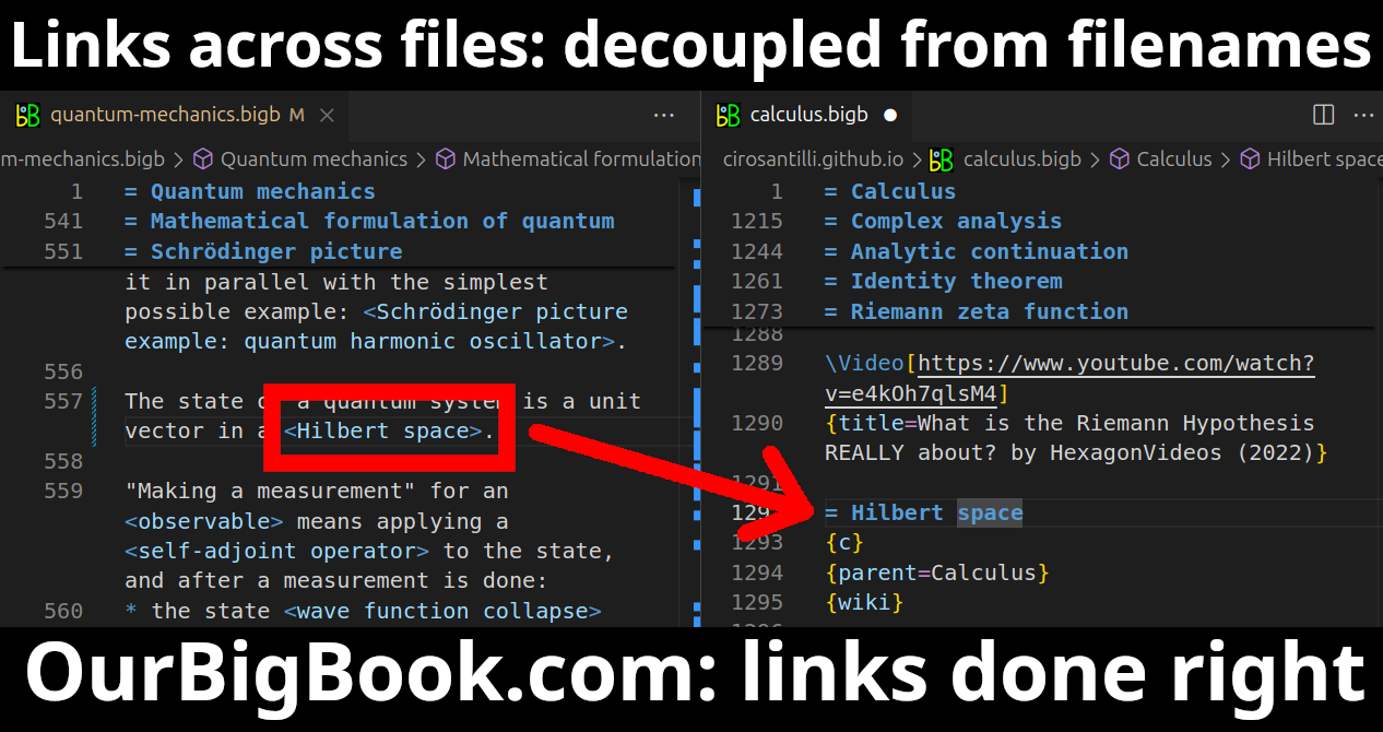
- Infinitely deep tables of contents:
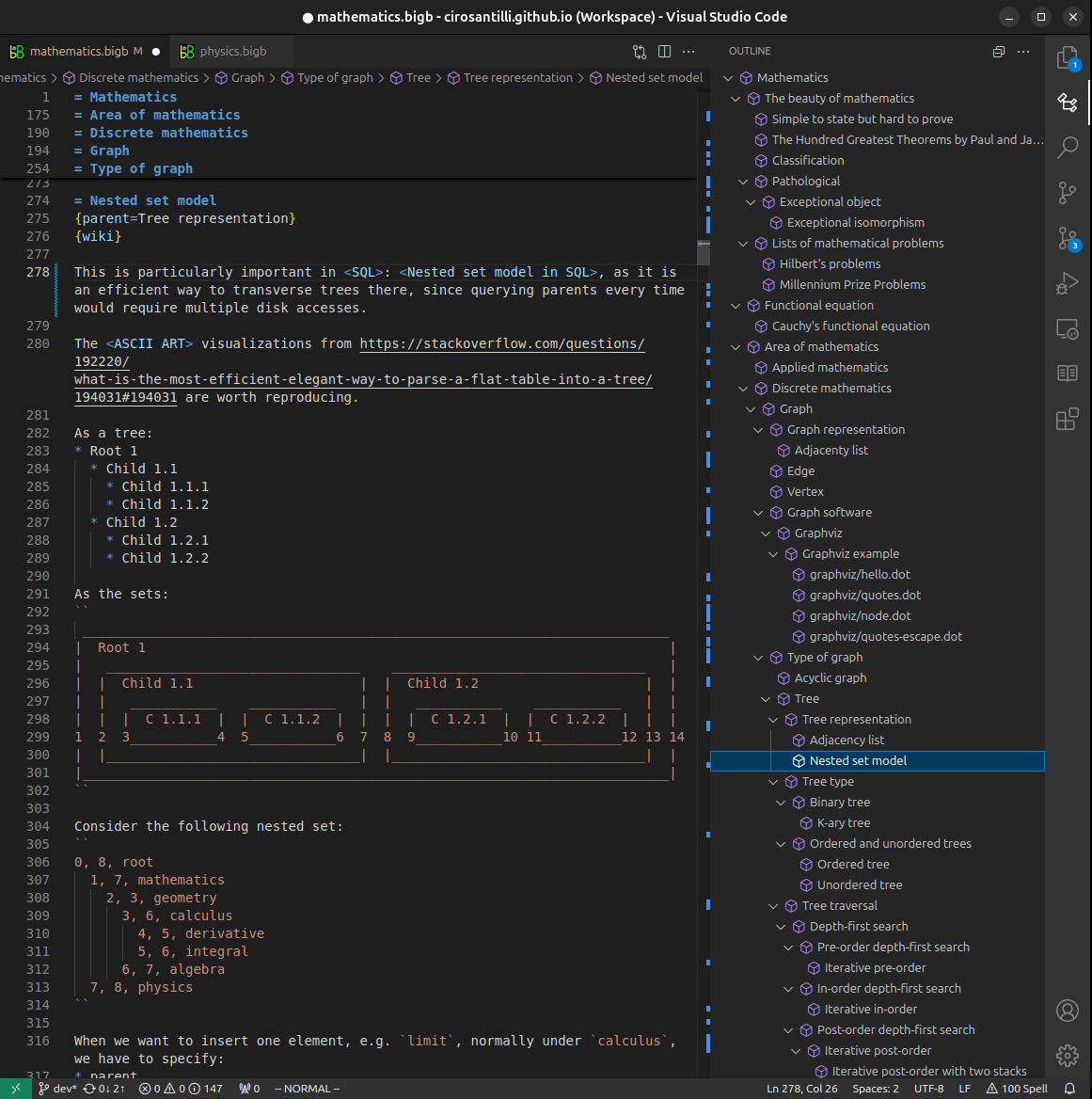
All our software is open source and hosted at: github.com/ourbigbook/ourbigbook
Further documentation can be found at: docs.ourbigbook.com
Feel free to reach our to us for any help or suggestions: docs.ourbigbook.com/#contact