Topics (384k) Articles (398k) Users (410) Discussions (242) Comments (383) Files (13k) New article

Scaffold degradation is the progressive alteration or breakdown of a tissue-engineering scaffold under its chemical and biological environment. For a degradable polymer, chain scission can change molecular size and mechanical behavior before a large amount of solid has disappeared. Polymer erosion, meaning loss of material from the scaffold, is therefore related to degradation but is not the same measurement.
A simple chain-counting example shows why. Suppose a specimen initially contains 10,000 identical repeat units arranged in 100 chains. Its mean chain length is 100 units. Cutting each chain once produces 200 chains and a mean of 50 units, while all 10,000 repeat units can still remain in the specimen. Chemical breakdown has occurred without mass leaving. The example neglects the small mass change from newly formed end groups; it is a bookkeeping model, not a measured degradation law.
Mechanical support and tissue formation must evolve together. As the original material weakens, cells can deposit extracellular matrix, but this does not imply that their mechanical contributions add as two independent springs. Geometry, bonding and local material changes affect the load paths. A scaffold that loses support before a functional matrix forms and one that retains obstructive remnants too long present different engineering problems.
The primary follow-up study of engineered bovine arteries examined culture time and scaffold type. It reported polymer remnants after eight weeks and a different smooth-muscle cell phenotype near those remnants.[ref]
Morphologic and mechanical characteristics of engineered bovine arteries by Laura E. Niklason and others. Printed p. 628, abstract and introduction
These observations motivate measuring local biological state alongside bulk mechanical performance; the association alone does not establish that every residual polymer fragment has the same effect.A useful laboratory comparison records retained dry mass, molecular-size change and a specified mechanical response at several times. An acellular incubation under matched medium conditions can help separate material changes from changes associated with the cells. Temperature, medium exchange, geometry and the analysis method should be stated, because a single percentage of mass remaining does not fully characterize scaffold degradation.
Cell seeding density specifies the number of biological cells introduced to, or initially retained on, a culture support relative to a stated area or volume. The denominator and observation time are part of the definition. A suspension concentration in cells per millilitre, a loading per projected scaffold area and a retained count per scaffold volume are different quantities.
For example, applying of a suspension containing cells per millilitre suppliesOver a projected area , the offered cell seeding density is . If a standardized wash and count finds attached cells, the attachment fraction is and the retained density is . These chosen values illustrate accounting, not a recommended culture condition. An internal porous area can be much larger than , so changing the area convention changes the reported density.
The distinction matters experimentally. Jinming Gao, Laura E. Niklason and Robert S. Langer studied surface hydrolysis of polyglycolic acid meshes and reported increased initial vascular smooth muscle cell seeding on the modified material. Their control experiments related protein adsorption to the increase.[ref]
Surface hydrolysis of poly(glycolic acid) meshes increases the seeding density of vascular smooth muscle cells by Jinming Gao and others. Printed p. 417, abstract
The experiment concerns an early cell–material interaction, not merely the nominal suspension concentration.A reproducible comparison states how many cells were offered, how unattached cells were removed, what was counted and when. Cell viability is another measurement: a retained cell count does not automatically specify the number of living cells. Later proliferation or cell loss can also change the density. Measuring both specimens at the same interval helps separate initial attachment from subsequent population change.
Nervous tissue, also called neural tissue, is the organized tissue of the nervous system, containing neurons and glial cells. Neurons specialize in communication, including action potentials and signaling between cells. Glial cells maintain the local environment, form insulating myelin in particular locations, and perform other active functions. The brain, spinal cord and peripheral nerves contain different arrangements of these components; a drawing of one isolated neuron is not a complete picture of nervous tissue.[ref]
Anatomy and Physiology 2e by J. Gordon Betts and others. Section12.2, Nervous Tissue: neurons, glial cells and their organization
A useful example is the median eminence, where hormone-releasing axon endings interact with tanycytes and blood-vessel endothelium. In a 2004 experiment, investigators compared female-rat tissue explants after 30 minutes with or without 500 micromolar arginine, a precursor for nitric oxide production. Electron microscopy showed identified gonadotropin-releasing hormone terminals close to the vascular boundary after treatment, with fewer intervening tanycyte processes than in controls.[ref]
Vascular endothelial cells promote acute plasticity in ependymoglial cells of the neuroendocrine brain by Sandrine De Seranno and others. Figure7 and Results: treated versus untreated hypothalamic explants
The observation makes tissue organization experimentally meaningful: changes in the position of a glial cell process can alter a neuron's access to a vascular interface. This specialized arrangement should not be mistaken for the structure of every synapse or every region of nervous tissue.
Mineralized tissue is biological tissue in which mineral forms a substantial structural component of an organic matrix. Bone, dentine and enamel are familiar examples, but they differ markedly in composition and architecture. In bone, mineral based on hydroxyapatite is associated with a collagen-rich extracellular matrix; biological cells form and maintain that matrix. Mineralization describes a material feature, not the absence of living activity.[ref]
A mineralized tissue is therefore also a biological composite material. Mineral amount, organic matrix, water, porosity and orientation can each affect the response. The Young's modulus of an entire specimen cannot be inferred from mineral content alone: constituent geometry changes load sharing, just as it does in the isostress rule of mixtures and the longitudinal rule of mixtures. These ideal mixture models clarify the role of geometry without being exact models of a real bone.
Even the phrase “mineral fraction” needs a specified measurement. Consider a hypothetical dry two-component specimen with of mineral of density and of organic material of density . Its mineral mass fraction is , but its mineral volume fraction isThese chosen numbers describe a bookkeeping example, not a measured tissue composition. Adding water or pores changes which total volume belongs in the denominator. A mechanical mixture calculation needs the appropriate volume fractions, not a mass percentage silently substituted for them.
An instructive primary structural study examined elephant ivory, a form of dentine, with complementary X-ray scattering, polarized-light microscopy and scanning electron microscopy. The investigators found that mineralized collagen orientations differed between the outer and inner parts of the tusk, including an outer wrapping orientation and a predominantly axial, plywood-like arrangement farther inside.[ref]
The three-dimensional arrangement of the mineralized collagen fibers in elephant ivory and its relation to mechanical and optical properties by M. Albéric and others. Primary abstract, complementary methods and region-dependent fibre orientations
The result motivates combining methods: a bright pattern in polarized light, an electron-microscope image and a scattering pattern provide different evidence about the same organized mineralized tissue. Observing orientation is not by itself a direct measurement of its full mechanical constitutive equation.A functional time series is a time series whose observation at each time is a whole function, often modeled as a random element of the Hilbert space . For example, might be the temperature profile within day , with marking time of day. Dependence across days is different from dependence between nearby positions within one curve.
For a simple model, let , where is a stationary bivariate time series. Its mean curve is . The covariance between curves at lag is a kernel , retaining both within-curve coordinates.
Estimating by resembles scalar averaging, but its uncertainty depends on serial covariance operators rather than only the variance of one curve. Treating every densely measured point as an independent observation can severely overstate precision.[ref]
A time-of-flight timing offset, , is an additive time-origin parameter in a model connecting recorded ion arrival time with physical flight time. In a simple time-of-flight mass spectrometer calibration,where is the numerical mass-to-charge ratio in declared units. A 2017 instrument-analysis paper uses this equation and determines instrument-dependent constants from identified isotope peaks.[ref]
An original error calculation shows why the offset matters. Suppose the physical flight contribution is and , so the recorded time is . With held fixed, ignoring the offset multiplies the inferred coordinate bya overestimate. This deliberately large example isolates the effect; it does not describe a measured instrument error.
With fixed, a small error in the fitted offset propagates approximately asAn offset uncertainty of at a corrected flight time of therefore contributes about relative uncertainty in this approximation. The same absolute offset error has greater relative impact on earlier arrivals.
This parameter concerns timing, whereas time-lag focusing is a physical delayed-extraction technique affecting the ion packet. Fitting an offset does not by itself sharpen a peak. More detailed instruments can require additional calibration terms; the stated equation is a model, not a universal transfer function.
A unit of time is a reference duration used to express a time interval. Writing changes the numerical representation, not the elapsed time in physics. A date labels a point on a time scale; a duration measures separation between two such points.
The SI base unit is the second, defined by fixing the unperturbed ground-state hyperfine transition frequency of the caesium-133 atom to exactly . A clock realizes that definition by comparing an oscillator with the atomic response; practical imperfections still create measurement uncertainty.[ref]
The International System of Units (SI), ninth edition, SI Brochure. Section 2.3.1, definition of the second
A minute is exactly 60 seconds, an hour 3600, and a day used as a duration unit 86400. A calendar day need not coincide with a uniform 86400-second interval on every civil time scale. Specifying the time convention matters when combining clock measurements, astronomical observations and Julian years.
The line height governs the vertical spacing allocated to a line of text. For a simple repeated run with fixed metrics, it sets the distance from one text baseline to the next. The gap between visible ink is a separate quantity, because different glyphs need not fill their allocated vertical region.
In an elementary single-font model, let be the positive font ascent, the positive magnitude of the font descent, and an added font line gap. ThenFor hypothetical metrics , and font units, font units. With 1000 font units per em and 20 pixels per em, the text baselines are 24 pixels apart. The line height can thus exceed the nominal font size.
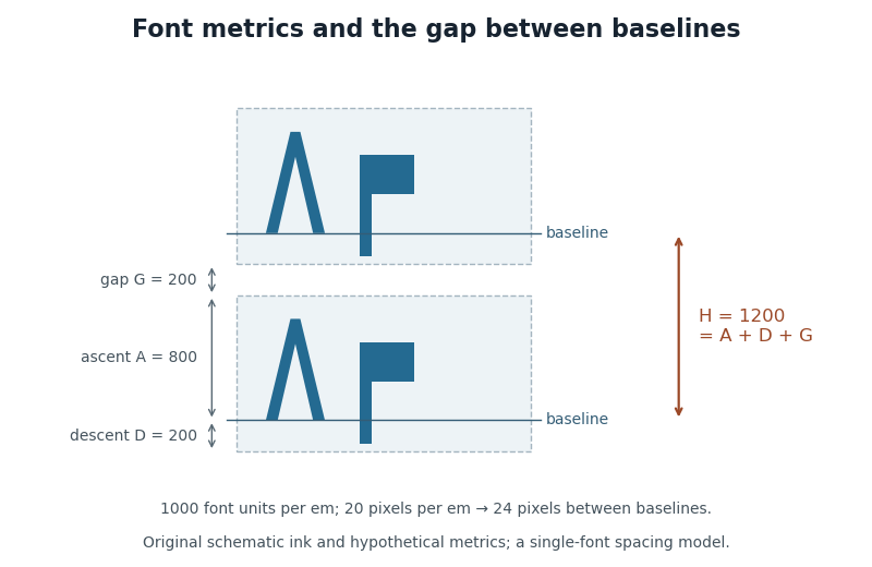
CSS lays out inline content through line boxes. Its
line-height participates in that calculation, and a block's declared value sets a minimum rather than guaranteeing that every mixed-content line has exactly that height. A taller inline object or a different alignment can enlarge a line box. The specification also describes how extra typographic leading is split above and below the chosen font metrics.[ref]Visual formatting model details in CSS 2.2 by W3C. Sections10.8–10.8.1: line-box construction, leading and the block-container minimum
The useful distinction is between the nominal font size, the font ascent and font descent used for layout, and the actual ink bounding boxes. They answer different geometric questions even when they are expressed in the same units.
A TeX control word is a TeX control sequence scanned from an escape followed by a maximal run of characters with TeX category code 11. The scanner then enters the space-skipping TeX input state S.[ref]
For example, after
\def\word{Q}, the input \word X yields QX; the spaces terminate the name and are skipped. The input \word X does the same. By contrast, \wordX names a single different command, so removing the separator does not preserve the token sequence.Token identity and current meaning are separate. Define
\def\call{\word}, then redefine \word to yield R. Expanding \call now yields R, because its stored command token still names word. This is useful when reading TeX macros: the spelling identifies the command, while its current definition controls expansion.A TeX control symbol is a TeX control sequence formed by an escape followed by one character whose TeX category code is not letter. Under ordinary conventions,
\% is one such command.[ref]For an invented example, define
\def\!{Q} and then read \! X. The resulting replacement starts with Q, one ordinary space token and X. The nonletter ends the command name immediately; the first following space is processed in state M and survives. Compare \def\word{Q} followed by \word X, which produces QX.Control space, written as an escape followed by a space, has special handling: it enters state S. Thus a rule that discards every space after every command would predict the wrong result for the
\! example.[ref]TeX source code by Donald E. Knuth. Scan a control sequence: state selection for spacer and letter; get_next whitespace cases
A tetrazolium cell-free control measures the reagent system without cells while preserving the relevant medium, test compound and processing conditions. It tests whether chemistry or optical background outside living cells can contribute to a tetrazolium readout. It does not measure cellular survival.
A primary study reported formazan formation in cell-free mixtures containing MTT and selected test compounds, alongside a disagreement between cell-number and MTT readouts.[ref]
Interference of plant extracts, phytoestrogens and antioxidants with the MTT tetrazolium assay by Regina Bruggisser and others. Abstract, direct cell-free reduction and cell-number comparison
This makes the absence of cells an experimental control on one proposed source of signal, rather than a purely mathematical blank.Controls with and without reagent answer different questions. Let and be absorbances with and without tetrazolium in the presence of the test compound; let and be the corresponding vehicle controls. An original cell-free tetrazolium interaction contrast isIt subtracts the compound's pre-existing optical contribution and the vehicle/reagent contribution under an additive model.
For chosen readings in that order, . Subtracting only the reagent-containing vehicle value would instead give and combine distinct contributions. A positive contrast identifies a reagent-dependent cell-free change, but product identity can require further spectral or chemical evidence.
The comparison should retain the intended compound concentration, medium, reagent amount, observation interval, temperature and product-handling conditions. A reference measured under a different chemical environment can fail to reproduce the interference present in the actual assay.
A negative cell-free control does not establish that a treatment leaves the per-cell reduction rate unchanged. Cells can transform compounds or alter the chemical environment in ways absent from the control. Likewise, a positive control does not quantify how much of the cell-containing signal has the same origin unless the model supports that transfer. Appropriate cellular controls and an independent readout remain needed for a cell viability interpretation.
A Sertoli cell is a supporting somatic biological cell within a seminiferous tubule of the testis. It helps maintain the local environment in which germ cells develop through spermatogenesis. A Sertoli cell supports the sperm-producing lineage; it is not itself a developing sperm.
A 2014 experiment tested whether these cells were required only during development or also in the adult. Investigators engineered adult MICE so that Sertoli cells could be selectively removed after exposure to a toxin. Subsequent examination showed loss of germ-cell populations, while the overall tubular framework persisted. Leydig cell numbers also fell substantially. Removing one supporting population thus disrupted both sperm production and another cell population elsewhere in the testis.[ref]
Sertoli cells maintain Leydig cell number and peritubular myoid cell activity in the adult mouse testis by Diane Rebourcet and others. Abstract; Results and Figures1,2,6: selective ablation and cell-population changes
The interpretation depends on comparing cellular function with gross appearance. A tubule can remain recognizable in a section while losing the cells and interactions needed for normal spermatogenesis. The experiment establishes a continuing requirement for Sertoli cells in this adult mouse model; it does not show that every affected neighboring cell normally contacts a Sertoli cell directly.
A seminiferous tubule is a coiled epithelial tube within the testis in which germ cells develop through spermatogenesis, supported by Sertoli cells. Developing germ cells occupy different positions within the epithelium, and released sperm enter the lumen. Leydig cells lie outside the tubules in the intervening tissue.[ref]
Anatomy and Physiology 2e by J. Gordon Betts and others. Section27.1: seminiferous-tubule organization and spermatogenesis
Specialized contacts between neighboring Sertoli cells contribute to the blood-testis barrier, helping organize the local environment of the developing germ cells. The wall is therefore an active cellular interface, rather than simply a pipe through which mature sperm flow.
A useful histology exercise compares a section with a three-dimensional reconstruction. Several circular profiles can be different cuts through one coiled seminiferous tubule; counting profiles does not directly count independent tubes. Likewise, preservation of a recognizable tubular outline does not establish normal germ-cell development. In a 2014 mouse experiment, removing Sertoli cells caused extensive germ-cell loss while much of the tubular framework remained identifiable.[ref]
Sertoli cells maintain Leydig cell number and peritubular myoid cell activity in the adult mouse testis by Diane Rebourcet and others. Results,Figure2: germ-cell loss and retained tubular architecture
Pinned article: Introduction to the OurBigBook Project
Welcome to the OurBigBook Project! Our goal is to create the perfect publishing platform for STEM subjects, and get university-level students to write the best free STEM tutorials ever.
Everyone is welcome to create an account and play with the site: ourbigbook.com/go/register. We belive that students themselves can write amazing tutorials, but teachers are welcome too. You can write about anything you want, it doesn't have to be STEM or even educational. Silly test content is very welcome and you won't be penalized in any way. Just keep it legal!
Intro to OurBigBook
. Source. We have two killer features:
- topics: topics group articles by different users with the same title, e.g. here is the topic for the "Fundamental Theorem of Calculus" ourbigbook.com/go/topic/fundamental-theorem-of-calculusArticles of different users are sorted by upvote within each article page. This feature is a bit like:
- a Wikipedia where each user can have their own version of each article
- a Q&A website like Stack Overflow, where multiple people can give their views on a given topic, and the best ones are sorted by upvote. Except you don't need to wait for someone to ask first, and any topic goes, no matter how narrow or broad
This feature makes it possible for readers to find better explanations of any topic created by other writers. And it allows writers to create an explanation in a place that readers might actually find it.
Figure 1. Screenshot of the "Derivative" topic page. View it live at: ourbigbook.com/go/topic/derivativeVideo 2. OurBigBook Web topics demo. Source. - local editing: you can store all your personal knowledge base content locally in a plaintext markup format that can be edited locally and published either:This way you can be sure that even if OurBigBook.com were to go down one day (which we have no plans to do as it is quite cheap to host!), your content will still be perfectly readable as a static site.
- to OurBigBook.com to get awesome multi-user features like topics and likes
- as HTML files to a static website, which you can host yourself for free on many external providers like GitHub Pages, and remain in full control

Figure 2. You can publish local OurBigBook lightweight markup files to either OurBigBook.com or as a static website.
Figure 3. Visual Studio Code extension installation.
Figure 5. . You can also edit articles on the Web editor without installing anything locally. Video 3. Edit locally and publish demo. Source. This shows editing OurBigBook Markup and publishing it using the Visual Studio Code extension. 
- Infinitely deep tables of contents:



All our software is open source and hosted at: github.com/ourbigbook/ourbigbook
Further documentation can be found at: docs.ourbigbook.com
Feel free to reach our to us for any help or suggestions: docs.ourbigbook.com/#contact