Topics (384k) Articles (398k) Users (410) Discussions (242) Comments (383) Files (13k) New article

The lumbosacral plexus is the connected system of lumbar plexus and sacral plexus pathways formed from anterior spinal nerve rami of lower spinal nerves. It redistributes sensory and motor axons into peripheral nerves supplying the pelvis and lower limb. Its connecting lumbosacral trunk usually carries contributions from L4 and L5 into the sacral plexus.[ref]
For example, the femoral nerve draws mainly on upper lumbar levels, while the sciatic nerve combines lower lumbar and sacral contributions. A named peripheral nerve consequently does not correspond one-to-one with a spinal nerve. The nerve plexus reroutes existing axons; its intersections do not imply that the fibres form synapses.
The sciatic nerve is a large mixed nerve connecting the lumbosacral plexus with the lower limb. It passes through the posterior hip region into the thigh and contains the components that continue as the tibial nerve and common fibular nerve. These pathways carry motor signals to skeletal muscles and sensory information from substantial regions of the leg and foot.
Its course relative to the piriformis muscle varies among individuals. A retrospective magnetic resonance imaging study compared common and variant anatomical arrangements and did not find a statistically significant difference in the recorded diagnoses involving pain.[ref]
Is it painful to be different? Sciatic nerve anatomical variants on MRI and their relationship to piriformis syndrome by Adam L. Bartret and others. European Radiology 28, pp. 4681–4686; MRI classification and retrospective outcome comparison
An unusual anatomical arrangement therefore does not by itself establish that the arrangement is causing symptoms; structure and physiological consequence require separate evidence.The Loop of Henle is the hairpin portion of a nephron's renal tubule, carrying fluid from the proximal tubule toward the renal medulla and back toward the renal cortex. Its descending and ascending portions have different transport properties. In particular, the thin descending limb can be highly water-permeable, while the thick ascending limb reabsorbs sodium chloride with little accompanying water. These differences support countercurrent multiplication rather than mere circulation around a bend.[ref]
Anatomy and Physiology 2e by J. Gordon Betts and others. Sections25.4 and25.6: nephron loop and tubular transport
In a 1970 experiment, isolated rabbit thin descending limb segments were perfused inside a separate surrounding bath. Raising bath osmolality concentrated the collected tubular fluid mainly through water leaving, with little solute entry. A separate 1973 study of isolated rabbit thick ascending limb segments found salt removal with very low osmotic water transport. The experimental comparison connects different functions to distinct parts of one continuous tubule.[ref]
Sodium chloride and water transport in the descending limb of Henle by Juha P. Kokko. Abstract: isolated segments, imposed bath gradients and water efflux
[ref]A simple mass balance clarifies why both observations matter. If a fluid initially contains 30 arbitrary units of solute in 10 units of water, removing 5 water units raises its concentration from 3 to 6. Removing 15 solute units at unchanged water content instead lowers it from 3 to 1.5. These are illustrative limiting cases, not complete measurements of a nephron. Concentrating and diluting fluid need not require the same kind of transport in reverse.
A renal corpuscle is the initial filtration structure of a nephron, consisting of a glomerulus enclosed by a glomerular capsule, also called Bowman's capsule. Blood flows through the glomerular capillaries, while filtered fluid enters the surrounding capsular space and then the proximal tubule. The blood pathway and the tubular-fluid pathway are adjacent but distinct.[ref]
Kidneys by the National Cancer Institute. nephron organization: glomerulus and surrounding epithelial capsule
The glomerular filtration barrier includes capillary endothelium, a basement membrane and the specialized surface formed by podocytes. These biological cells extend processes around the capillaries. In a 1999 study, antibody-based localization placed the membrane protein nephrin at the slit diaphragm between podocyte processes, connecting a named molecular component to the physical filtration interface.[ref]
Nephrin is specifically located at the slit diaphragm of glomerular podocytes by V. Ruotsalainen and others. Abstract and immunolocalization results
A section supplies a useful scale comparison: the rounded structure containing a capillary tuft and its surrounding capsule is the renal corpuscle, whereas the glomerulus is the tuft inside it. Counting those as separate complete filtration units would double-count parts of the same unit. Filtered water and small solutes still undergo later processing, so the fluid first leaving a corpuscle is not identical to final urine.
A juxtamedullary nephron is a nephron whose renal corpuscle lies in the deep renal cortex, near its boundary with the renal medulla, and whose long Loop of Henle extends into the medulla. “Juxtamedullary” describes the location of the corpuscle relative to that boundary; it does not place the corpuscle at the renal papilla. A more superficial cortical nephron generally has a shorter loop.[ref]
Anatomy and Physiology 2e by J. Gordon Betts and others. Section25.4: cortical and juxtamedullary nephrons
The long loop brings tubular fluid through different levels of the medullary concentrating environment. The associated renal vasa recta supply blood while supporting countercurrent exchange, and the loop's segment-specific transport contributes to countercurrent multiplication. The same anatomical arrangement connects local membrane transport to the whole kidney's ability to conserve water.
For a reconstruction exercise, mark the position of one renal corpuscle, then follow its tubule rather than classifying it from a single cross-section. A deep-running loop can belong to a corpuscle that remains in cortical tissue. Conversely, a superficial tubular sample alone does not tell an experimenter how deeply the loop runs. Anatomical tracing therefore supplies information that a concentration measurement at one site cannot.
A cortical nephron is a nephron whose renal corpuscle lies relatively superficially in the renal cortex and whose Loop of Henle is usually shorter than that of a juxtamedullary nephron. The contrast concerns the position of the corpuscle and the depth of the loop. It does not mean that every part of a cortical nephron is confined to cortical tissue.[ref]
Anatomy and Physiology 2e by J. Gordon Betts and others. Section25.4: cortical and juxtamedullary nephrons
A useful reconstruction starts at a glomerulus and follows the continuous tubule. A loop can enter the outer renal medulla and return to the cortex even though its corpuscle is superficial. A single transverse section through a tubule does not reveal either its complete path or the depth of its originating corpuscle. Thus anatomical tracing provides information beyond simply counting circular profiles in a histology image.
Both nephron classes perform glomerular filtration and tubular processing. Their different arrangements contribute differently to the medullary concentrating system, rather than dividing the kidney into a filtration-only class and a concentration-only class. Studies sampling visible surface tubules must also identify which nephrons are accessible before treating those samples as representative of all nephron types.[ref]
Micropuncture study of the mammalian urinary concentrating mechanism: evidence for the countercurrent hypothesis by Carl W. Gottschalk and Margaret Mylle. Methods,p928: identity and short-loop organization of sampled surface nephrons
Nail-plate material age from growth marks estimates how long a marked part of the nail plate has travelled since crossing a specified reference. It is a transport clock, not a direct observation of the instant when the material first formed in the nail matrix. The anatomical reference and any hidden travel before reaching it must be specified.
In a primary growth experiment, 22 young adults made file marks near the proximal fold, recorded their positions and repeated the measurements one to three months later.[ref]
Growth rate of human fingernails and toenails in healthy American young adults by S. Yaemsiri and others. Abstract, Methods
This measures average displacement during the observation interval; it does not by itself recover every earlier growth rate.For an original one-dimensional advection model, let a material element cross the reference at time . If plate advance is spatially uniform with speed , its later position isA known growth history permits inversion for . A current speed alone need not. For example, 30 days at mm/day followed by 30 days at mm/day gives displacement 6 mm and age 60 days. Dividing by the final speed gives only 50 days, because it substitutes today's speed for the earlier history.
If only bounds are justified throughout the travel, thenFor 6 mm and stipulated speeds between 0.08 and 0.12 mm/day, the reference-crossing age lies between 50 and 75 days. These bounds describe an original model, not published population limits.
A clipping also spans material positions. Even at a perfectly known constant speed of 0.10 mm/day, a 3 mm longitudinal segment spans 30 days of reference-crossing times. Temporal averaging in nail clippings therefore remains after instrument precision improves. External contamination or chemical exchange can further separate a measured chemical signal from the time of matrix production. Record mark position, reference fold, clipping extent and observation dates before treating a nail plate sample as a dated exposure record.
Fluorescence contamination in nail-transmission measurements occurs when light emitted by a specimen contributes to a detector signal intended to quantify transmitted illumination. Emission can reach the detector at wavelengths different from the selected illumination wavelength, where detector spectral responsivity may also differ. Total current therefore need not be proportional to the desired transmitted power alone.
The primary human-nail experiment explicitly included a correction for nail fluorescence in its monochromator, integrating sphere and photodiode measurement.[ref]
The transmission of optical radiation through human nails by S. G. Parker and B. L. Diffey. Abstract, fluorescence correction
It supplies an experimental motivation for distinguishing optical transmission from all detected light.An original two-channel calibration model makes the separation explicit. Let be transmitted excitation-band power and an emitted component with a specified spectral shape. After dark-signal subtraction, calibrated channel readings satisfyThe coefficients incorporate filters, detector spectral responsivity and collection efficiency. Separation requires ; two identical mixtures provide no additional information.
For stipulated dimensionless coefficients and , power-equivalent readings 0.100 and 0.024 mW giveAt incident power 1 mW, the corrected excitation-band transmittance is 8%; treating the first channel as transmission alone gives 10%. These are illustrative signals, not reported nail data.
Excitation-band selectivity in nail fluorescence corrections can be tested by changing the detection spectral band while keeping excitation and geometry fixed. The calibration still depends on the assumed emission spectrum: a different spectral shape can change the coefficients. A filter can also reject some desired transmitted light, requiring its own calibration.
Collection geometry in nail-transmission measurements specifies which light emerging from a nail plate reaches the detector. A narrow forward detector and an integrating sphere can report different signals from the same specimen because light scattering redistributes directions. The reported transmittance must identify its collection conditions.
The primary 1983 nail study measured total transmitted radiation at fifteen wavelengths from 300 to 600 nm through thirteen autopsy toenails, using a monochromator, integrating sphere and photodiode.[ref]
The transmission of optical radiation through human nails by S. G. Parker and B. L. Diffey. Abstract, specimen set and apparatus
The angular collection is part of the measurement, alongside the wavelength and the separate correction for fluorescence.An original angular model separates on-axis direct transmission from a diffuse transmitted fraction . Suppose the diffuse component has constant radiance over the outgoing hemisphere. A detector accepting all azimuths up to angle from the surface normal collects the fractionThe cosine accounts for projected emitting area. Assuming the direct beam is entirely collected, . This is a constructed angular distribution, not a measured scattering law for nails.
At stipulated , and degrees, and the detected fraction is 0.06 although total transmission is 0.12. Aperture-dependent diffuse transmission in nail measurements also depends on distance. For a small emitting region facing a circular aperture of radius at distance , in this model. With mm, distances 10 and 20 mm give approximately 0.0560 and 0.0447 for measured transmission, respectively, without a specimen change.
An integrating sphere collects a much broader angular distribution, but its ports, coating and detector coupling still require characterization. A useful comparison holds the specimen, incidence and spectral band fixed while changing collection aperture or recording angular response. Correcting fluorescence does not recover scattered light that was never collected, and a calibration made in one geometry does not automatically apply in another.
Matrix growth and plate-advection balance relates newly supplied nail material to movement and changing geometry of the nail plate. The nail matrix contains the growth-producing region, whereas the mature nail plate is transported material.[ref]
Anatomy and Physiology 2e by J. Gordon Betts and others. §5.2, Nails: proliferating basal cells at the root
Measured advance is consequently a material-flow observation, not a direct count of cell divisions.For an original one-dimensional conservation of mass model, define line density , where is plate cross-sectional area, its density and its advance speed. A local production rate per unit length gives the continuity equationIf the chosen downstream segment has negligible production or loss, . At steady state its mass flux is constant. Uniform density then makes the volume flux constant, recovering the simple width-times-thickness model.
For stipulated , cross-sectional areas of 3 and require speeds 0.10 and 0.15 mm/day. Equal production can therefore accompany unequal advance when geometry changes. Conversely, equal speeds do not establish equal production without knowing area and density.
For a fixed segment , integrating the continuity equation givesIf inflow exceeds outflow, material accumulates rather than vanishing from the accounting. Growth, hydration-related geometry change and clipping must be assigned to appropriate terms before interpreting an apparent imbalance.
The nail-marking study measured displacement over known time intervals in 22 young adults.[ref]
Growth rate of human fingernails and toenails in healthy American young adults by S. Yaemsiri and others. Abstract, Methods
Extending that design with repeated width and thickness measurements would test a nail-plate storage balance, rather than assuming steady geometry. Inferring a mitotic rate would require further information about cell size and conversion into mature material.The optical contribution of the nail bed is the portion of measured light associated with tissue beneath the nail plate, after accounting for illumination, the covering plate and collection geometry. The vascular tissue beneath the plate contributes to its ordinary pink appearance, while the compact plate itself consists of dead keratinocytes.[ref] A colour measurement combines optical transport and anatomy rather than directly reporting a single blood quantity.
An original simplified reflectance model iswhere represents light returned by the plate and interfaces, is single-pass plate transmittance and is the fraction returned from the underlying region. The square represents outward and return passage through the plate. This deliberately omits detailed multiple scattering; it is an explanatory model, not a clinical calibration.
Even with known , can arise from or . One measurement cannot distinguish those pairs. For small changes with ,Thus a 1% relative change in plate transmittance contributes approximately a 2% relative signal change without any change in underlying reflectance. This is a concrete inverse problem.
A primary experiment separated one optical component physically: thirteen autopsy toenails were measured at fifteen wavelengths between 300 and 600 nm using a monochromator, integrating sphere and photodiode, with a correction for nail fluorescence.[ref]
The transmission of optical radiation through human nails by S. G. Parker and B. L. Diffey. Abstract; British Journal of Dermatology 108, 11–16 (1983), DOI 10.1111/j.1365-2133.1983.tb04573.x
Measuring an isolated plate constrains its transmission; it does not measure living nail-bed blood flow.Plate-bed optical identifiability requires additional constraints: independent plate measurements, a specified spectral model or multiple collection geometries. More wavelengths help only if the added data constrain the unknowns; allowing an arbitrary new and at every wavelength preserves the ambiguity.
Pinned article: Introduction to the OurBigBook Project
Welcome to the OurBigBook Project! Our goal is to create the perfect publishing platform for STEM subjects, and get university-level students to write the best free STEM tutorials ever.
Everyone is welcome to create an account and play with the site: ourbigbook.com/go/register. We belive that students themselves can write amazing tutorials, but teachers are welcome too. You can write about anything you want, it doesn't have to be STEM or even educational. Silly test content is very welcome and you won't be penalized in any way. Just keep it legal!
Intro to OurBigBook
. Source. We have two killer features:
- topics: topics group articles by different users with the same title, e.g. here is the topic for the "Fundamental Theorem of Calculus" ourbigbook.com/go/topic/fundamental-theorem-of-calculusArticles of different users are sorted by upvote within each article page. This feature is a bit like:
- a Wikipedia where each user can have their own version of each article
- a Q&A website like Stack Overflow, where multiple people can give their views on a given topic, and the best ones are sorted by upvote. Except you don't need to wait for someone to ask first, and any topic goes, no matter how narrow or broad
This feature makes it possible for readers to find better explanations of any topic created by other writers. And it allows writers to create an explanation in a place that readers might actually find it.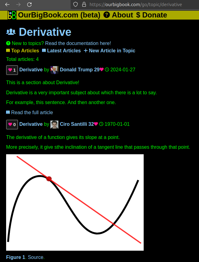
Figure 1. Screenshot of the "Derivative" topic page. View it live at: ourbigbook.com/go/topic/derivativeVideo 2. OurBigBook Web topics demo. Source. - local editing: you can store all your personal knowledge base content locally in a plaintext markup format that can be edited locally and published either:This way you can be sure that even if OurBigBook.com were to go down one day (which we have no plans to do as it is quite cheap to host!), your content will still be perfectly readable as a static site.
- to OurBigBook.com to get awesome multi-user features like topics and likes
- as HTML files to a static website, which you can host yourself for free on many external providers like GitHub Pages, and remain in full control

Figure 2. You can publish local OurBigBook lightweight markup files to either OurBigBook.com or as a static website.
Figure 3. Visual Studio Code extension installation.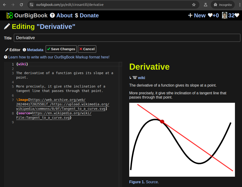
Figure 5. . You can also edit articles on the Web editor without installing anything locally. Video 3. Edit locally and publish demo. Source. This shows editing OurBigBook Markup and publishing it using the Visual Studio Code extension. 
- Infinitely deep tables of contents:
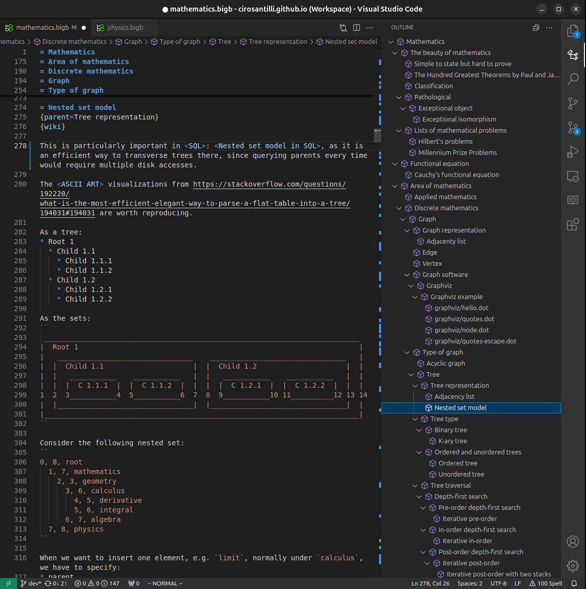
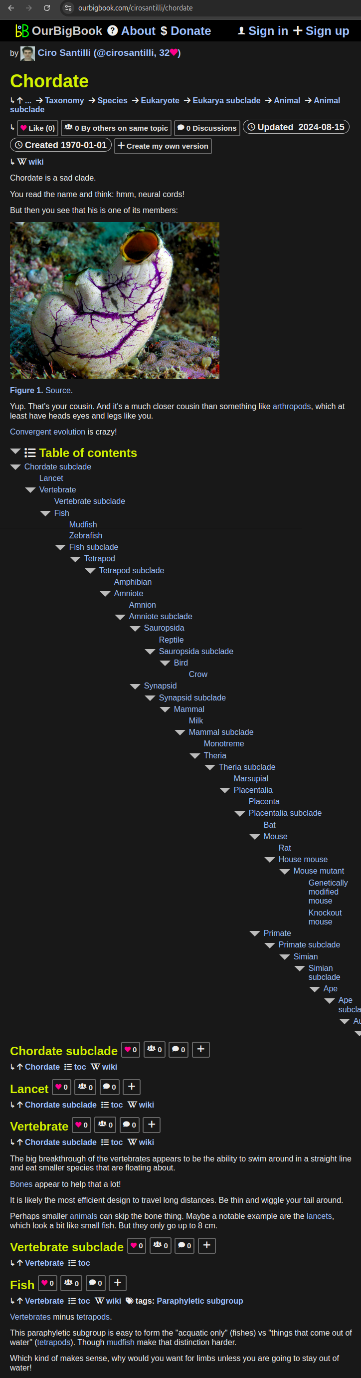
All our software is open source and hosted at: github.com/ourbigbook/ourbigbook
Further documentation can be found at: docs.ourbigbook.com
Feel free to reach our to us for any help or suggestions: docs.ourbigbook.com/#contact