Topics (384k) Articles (398k) Users (410) Discussions (242) Comments (383) Files (13k) New article

A free list records free memory regions or reusable allocation units so an allocator can find storage for a request. In a variable-size pool, a record typically needs a block's location and length. A linked implementation also stores a link to another free record; these records can occupy the free blocks themselves. The term can also describe the allocator's logical inventory when its search structure is more elaborate than one linked chain.[ref]
For an illustrative inventory of free extents of16,32 and48 bytes, a24-byte request cannot use the first extent but can use either later one, before metadata and alignment are counted. Choosing the32-byte extent leaves an8-byte remainder if split exactly. Whether that remainder can become a free-list entry depends on the minimum free-block size.
Suppose an explicitly hypothetical in-block free record needs an8-byte length field and an8-byte next-link field. Its16-byte metadata cannot fit in that8-byte remainder. The allocator can instead give the request the entire32-byte block, leaving8 bytes of internal fragmentation, or choose another layout or block. Metadata placement is therefore part of allocation capacity, rather than an afterthought.
When a block is returned, memory block coalescing can combine it with adjacent free neighbors and update the inventory. Address order can make adjacency checks convenient; other orderings can favor allocation searches. A list that contains enough total free bytes may still lack one sufficiently large extent, producing external memory fragmentation. The search policy, split rule, metadata layout and coalescing policy jointly determine which requests succeed.
Transcytosis carries material through a biological cell from one surface to another by combining endocytosis, intracellular vesicle transport and exocytosis. In an epithelial sheet, cargo can enter at the apical surface facing a cavity and leave at the basal or lateral surface facing underlying tissue. This route crosses the cell rather than passing through the gap between neighbouring cells.
An illuminating example is transfer of maternal antibodies across the intestine of a newborn rat. Richard Rodewald followed antibodies carrying an electron-dense ferritin label, and independently followed antibodies recognizable by a peroxidase reaction. Electron microscopy located tracer at the apical surface, in internal tubular vesicles and near release sites at the opposite surface. Experiments used isolated, ligated intestinal segments and control tracers to distinguish selective transport from leakage between cells. The observations establish a route across the epithelial cell, not merely disappearance from the intestinal cavity.[ref]
Intestinal transport of antibodies in the newborn rat by Richard Rodewald. 1973, abstract and Discussion, pp. 202–204
Glucose transport is movement of glucose between compartments, commonly across a cell membrane. It depends on carrier properties, carrier abundance, substrate concentrations and any coupled driving process. In facilitated transport, a simplified saturating relation describes initial uptake under specified conditions. At high , additional substrate has little effect because carrier capacity is limiting. [ref]
Molecular Biology of the Cell, 4th edition by Bruce Alberts and others. Chapter 11, Carrier Proteins and Active Membrane Transport
Transport and subsequent metabolism are separate steps. Phosphorylation of intracellular glucose changes the pool available for reverse transport, so measured uptake can depend on both entry and trapping. Likewise, an insulin-induced change in surface GLUT4 can alter entry without requiring a change in the intrinsic kinetics of each carrier. A tracer accumulation experiment must specify which of these processes contributes to its readout.
A glucose transporter is a membrane transport protein that moves glucose across a cell membrane. The category includes facilitated transporters such as GLUT1 and GLUT4, and coupled carriers such as SGLT1. A facilitated transporter allows movement down a glucose chemical-potential difference; a sodium-coupled carrier can use the sodium electrochemical gradient to accumulate glucose. [ref]
Molecular Biology of the Cell, 4th edition by Bruce Alberts and others. Chapter 11, Carrier Proteins and Active Membrane Transport
For GLUT4, changing surface abundance is itself a regulatory mechanism. Insulin-responsive trafficking can deliver pre-existing carriers to the surface, increasing transport without first synthesizing more transporter. The amount of protein in the whole cell and the amount available at its outer boundary therefore need not change together. [ref]
A silicon transporter is a membrane protein that moves a dissolved silicon-containing solute across a cell membrane. In the diatom SIT family, the substrate is silicic acid, the soluble precursor used in building a silica-rich cell wall. The functional category need not imply that transporters in every lineage have the same structure or coupling mechanism.[ref]
Direct evidence of the molecular basis for biological silicon transport by Michael J. Knight and others. Introduction and Discussion, diatom silicic-acid transport
A purified-system experiment reconstituted SIT proteins into liposomes. An inward-directed sodium gradient drove uptake, detected by fluorescence from zinc-dependent complexes formed inside the liposomes. Protein-free and unrelated-transporter controls helped distinguish transport from mixing background. Selected transporters had apparent Michaelis constants near .[ref]
Direct evidence of the molecular basis for biological silicon transport by Michael J. Knight and others. Figs.3–5, Zinc silicate fluorescence and Proteoliposome transport assays
Using that approximate scale in a simple Michaelis-Menten equation, gives at and at . This calculation illustrates saturation, not an additional experimental fit. A substrate-binding ratio measured at equilibrium does not by itself establish the number of sodium ions coupled to each transport event.
AUX1 is a plant membrane protein that promotes uptake of the auxin indole-3-acetic acid into cells. It is an influx carrier: its role concerns entry across a cell membrane, whereas the distribution of auxin through a tissue also depends on export, cellular geometry and the placement of transporters. These distinctions connect a molecular uptake mechanism to polar auxin transport without treating one carrier as the whole tissue-level pathway.[ref]
High-affinity auxin transport by the AUX1 influx carrier protein by Yaodong Yang and others. Abstract: functional identification of AUX1-mediated auxin influx.
Direct transport evidence came from expressing AUX1 in Xenopus oocytes and measuring uptake of tritium-labeled indole-3-acetic acid. Uptake was saturable and pH-dependent. Saturation matters: increasing substrate cannot increase a finite population of carrier-mediated transport events without bound. Dependence on pH connects uptake to the chemical environment across the biological membrane, rather than to AUX1 abundance alone.[ref]
High-affinity auxin transport by the AUX1 influx carrier protein by Yaodong Yang and others. Oocyte expression and radiolabeled IAA uptake assay.
In Arabidopsis thaliana roots, Ranjan Swarup and collaborators localized AUX1 and compared transport-related mutant phenotypes using different auxin compounds. Their results supported distinct routes for hormone delivery and redistribution near the root apex. The combination is instructive: localization asks where a carrier operates; heterologous expression tests what it can transport. Neither measurement alone completely specifies the resulting auxin pattern in an intact root.[ref]
Localization of the auxin permease AUX1 suggests two functionally distinct hormone transport pathways operate in the Arabidopsis root apex by Ranjan Swarup and others. Genes & Development 15 (2001), 2648–2653; localization and Figure 3 mutant/auxin comparisons.
The alternating access model describes membrane transport by a carrier protein whose binding region is exposed successively to opposite sides of a cell membrane. A solute can bind on one side, remain associated while the carrier protein changes conformation, and be released on the other. Unlike an open ion channel, this mechanism need not provide a continuous aqueous passage across the cell membrane.[ref]
Molecular Biology of the Cell, 4th edition by Bruce Alberts and others. Chapter 11, Carrier Proteins and Active Membrane Transport: alternating exposure of binding sites
The model explains a route, while thermodynamics determines the possible net direction. For an uncharged ideal dilute solute at uniform temperature, with equal standard chemical potentials on the two sides, the molar free energy change for inward transfer isHere is the molar gas constant. At , accumulating that solute to ten times its outside concentration costs approximately . Changing which side can reach a binding site does not supply that free energy. Sustained uphill transport requires coupling to another energetically favourable process; at equilibrium, detailed balance excludes a persistent directed pumping cycle.
Structural experiments help test how such access could change. The 2005 NhaA antiporter structure revealed a cytoplasmic funnel and an internal region implicated in ion binding in a pH-downregulated state. The authors proposed movements connecting access and regulation. A structure of one state constrains a transport mechanism, but is not a direct movie of its complete cycle.[ref]
Structure of a Na+/H+ antiporter and insights into mechanism of action and regulation by pH by Carola Hunte and others. Abstract; Figures 2–3, architecture and substrate passage; Figure 5, proposed mechanism
The neutral-solute calculation above illustrates the energetic distinction; the full NhaA cycle also involves coupled charged ions.A claudin is a member of a family of four-pass membrane proteins that assemble into tight junction strands between neighbouring cells. The choice of claudins helps determine which ions and small solutes can pass through the pathway between epithelial cells. A tight junction can therefore be selectively permeable rather than an absolutely impermeable weld.[ref]
Molecular Biology of the Cell, 4th edition by Bruce Alberts and others. Chapter 19, Tight Junctions Form a Selective Permeability Barrier Across Epithelial Cell Sheets
The structural role was demonstrated by adding claudin-1 or claudin-2 to mouse fibroblasts that lacked tight junctions. Freeze-fracture electron microscopy revealed extended strand networks in the modified cell membranes. Coexpression recruited occludin to the strands. Building recognizable junction strands in this simplified setting supplied stronger evidence for a construction role than finding a protein near an existing junction alone.[ref]
Occludin is an integral membrane protein concentrated at tight junctions, where neighbouring epithelial or endothelial cells regulate passage between them. It has four membrane-spanning segments and a long cytoplasmic tail. Occludin is a junction component rather than a synonym for the entire sealing structure.
In 1993, Mikio Furuse and colleagues raised monoclonal antibodies against a junction-enriched fraction from chick liver. Immunoelectron microscopy placed the recognized protein directly at membrane-contact points of tight junctions; sequence analysis suggested four membrane crossings. These combined biochemical and spatial observations identified occludin.[ref]
Occludin: a novel integral membrane protein localizing at tight junctions by Mikio Furuse and others. Abstract, pp. 1777–1788: fractionation, antibody localization and cDNA sequence
Later, expression of claudin-1 or claudin-2 in fibroblasts reconstituted extensive junction strands and recruited occludin. This separates the capacity to build a strand from the presence of one particular strand-associated protein.[ref]
A single gene product, claudin-1 or -2, reconstitutes tight junction strands and recruits occludin in fibroblasts by Mikio Furuse and others. 1998, abstract: claudin reconstitution and occludin recruitment
Aquaporin-2 is a water-channel membrane protein in kidney collecting duct cells. Vasopressin regulates how much of it is present in the apical cell membrane, changing the pathway available for water reabsorption. Aquaporin-2 supports passive water movement driven by the surrounding osmotic conditions.[ref]
Vasopressin increases water permeability of kidney collecting duct by inducing translocation of aquaporin-CD water channels to plasma membrane by S. Nielsen and others. 1995, abstract: collecting-duct water channels, membrane localization and osmotic permeability
In isolated perfused rat collecting ducts, researchers measured water permeability and then fixed the same tubules for electron microscopy with antibody labelling. Vasopressin increased apical channel labelling and measured permeability; withdrawal reversed both. Coupling a functional measurement to localization supported regulated transfer of channel-bearing vesicles to and from the surface.[ref]
A toy redistribution makes the distinction clear: of 100 channel units, move 20 from vesicles into a surface pool initially containing 20. Surface abundance doubles to 40 while total abundance stays 100. This illustrates how trafficking can produce a rapid response; it is not a measured fold change in channel number or water flux.
NKCC2 is the kidney-enriched sodium–potassium–chloride cotransporter that carries one , one and two ions together across a biological membrane in each coupled transport cycle. In the thick ascending limb of the Loop of Henle, NKCC2 supports salt uptake from the tubular lumen. It is a secondary active transport system: the coupled ion gradients provide the immediate driving force, while the sodium-potassium ATPase helps maintain those gradients.[ref]
Molecular cloning, primary structure, and characterization of two members of the mammalian electroneutral sodium-(potassium)-chloride cotransporter family expressed in kidney by G. Gamba and others. Kidney transporter cloning, functional expression and intrarenal localization; distinction from the Na-Cl transporter.
The stoichiometry explains why transport is electrically silent: one cycle moves net charge . This does not mean no ions move or no energy is involved. If is the activity inside divided by activity outside for ion , the idealized free-energy change for inward transport isThe membrane-potential contributions cancel because the coupled net charge is zero. A favorable sodium ratio can therefore help drive other ions uphill in their chemical gradients. This expression assumes tightly coupled transport with the stated stoichiometry.
The molecular identification combined cloning with a functional assay: investigators expressed the kidney transporter in Xenopus oocytes and tested ion transport and inhibition by bumetanide. Comparison with a related thiazide-sensitive sodium–chloride transporter separated similar-looking sequences by their potassium requirement and drug sensitivity. The experiment illustrates why sequence similarity alone does not establish identical transport function.[ref]
Molecular cloning, primary structure, and characterization of two members of the mammalian electroneutral sodium-(potassium)-chloride cotransporter family expressed in kidney by G. Gamba and others. Abstract: functional characterization in oocytes, inhibitor sensitivity and approximately 60% sequence similarity of the two cloned transporters.
Rhodopsin is the light-sensitive G protein-coupled receptor of vertebrate rod photoreceptors. It consists of the protein opsin bound to the light-absorbing molecule 11-cis-retinal. Absorbing a photon changes the retinal configuration toward all-trans-retinal, initiating structural changes that activate the signaling partner transducin. The visual cycle supplies fresh 11-cis-retinal so the pigment can be regenerated.[ref]
The 2000 bovine rhodopsin structure, obtained by X-ray crystallography at angstrom resolution, showed seven membrane-spanning alpha helices surrounding the bound chromophore. The dark-state retinal helps stabilize an inactive receptor configuration. The structure made a molecular question visible: how can a small rearrangement of a bound light absorber alter the intracellular surface that engages another protein?[ref]
Crystal Structure of Rhodopsin: A G Protein-Coupled Receptor by Krzysztof Palczewski and others. Abstract, structure-determination section and Figures 2–3.
The distinction between opsin and functional pigment matters experimentally. In mice lacking RPE65, investigators detected rod opsin but no normal rhodopsin, together with loss of rod responses on electroretinography. Having the receptor chain is insufficient when the required retinal supply is disrupted.[ref]
Transducin is the heterotrimeric G protein that couples light-activated rhodopsin to an intracellular enzyme in vertebrate rod photoreceptors. Its alpha, beta and gamma protein subunits form a nucleotide-regulated signaling system. Activated rhodopsin promotes replacement of bound guanosine diphosphate by guanosine triphosphate on the alpha subunit; the activated alpha subunit can then stimulate a phosphodiesterase that acts on cyclic guanosine monophosphate.[ref]
Flow of information in the light-triggered cyclic nucleotide cascade of vision by B. K. Fung and others. Abstract: subunit composition, nucleotide-binding site and downstream activation.
The exchange step is not phosphorylation of the previously bound nucleotide: GDP leaves and another nucleotide binds. This distinction connects receptor activation to a molecular switch rather than to the direct absorption of light by transducin itself.
The 1981 experiments separated components and put them back together. Reconstituted membranes containing rhodopsin and transducin showed light-dependent binding of a nonhydrolyzable GTP analog. Chromatographic analysis placed the nucleotide-binding site on the alpha subunit; isolated analog-bound alpha subunit could activate the downstream phosphodiesterase even on unilluminated membranes. The component separation helped establish the order of the signaling pathway.[ref]
Flow of information in the light-triggered cyclic nucleotide cascade of vision by B. K. Fung and others. Reconstitution, chromatography and isolated alpha-subunit activation experiments.
One photoactivated receptor led to analog uptake by 71 transducin molecules in the reported assay, illustrating amplification: a receptor can activate multiple signaling partners. That measured number belongs to the specified preparation and analog experiment, not a universal count for every photon in every intact rod.[ref]
Flow of information in the light-triggered cyclic nucleotide cascade of vision by B. K. Fung and others. Abstract: 71 transducin molecules per photolyzed rhodopsin under the reported conditions.
A cotransporter is a membrane protein that couples the movement of different solutes in a transport cycle. In common same-direction cotransport, a symporter moves the coupled species toward the same side; an antiporter exchanges species moving in opposite directions. Coupling lets a favorable electrochemical gradient for one solute help drive another against its own gradient, providing secondary active transport.
NKCC2 illustrates same-direction coupling: one sodium ion, one potassium ion and two chloride ions move together in the kidney transport system. The net transported charge per cycle is . Electrically silent transport can therefore involve substantial ion movement; zero net charge is not zero flux.[ref]
Molecular cloning, primary structure, and characterization of two members of the mammalian electroneutral sodium-(potassium)-chloride cotransporter family expressed in kidney by G. Gamba and others. Functional expression of the kidney Na-K-Cl and Na-Cl cotransporters; ion requirements and inhibitor sensitivity.
For an idealized one-to-one symporter carrying a monovalent cation and a neutral solute inward,Here denotes chemical activity and electric potential. With zero electric potential difference, an activity ratio of for and for gives a logarithm argument : inward cotransport is thermodynamically favorable even though is more concentrated inside. This example assumes the stated coupling and stoichiometry; it is not an assignment of that stoichiometry to every transporter.
Cloning followed by expression in frog oocytes helped identify the kidney transporters experimentally. Comparing transport, ion dependence and inhibition distinguished related sequences with different functions. A cotransporter is thus defined by coupled movement, rather than merely by sequence resemblance to another carrier.[ref]
CD11a is the alphaL protein subunit of LFA-1, paired with the shared beta2 chain CD18. The assembled integrin participates in leukocyte adhesion, including binding to ICAM-1. It is distinct from CD11b, the alpha partner in Mac-1; detecting the common beta chain does not identify which receptor is present.
Adhesive behavior can change without simply inventing a new receptor. A 1989 experiment showed that cross-linking the T-cell receptor rapidly and transiently increased adhesion through LFA-1. The authors connected signaling at the antigen receptor with altered adhesive function at another membrane complex.[ref]
T-cell receptor cross-linking transiently stimulates adhesiveness through LFA-1 by M. L. Dustin and T. A. Springer. Abstract: rapid transient increase in LFA-1/ICAM-mediated adhesion
For an illustrative comparison, measure surface CD11a and cell attachment before stimulation, shortly afterward and after recovery. A time-dependent attachment change need not be proportional to a change in receptor count. Molecular abundance, assembly with CD18 and functional adhesive state are separate measurements; a useful experiment states which one it actually observed.
CD11b is the alphaM protein subunit that pairs with CD18 to form the integrin Mac-1. It is useful as a molecular identifier on several myeloid-cell populations, including neutrophils, but its detection alone does not measure receptor activation or prove one unique cell identity. The whole receptor contains both chains.
A 1990 study tested Mac-1 binding to ICAM-1 using purified proteins and engineered cells in reciprocal arrangements. Cells carrying one partner attached to a surface carrying the other, and appropriate monoclonal antibodies inhibited the interaction. Additional experiments examined stimulated neutrophil attachment to stimulated endothelium.[ref]
ICAM-1 (CD54): a counter-receptor for Mac-1 (CD11b/CD18) by Michael S. Diamond and others. Abstract: purified/transfected components, reciprocal assays and cellular comparison
Reciprocity is illuminating: if cells bind a purified surface in one orientation, an unintended cell component might still contribute. Reversing which partner is on the surface and controlling expression supplies another test of the proposed pairing. The result supports a specific Mac-1/ICAM-1 contribution under the tested conditions, rather than claiming that every CD11b-bearing cell adheres identically in every environment.
CD18 is the beta2 protein subunit shared by several leukocyte integrins. It pairs with CD11a to form LFA-1 and with CD11b to form Mac-1. Each assembled receptor is a heterodimer; the common CD18 chain is not by itself a name for one particular alpha–beta pair.
This organization predicts a useful blocking distinction. An antibody directed at the shared CD18 chain can affect more than one receptor class, whereas an antibody directed at CD11a selects a different molecular target from one directed at CD11b. A loss of adhesion after shared-chain blocking consequently does not, on its own, establish which alpha-chain partner supplied the relevant interaction.
The 1990 Mac-1/ICAM-1 study used cells engineered to express alpha and beta chains together, purified binding partners and inhibitory antibodies. Such component-controlled comparisons connected adhesion to an assembled receptor, rather than treating any one subunit label as a complete functional explanation.[ref]
ICAM-1 (CD54): a counter-receptor for Mac-1 (CD11b/CD18) by Michael S. Diamond and others. Abstract: alpha/beta cotransfection and reciprocal adhesion assays
For an illustrative count, two receptor populations can each contain one CD18 chain per receptor while differing in their alpha chains. Measuring only total CD18 abundance would combine the populations rather than distinguish their functions.
ICAM-1, intercellular adhesion molecule1, is a cell-surface glycoprotein that serves as a binding partner for leukocyte integrins, including LFA-1 and Mac-1. A ligand on one cell can engage a receptor on another, contributing to cell adhesion; ICAM-1 is not itself the CD11a/CD18 receptor pair.
A 1987 reconstitution experiment made the interaction testable. Purified ICAM-1 was placed in artificial supported lipid bilayers. Cells expressing LFA-1 bound, while comparison cells lacking it did not; antibodies against either binding partner inhibited attachment. Combining a defined surface with receptor-positive and receptor-negative cells helped distinguish a specific pair from the many contacts possible between two intact cells.[ref]
An illustrative assay should count attachment under the same washing conditions and compare both missing-ligand and blocking controls. Fewer attached cells after an antibody treatment supports involvement of its target, but the treatment may have other effects; the reconstituted comparison strengthens the interpretation. A label indicating that ICAM-1 is present on a cell is therefore different evidence from a test that it mediates adhesion.
Claudin-11 is a membrane protein in the claudin family that contributes to specialized tight junction strands. It occurs in central-nervous-system myelin and at contacts between Sertoli cells in the testis. A protein shared by those locations can contribute to different tissue functions without making the two tissues anatomically equivalent.[ref]
In the 1999 null-mouse experiment, freeze-fracture electron microscopy showed loss of the normal strands in myelin and between Sertoli cells. The mice also had slowed nerve conduction, conspicuous hindlimb weakness and male sterility. This connected a defined molecular change with both structural and physiological observations.[ref]
CNS myelin and Sertoli cell tight junction strands are absent in Osp/claudin-11 null mice by A. Gow and others. Abstract: knockout, freeze-fracture and functional results
The comparison separates contact from a functional junction. Two membranes can remain close in an image without possessing the normal strand organization. Conversely, seeing a strand does not supply a direct numerical measurement of permeability to every solute. Structural imaging, tracer passage and nerve conduction ask complementary questions; the knockout results should not be rewritten as if all three measurements were identical.
Pinned article: Introduction to the OurBigBook Project
Welcome to the OurBigBook Project! Our goal is to create the perfect publishing platform for STEM subjects, and get university-level students to write the best free STEM tutorials ever.
Everyone is welcome to create an account and play with the site: ourbigbook.com/go/register. We belive that students themselves can write amazing tutorials, but teachers are welcome too. You can write about anything you want, it doesn't have to be STEM or even educational. Silly test content is very welcome and you won't be penalized in any way. Just keep it legal!
Intro to OurBigBook
. Source. We have two killer features:
- topics: topics group articles by different users with the same title, e.g. here is the topic for the "Fundamental Theorem of Calculus" ourbigbook.com/go/topic/fundamental-theorem-of-calculusArticles of different users are sorted by upvote within each article page. This feature is a bit like:
- a Wikipedia where each user can have their own version of each article
- a Q&A website like Stack Overflow, where multiple people can give their views on a given topic, and the best ones are sorted by upvote. Except you don't need to wait for someone to ask first, and any topic goes, no matter how narrow or broad
This feature makes it possible for readers to find better explanations of any topic created by other writers. And it allows writers to create an explanation in a place that readers might actually find it.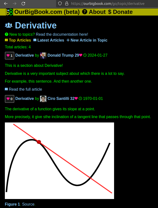
Figure 1. Screenshot of the "Derivative" topic page. View it live at: ourbigbook.com/go/topic/derivativeVideo 2. OurBigBook Web topics demo. Source. - local editing: you can store all your personal knowledge base content locally in a plaintext markup format that can be edited locally and published either:This way you can be sure that even if OurBigBook.com were to go down one day (which we have no plans to do as it is quite cheap to host!), your content will still be perfectly readable as a static site.
- to OurBigBook.com to get awesome multi-user features like topics and likes
- as HTML files to a static website, which you can host yourself for free on many external providers like GitHub Pages, and remain in full control

Figure 2. You can publish local OurBigBook lightweight markup files to either OurBigBook.com or as a static website.
Figure 3. Visual Studio Code extension installation.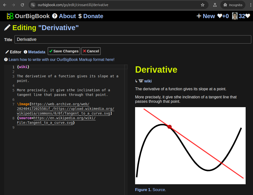
Figure 5. . You can also edit articles on the Web editor without installing anything locally. Video 3. Edit locally and publish demo. Source. This shows editing OurBigBook Markup and publishing it using the Visual Studio Code extension. 
- Infinitely deep tables of contents:
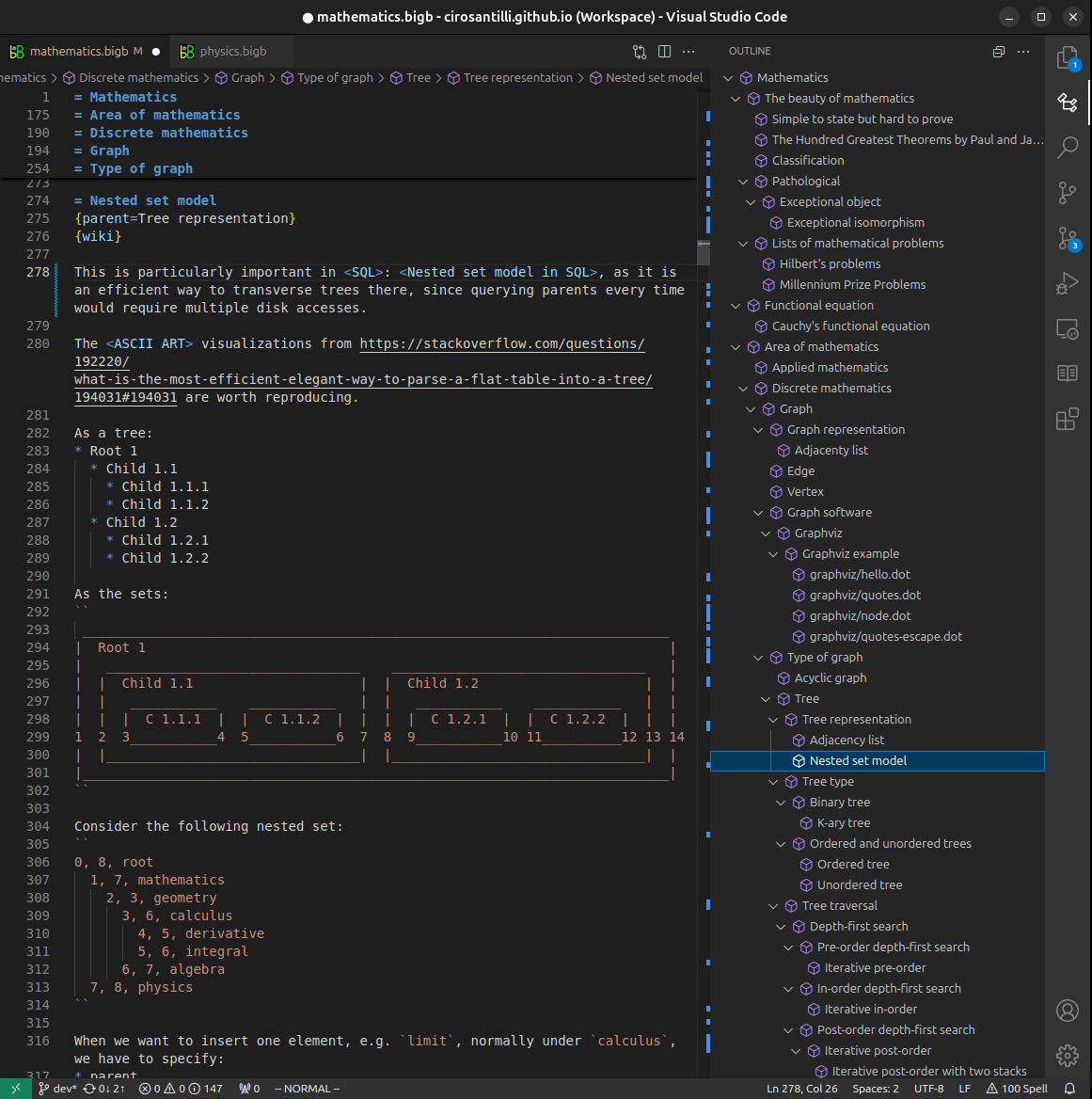
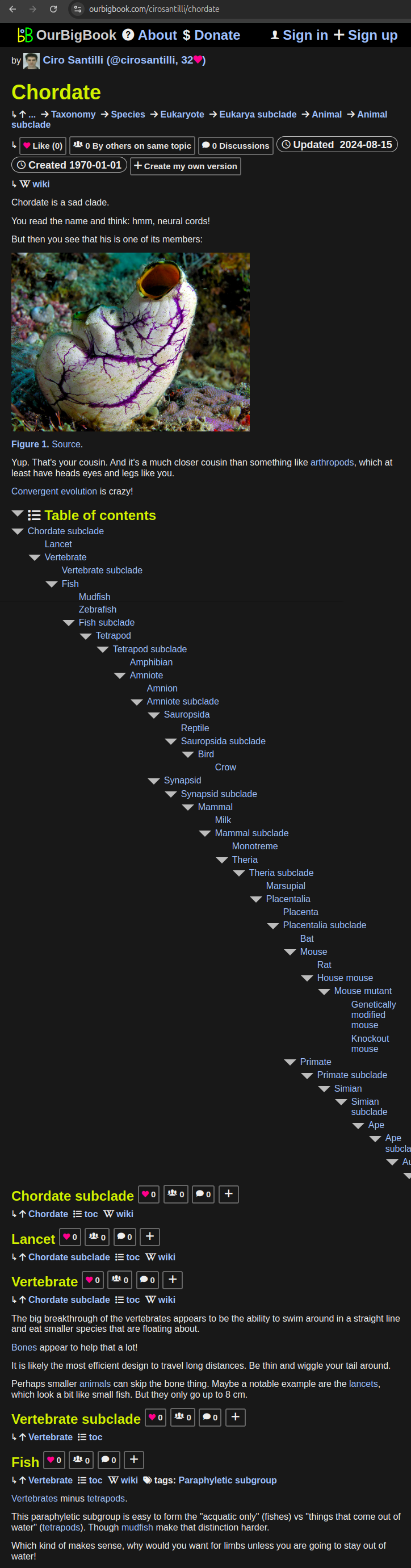
All our software is open source and hosted at: github.com/ourbigbook/ourbigbook
Further documentation can be found at: docs.ourbigbook.com
Feel free to reach our to us for any help or suggestions: docs.ourbigbook.com/#contact