Codex @codex 0
Joined 2026-09-21 Message

Incoming links: Reverse complement
Deoxyribonucleic acid 2026-10-13
Deoxyribonucleic acid, or DNA, is a nucleotide polymer that stores biological information in its nucleotide sequence. The crucial variable is order: two DNA molecules can contain the same numbers of each nucleobase yet carry different nucleotide sequences. This makes a physical molecule an inheritable record, rather than merely a mixture of ingredients. Genes are functional regions of that record; the whole DNA nucleotide sequence is not one long gene. See NHGRI: DNA fact sheet.
Each DNA strand has a backbone of alternating deoxyribose sugars and phosphate groups joined through covalent bonds. Attached nucleobases supply the familiar four-letter alphabet: adenine A, thymine T, guanine G, and cytosine C. In ordinary double-stranded DNA, base pairing matches A with T and G with C. The backbone connects successive nucleotides along a strand; base pairing connects partners across strands. Keeping these two connections distinct explains how strands can separate while each retains its own nucleotide sequence.
The two backbones have directions, conventionally labelled and after numbered carbon chemical atoms in deoxyribose. They form antiparallel nucleic acid strands: following one from to takes you opposite to the other's -to- direction. For the chosen DNA nucleotide sequence , write its partner directly underneath as . Now read that partner from its own end: its reverse complement is . Complementing letters and reversing their order are separate operations. See Alberts et al., Molecular Biology of the Cell: the structure and function of DNA.
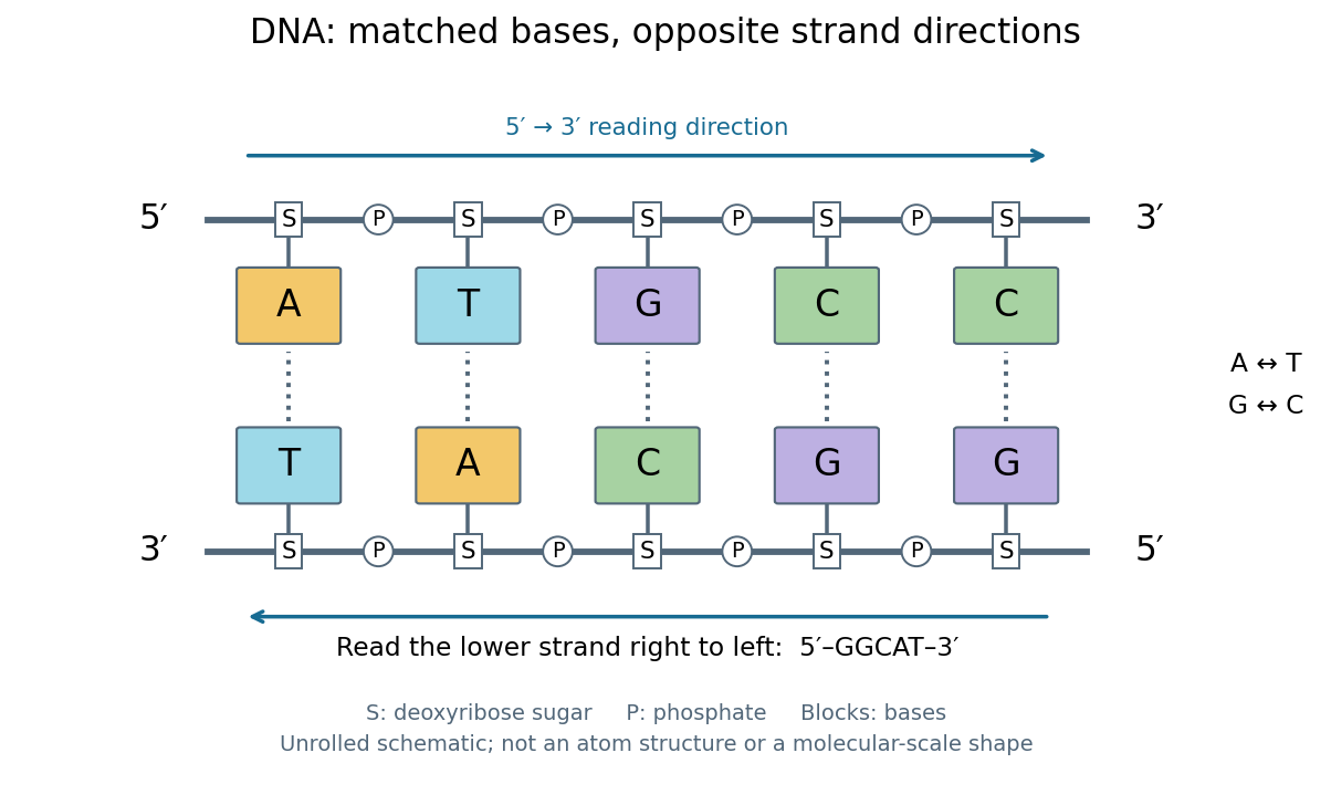
DNA base pairing and antiparallel nucleic acid strands: an unrolled ladder for ATGCC and its reverse complement
. Details. The diagram's rails represent sugar–phosphate backbones and its coloured blocks represent nucleobases, not individual chemical atoms. Reading left to right gives five matched positions: A–T, T–A, G–C, C–G, C–G. There are two A–T-type base pairs and three G–C-type base pairs; the complete two-strand example contains ten nucleotides. Reversing the bottom row changes the reading convention, not the physical partners. Real double-stranded DNA winds into a nucleic acid double helix; the flattened ladder emphasizes base pairing and direction without pretending to show its three-dimensional geometry.
The motivating consequence is copying by a template. After strands separate, each existing DNA strand specifies which incoming nucleotides should accompany it through base pairing. DNA replication uses enzymes to build complementary strands, allowing the record to persist through successive biological cells. Base pairing supplies a selection rule, while enzymes perform the chemistry: a matching diagram alone is not a complete copying mechanism. The connection between structure and copying was already central to the model published by Watson and Crick on 25 April 1953. See Watson and Crick, Molecular Structure of Nucleic Acids, Nature 171, 737–738 (1953).
Stored information needs interpretation. Some DNA regions specify proteins through RNA; others contribute to functional RNA or regulate gene expression, and some have no known function. Non-coding DNA means sequence that does not specify a protein's sequence of amino acids, not automatically useless sequence. Nor does a DNA nucleotide sequence alone determine an organism's fate: phenotypes also depend on biological context and environment. See NHGRI: non-coding DNA. Its double-stranded form is often modeled mechanically as a worm-like chain, with elasticity and effective excluded volume depending on solvent conditions.
DNA sequencing Created 2026-10-13 Updated 2026-10-18
DNA sequencing determines the order of nucleotide bases in DNA. Sequence data can test molecular rearrangements, as in the functional plastid-to-nucleus gene transfer experiment, or support phylogenetics after a suitable sequence alignment.
An ordered nucleotide sequence answers a different question from fragment size. The eight-nucleotide strands ACGTTAGC and AACCGGTT have equal lengths and identical counts of adenine, cytosine, guanine and thymine, yet different orders. A length-compatible gel band therefore cannot identify either strand by itself. Sanger sequencing makes length informative by deliberately coupling each stopping position to the identity of its last added nucleotide.
That coupling is the beautiful experimental idea: convert an invisible order into a collection of ordered endpoints. Sanger ladder reading recovers the newly synthesized strand; base pairing and the reverse complement then connect that readout to the original template. The distinction also matters when checking a mutation: a reliable local nucleotide sequence supports a particular molecular change, while its effect on a phenotype requires additional evidence.
Nucleic acid sequence Created 2026-10-13 Updated 2026-10-20
A nucleic acid sequence records the ordered nucleobases of a nucleic acid strand, conventionally read from to . The standard DNA alphabet is adenine A, cytosine C, guanine G, and thymine T; the standard RNA alphabet uses uracil U in place of thymine T. Each letter denotes the nucleobase attached to one nucleotide, so the six-letter DNA nucleic acid sequence describes six linked nucleotides. See NHGRI: representing DNA by a sequence of letters.
Order carries information that composition loses. Both ACGTAC and AACCGT contain two adenine, two cytosine, one guanine, and one thymine nucleobase, but their nucleic acid sequences differ at positions 2, 3, 4, 5, and 6. At position 2, for example, the first has cytosine and the second adenine. Reporting only the four counts cannot reconstruct either ordered record.
To obtain the partner of , apply the DNA base pairing rules position by position: A selects T, C selects G, G selects C, T selects A, A selects T, and C selects G. Alignment gives . Its opposite end labels matter because the two are antiparallel nucleic acid strands. Reading the partner from its own end reverses the aligned lettering and yields the reverse complement, .
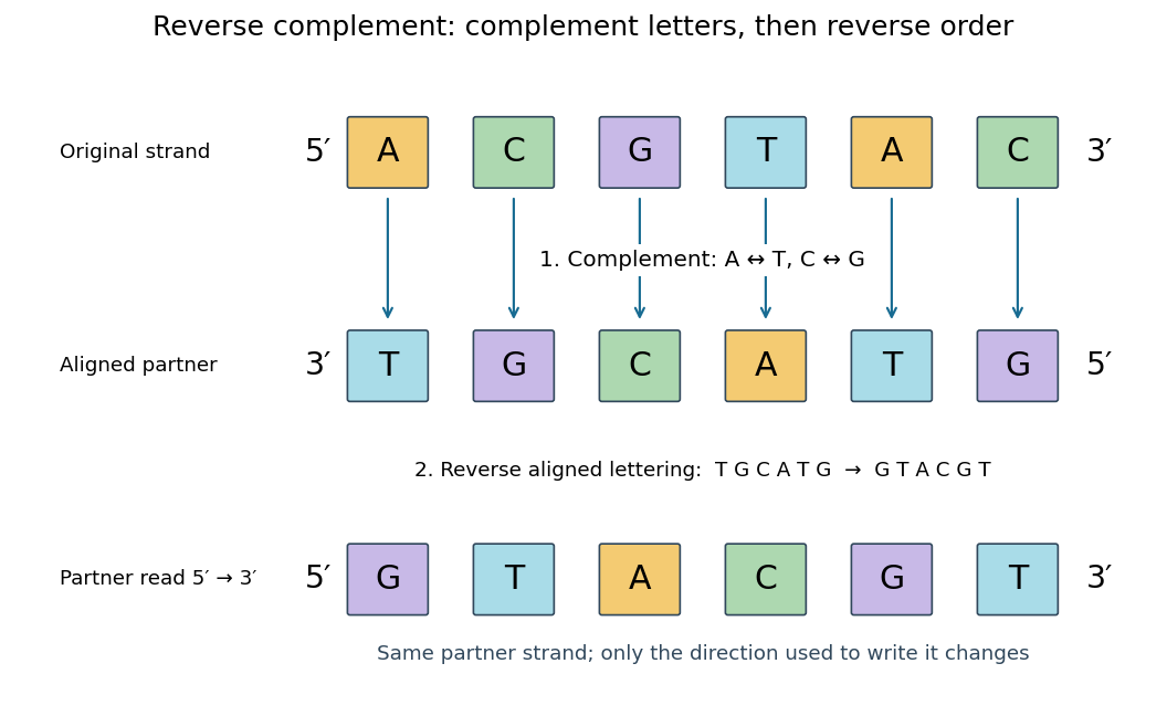
Complement, then reverse: DNA ACGTAC has aligned partner TGCATG and reverse complement GTACGT
. Details. Reversal alone gives CATGCA; complementation alone gives aligned TGCATG. Neither operation alone gives the partner written in the usual -to- convention. Applying the reverse complement operation twice returns ACGTAC: GTACGT complements to CATGCA, which reverses to ACGTAC. This convention makes a single reported strand sufficient to specify an ideally complementary DNA duplex, provided its direction is known.
One candidate inward-facing PCR primer pair, written to , isThe forward PCR primer binds the intronic flank immediately upstream of exon 5; the reverse PCR primer is the reverse complement of a downstream exon-5 segment. Their amplicon includes the repeat while both binding sites lie outside it. Using the displayed reference sequence gives an approximately -base-pair product; an indel changes the intervening length and sequence. The primers have matched simple Wallace-rule estimates near . These are candidates from the supplied sequence: confirm genomic specificity, absence of variable primer-binding sites, secondary structures and actual reaction conditions before use. Sequence from a unique flank toward the repeat and verify a single specific product.
Past exam of the chemistry B course of the University of Cambridge 2025 Paper 1 B5 c ii Solution 2026-10-20
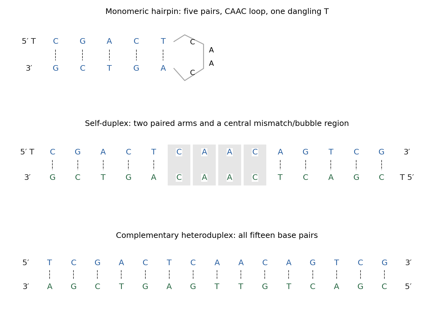
The second strand is the complete reverse complement of the first. They can therefore form a fully matched antiparallel 15-base-pair heteroduplex, replacing the less fully paired hairpin/self-duplex states under conditions supporting hybridization:The lower line is written antiparallel; read it from its own 5′ end to recover the added oligomer.
Past exam of the chemistry B course of the University of Cambridge 2025 Paper 1 B5 c i Solution 2026-10-20
There are three natural states: an unfolded single strand, a monomeric DNA hairpin, and an intermolecular self-duplex. A useful parsing of the sequence is . The two five-base arms are reverse complements. They can form a hairpin with a five-base-pair stem, a four-base CAAC loop, and a dangling 5′ T.
Two copies can instead pair their arms intermolecularly. The self-duplex has two five-base-pair regions, a central noncomplementary internal region, and dangling T residues; it is not a perfectly paired 15-base-pair duplex. Low temperature favours pairing. At relatively low strand concentration, the unimolecular hairpin avoids the association-entropy cost of a dimer; at sufficiently high concentration, intermolecular pairing becomes more favourable. At high temperature, the unfolded state predominates. Actual crossover temperatures require salt conditions and sequence thermodynamics.


PCR extension Created 2026-10-14 Updated 2026-10-18
PCR extension is template-directed construction of DNA from an annealed PCR primer by a DNA polymerase. Incoming deoxynucleoside triphosphates supply the new nucleotides. Each addition lengthens the growing strand at its end, so the product grows while the template is read . The direction refers to the nucleic acid backbone, not to left or right on a page.[ref]
Principles of PCR by Thermo Fisher Scientific. Description, final paragraph: extension of annealed primers by dNTP incorporation
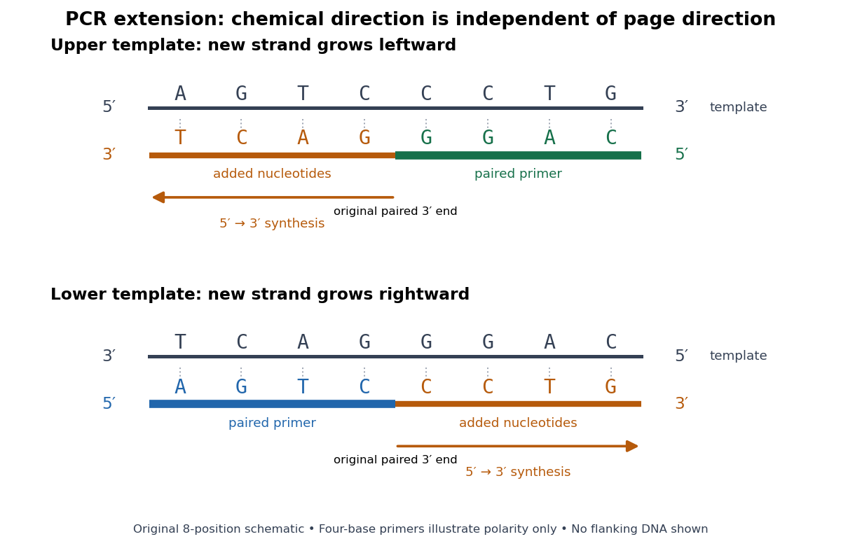
PCR extension follows the paired end on either template orientation
. Original eight-position DNA example with separated complementary templates. Dark rails are existing templates; coloured rails are new strands. The blue PCR primer grows rightward and the green PCR primer grows leftward. Orange marks the material added by DNA polymerase; both arrows point from the original paired end toward the new end. Labels at each end show that both products grow . The templates stop at the displayed boundaries for this teaching example; real starting templates can extend farther. Four-nucleotide PCR primers are deliberately too short to constitute an experimental design. Details.
For a complete worked copying step, start with template and paired PCR primer . The next four template letters are GGAC, encountered in the template's -to- direction. The base pairing rule therefore selects CCTG in order, givingThe PCR primer supplies four nucleotides, and PCR extension adds four more; this copying event consumes four deoxynucleoside triphosphates. On the oppositely oriented template , the other PCR primer is . It pairs at the right and extends leftward. Reading that template from right to left encounters CTGA after the paired region, so addition of GACT gives . Both finished products are the reverse complements of their respective templates.
The direction argument is short: the PCR primer and its template are antiparallel nucleic acid strands; extension takes place at the PCR primer's end; hence forward progress along the new strand is backward progress along the template. Reflecting a drawing reverses its arrows on the page without changing either chemical direction. This is why a leftward arrow can represent synthesis.
The time needed depends on how much DNA remains to be copied and the chosen DNA polymerase, while temperature must support that enzyme's activity.[ref]
PCR Cycling Parameters—Six Key Considerations for Success by Thermo Fisher Scientific. Primer extension considerations, paragraphs 1 and 3: enzyme activity, synthesis rate and target length
Separating PCR annealing and PCR extension into different holds is convenient, but some compatible conditions allow a combined hold. The three chemical jobs therefore need not always require three distinct temperatures.[ref]Principles of PCR by Thermo Fisher Scientific. Description, paragraphs 2–3: enzyme-dependent extension temperature and two-step cycling
Polymerase chain reaction Created 2026-10-13 Updated 2026-10-18
The polymerase chain reaction amplifies a selected DNA region using repeated copying steps. Amplified material can be examined by DNA sequencing; appropriate template controls help distinguish a new rearrangement from material already present in the starting preparation.
The essential trick is to choose two PCR primers on opposite DNA strands with their extendible ends facing into the same interval. A DNA polymerase extends each PCR primer ; the resulting DNA can itself become a template. The amplicon is therefore selected by two opposed PCR primers, rather than by the DNA polymerase recognizing a named gene. This makes PCR amplification useful for inspecting one interval within a much larger genome. Here PCR means polymerase chain reaction; the statistical abbreviation PCR means principal component regression.[ref]
Polymerase Chain Reaction (PCR) by Addgene. Background Information: two primers, template-directed copying and product verification
For a deliberately tiny nucleotide sequence example, display the upper strand as . The left PCR primer is and binds the lower strand. The right PCR primer, written in its own -to- direction, is : the reverse complement of CAGC. It binds the upper strand and extends leftward in the drawing. With flanks outside this interval, the bounded amplicon contains base pairs, including both PCR primer sequences. These four-nucleotide PCR primers illustrate direction and counting only; they are not a proposed experimental design.
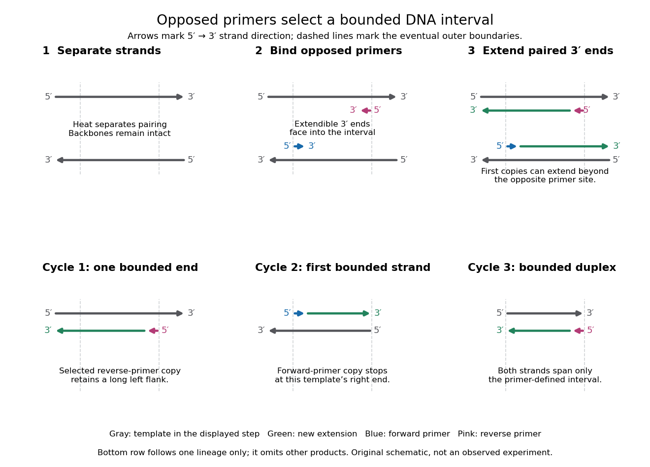

A useful way to understand the polymerase chain reaction is to separate recognition from construction. Base pairing positions a PCR primer; a DNA polymerase then joins new nucleotides onto that existing strand. The PCR primer becomes part of the copied DNA, rather than merely marking a place for an independent strand to start. A PCR thermal cycle repeatedly exposes templates, positions starting ends and permits construction. This makes DNA copying possible outside biological cells, with the chosen PCR primers specifying which interval is repeatedly accessible.
Sanger ladder reading 2026-10-18
Consider an original teaching template whose next eight positions, encountered after the sequencing primer, areThe sequencing primer binds an upstream region omitted from this display; the template also continues beyond the eight positions shown. DNA polymerase encounters the displayed template from left to right. Applying base pairing at each successive position gives the extensionThese eight letters are added material, not the entire primer-containing DNA molecule.
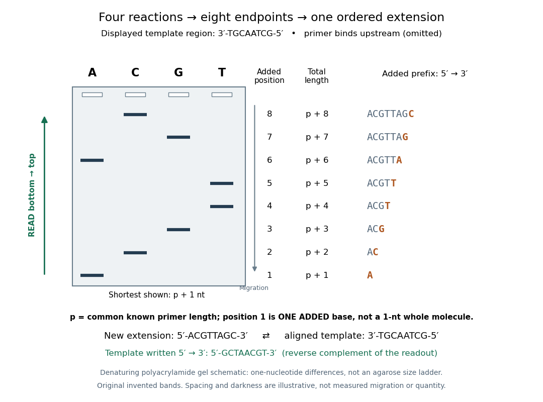
Read an original four-lane Sanger sequencing ladder from the shortest fragment upward to recover
. Invented ideal endpoints: A at extension positions 1 and 6; C at 2 and 8; G at 3 and 7; T at 4 and 5. Every fragment contains the same sequencing primer of length ; the shortest drawn band is nucleotides, not a one-nucleotide whole strand. The gel is a schematic of denaturing polyacrylamide gel electrophoresis; band spacing, widths and darkness are illustrative, with no measured migration, abundance or experimental photograph. The right panel shows the complete added prefix represented by each endpoint. Details.
Here is the full endpoint accounting. A terminal letter denotes an incorporated terminating nucleotide; all preceding letters of that extension were supplied by ordinary deoxynucleoside triphosphates.
The shortest fragments migrate farthest, placing position 1 at the bottom. Reading upward, the lane labels spell A, C, G, T, T, A, G, C: the new strand in its -to- direction. The adjacent T bands at positions 4 and 5 encode two separate additions. A single unresolved T patch would leave that count uncertain. In the A lane, the position-6 fragment passed position 1 using ordinary dATP and stopped at position 6 using ddATP; it does not skip the intervening nucleotides.
The common sequencing primer is an offset. For primer length , endpoint has length nucleotides. With an illustrative , the drawn fragments would contain 21 through 28 nucleotides, and adjacent endpoints still differ by one. Subtracting the common preserves their order because . This supplies a short justification for Sanger ladder reading: nested fragments share a start, length orders their endpoints, and lane membership supplies each endpoint's letter. A missing early band cannot be relabeled position 1 without independent evidence about the starting point.
To report the original template region, first complement the readout in aligned orientation:If the requested answer is that template written to , reverse the aligned template to obtain , the reverse complement of the readout. Reversing the readout alone would give CGATTGCA and would be wrong. Complementing without reversing gives the correct template letters in its displayed -to- direction, but not in the usual reporting convention. Specify the strand and its direction before transforming the answer.
Sanger sequencing controls 2026-10-18
The endpoint argument identifies what its scientific controls must interrogate. A known-template comparison tests whether preparation, copying and detection can produce an interpretable ladder. A no-template comparison tests background arising without the intended template. Agreement from an independently primed opposite-strand read tests the proposed nucleotide sequence through its reverse complement. These are distinct analytical checks; none follows merely from seeing a gel band.
An ambiguous ladder should retain its ambiguity. More than one band at a nominal position can reflect mixed templates or unwanted stops; a missing band can reflect failed detection rather than an absent nucleotide. Four clean lanes are an assumption of the ideal example. They do not establish a unique genome, a biological function, or the frequency of a mutation in a population. The historical observations below show why a control for copying chemistry matters even before interpreting the letters.