Biological translation uses a ribosome to read messenger RNA codons and assemble the corresponding amino acid chain. Transfer RNA adaptors match codons through base pairing and carry the amino acids; the genetic code supplies the correspondence. The chain grows from the N-terminus toward the C-terminus, by adding each new residue at the C-terminus. See Clark, Douglas and Choi, Biology 2e: ribosomes, transfer RNA and protein synthesis.
In the gene expression diagram, read in the chosen reading frame. Under the standard genetic code, AUG specifies methionine and GCU specifies alanine; UAA is a stop codon. The result before any subsequent processing is the two-residue chain N–methionine–alanine–C, followed by termination. The stop codon contributes zero amino acids. See Clark, Douglas and Choi, Biology 2e: the standard genetic code. This example assumes appropriate initiation at the displayed AUG in a suitable context in a eukaryote; the diagram displays the coding segment, not a complete independently translatable messenger RNA.
Initiation establishes the starting position and reading frame, using a ribosome, an initiating transfer RNA and additional factors. At a standard stop codon, a release factor promotes release of the chain rather than delivery of another amino acid by transfer RNA. See Clark, Douglas and Choi: initiation and termination of translation. A short two-residue example illustrates decoding, not the size or folding of a typical functional protein.
The useful connection is between two different alphabets: nucleotides specify the order of amino acids through the genetic code. Base pairing helps select transfer RNA, while the ribosome catalyzes joining the delivered amino acids. The messenger RNA remains a template rather than being consumed as the chain's building material. Separating template, adaptor and product makes clear why transcription and biological translation are distinct mechanisms.
Reduction of protein production from an RNA without requiring an initial decrease in its abundance. RNA stability and translation measurements are needed to distinguish coupled effects.
Assembly and positioning of the biological translation machinery at a selected start. ribosome recruitment, scanning and start-codon recognition are distinguishable processes.
A biological translation inhibitor with preferential effects on early ribosomes, used in initiation-site profiling. Peak size need not equal initiation flux.
A biological translation inhibitor used to enrich ribosomes near initiation sites while much ongoing elongation runs off. Exposure time and trapping kinetics affect inferred initiation positions.
Initiation after a preceding biological translation event on the same RNA, often involving retention of a small subunit and reacquisition of required factors. Upstream reading-frame effects depend on these kinetics.
Bypass of a potential initiation site by a scanning small-subunit complex, allowing initiation farther downstream. Local context and factor availability affect the probability of selection.
A reagent that can release nascent chains by accepting the growing peptide, useful as a control for translation-site signals. Global treatment also perturbs cellular physiology.
Continuation of biological translation past a stop codon through incorporation of an amino acid instead of normal termination.
Movement of the messenger-RNA–tRNA complex relative to the ribosome after peptide-bond formation, advancing the coding position and repositioning tRNAs.
A bacterial GTPase assisting recycling of class-I release factors. The release factor catalyses peptidyl-tRNA hydrolysis; GTP cleavage is not itself the peptide-release reaction.
A bacterial GTPase promoting ribosomal translocation. Its nucleotide-exchange kinetics differ from those of EF-Tu, so it does not require EF-Ts for recycling.
The eukaryotic elongation GTPase delivering aminoacyl-tRNA to the ribosome. Its nucleotide cycle is regenerated by eEF1B exchange activity.
Increased discrimination through successive kinetically controlled selection steps coupled to an energy-consuming reaction. Ribosomal tRNA selection includes rejection before and after GTP hydrolysis, improving fidelity beyond a single equilibrium binding comparison.
In a generic two-stage selection model with correct and incorrect dissociation rates and , the displayed error ratio increases with either common forward rate. Slow forward transitions allow rejection at both stages, approaching discrimination , but zero forward rate gives zero output. A high-yield window can instead lie between well-separated correct and incorrect rejection rates. A finite useful optimum needs a throughput or yield objective as well as an error ratio.
A bacterial translational GTPase delivering aminoacyl-tRNA to the ribosome. Correct decoding promotes its hydrolysis cycle; EF-Ts accelerates nucleotide exchange for reuse.
Translational coupling makes translation of one coding region influence translation of another on the same transcript. One mechanism is unfolding of a structure masking the downstream initiation region by an upstream translating ribosome.
A Shine-Dalgarno sequence is a bacterial messenger-RNA element that can pair with the anti-Shine-Dalgarno region of small-subunit ribosomal RNA to position initiation. Base pairing within the messenger RNA can make the element unavailable.
A ribosome-binding site is a transcript region involved in recruiting or positioning a ribosome for initiation. In many bacteria it includes a Shine-Dalgarno sequence, but accessibility and start-codon context matter and not all bacterial initiation uses that motif.
A cap-associated initiation-factor complex including eIF4E and eIF4G, supporting recruitment and initiation on many messenger RNAs.
An initiation-factor scaffold participating in assembly of eIF4F.
An eIF4E-binding regulator. Hypophosphorylated forms can restrict eIF4F assembly; phosphorylation downstream of mTORC1 reduces this inhibition.
A translated reading region upstream of a main coding region in a messenger RNA. Its effects depend on initiation, termination, scanning and reinitiation, rather than its mere presence.
The nucleotide-exchange factor that promotes replacement of GDP by GTP on eIF2. Inhibition of this exchange reduces the supply of initiation-competent ternary complex.
A eukaryotic initiation factor carrying initiating methionyl-tRNA in a GTP-bound ternary complex. Its alpha-subunit phosphorylation can inhibit nucleotide exchange by eIF2B and lower initiation capacity.
Processes recognizing problematic stalled ribosomes and managing their incomplete polypeptides and associated components.
Insufficient availability of one or more amino acids relative to cellular requirements. Its translational consequences include substrate limitation and regulated changes in initiation and stress adaptation.
The genetic code assigns RNA codons to amino acids or termination signals. Four possible nucleobases at each of three positions give codons. In the standard genetic code, 61 specify amino acids and three are stop codons; several codons can specify the same amino acid. Variants exist, so state which genetic code is being used. See Clark, Douglas and Choi: genetic code assignments and variants.
A codon is a three-nucleotide unit read in a specified reading frame during biological translation. For example, GCU specifies alanine in the standard genetic code. Its letters are read .
For an original example, choose the messenger RNA segment and stipulate initiation at the displayed AUG in a suitable eukaryotic context. Under the standard genetic code, its first three codons specify methionine, alanine and phenylalanine; the fourth is a stop codon. The resulting peptide before processing is N–methionine–alanine–phenylalanine–C. Twelve nucleotides therefore specify three amino acid residues and termination. The displayed segment omits the surrounding initiation signals; its nucleotide sequence alone is not a complete expression system.
A codon belongs to an oriented nucleotide sequence: GCU and UCG are different ordered triplets. Four possible nucleobases, adenine, cytosine, guanine and uracil, allow triplets because each position can be chosen independently when enumerating possible sequences. This is a counting argument, not a claim that nucleobases occur independently in real RNA. Sixty-one triplets are sense codons, meaning amino acid assignments, and UAA, UAG and UGA are stop codons in the standard genetic code.[ref]
Biology 2e by Mary Ann Clark and others. §15.1, The Genetic Code, Figure 15.4 and following paragraph: triplets and standard assignments
The useful unit is the ordered triplet together with its reading frame and genetic code, rather than three letters viewed in isolation. That combination lets DNA sequencing data support predictions about protein sequences after identifying the relevant messenger RNA. The Nirenberg and Matthaei experiment illustrates how the correspondence became experimentally accessible.
A codon used to begin a translated reading frame. AUG is common, but initiation at near-cognate codons can occur in an appropriate context.
A codon position synthesized as a nucleotide mixture, producing several codons across a library. Equal nucleotide mixture proportions do not generally produce equal amino-acid frequencies.
A library codon with any base at the first two positions and G or C at the third. Its 32 codons represent the 20 standard amino acids unequally and include one stop codon.
A library codon with any base at the first two positions and G or T at the third. Its 32 codons represent the 20 standard amino acids unequally and include one stop codon.
In the standard genetic code, UAA, UAG and UGA are stop codons: they signal termination of biological translation rather than an amino acid assignment. See Clark, Douglas and Choi: stop codons.
A stop codon supplies no additional amino acid residue. In the constructed AUG GCU UUC UGA example, replacing UGA by UAA preserves termination after the same three amino acid residues. Replacing UUC by UAA instead terminates after methionine and alanine. These are consequences of the stated genetic code, not measurements of expression efficiency.
At a standard stop codon, a release factor promotes release of the polypeptide from the ribosome. Ordinary delivery of an amino acid by transfer RNA does not supply a “stop amino acid”.[ref]
Biology 2e by Mary Ann Clark and others. §15.5, Translation, Elongation, and Termination, paragraph beginning “Termination of translation”: stop codons and release factors
A release factor is a protein involved in terminating biological translation and releasing the completed polypeptide at a stop codon.[ref]
Biology 2e by Mary Ann Clark and others. §15.5, Translation, Elongation, and Termination, paragraph beginning “Termination of translation”
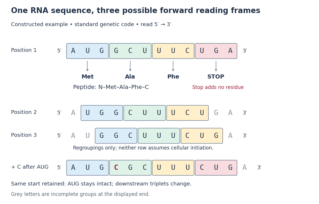
A reading frame specifies how consecutive nucleotides are grouped into codons. Starting at position 1 of AUGGCUUAA gives AUG / GCU / UAA; starting at position 2 gives UGG / CUU with AA left over. The letters are unchanged but the groups differ. A possible grouping alone does not establish an actual biological translation initiation site.
For the twelve-nucleotide example, number positions from the end. The three possible forward reading frames start at positions 1, 2 and 3:Parentheses enclose incomplete trailing groups. The second and third rows do not assert initiation of biological translation in a biological cell. A chosen start fixes which positions belong together; the ribosome then advances through consecutive codons rather than searching each three-letter window independently.
A sequence interpreted in a particular codon frame without an internal in-frame stop, with the boundary convention stated. Prediction of an open reading frame alone does not demonstrate biological translation or a stable functional product.
A relatively short potential coding region under an explicitly chosen length convention. Evidence of biological translation and evidence for a stable functional peptide are different.
A change of reading frame during biological translation. It need not change the underlying DNA sequence, unlike a frameshift mutation. It can be programmed by a recoding signal or occur as a translation error.
Programmed ribosomal frameshifting changes the reading frame during translation at a specific recoding signal. A slippery sequence and a downstream RNA structure can bias the event, allowing one transcript to produce different polypeptides in controlled proportions.
A frameshift mutation changes downstream grouping into codons through insertion or deletion of a number of nucleotides not divisible by three. In the constructed messenger RNA example, insert C immediately after AUG while retaining the same initiation position: . The first codon is preserved, but the original downstream groups GCU, UUC and UGA disappear. The displayed fragment now has no complete standard stop codon; what happens later depends on its continuation.
The short proof is positional: every downstream nucleotide moves by positions after an insertion of length . Boundaries recur every three positions, so they retain the original phase exactly when is divisible by three. An insertion of three nucleotides can still change the peptide, but it does not shift the downstream reading frame. This distinction helps interpret genetic variants without predicting a particular phenotype from grouping alone.[ref]
Biology 2e by Mary Ann Clark and others. §15.1, The Genetic Code, Figure 15.5: nucleotide deletion and shifted grouping
Degeneracy of the genetic code means that distinct codons can specify the same amino acid. For example, UUU and UUC both specify phenylalanine in the standard genetic code.[ref] In the original AUG GCU UUC UGA example, changing UUC to UUU preserves the predicted amino acid order. This illustrates redundancy of assignment, not equality of every possible effect on gene expression.
Degeneracy of the genetic code differs from ambiguity: multiple inputs share one output, whereas an ambiguous assignment would give a single codon more than one meaning in the same specified decoding context. The standard genetic code table has one listed assignment per triplet. Alternative genetic codes, including those used in some mitochondria, and engineered decoding contexts must be specified separately; the standard table is not a universal statement about all organelles or synthetic systems.
In 1961, Marshall Warren Nirenberg and J. Heinrich Matthaei ground washed Escherichia coli and used centrifugation to prepare a cell-free extract. Reactions at combined ribosomes, soluble components, ATP regeneration and phenylalanine carrying a isotope label, with or without polyuridylic acid, an RNA containing only U. After incubation, trichloroacetic acid precipitation, washing and counting radioactive decay detected label in insoluble material. Added polyuridylic acid strongly increased incorporation; removing ribosomes or ATP regeneration suppressed it. Product characterization supported a phenylalanine polypeptide.[ref]
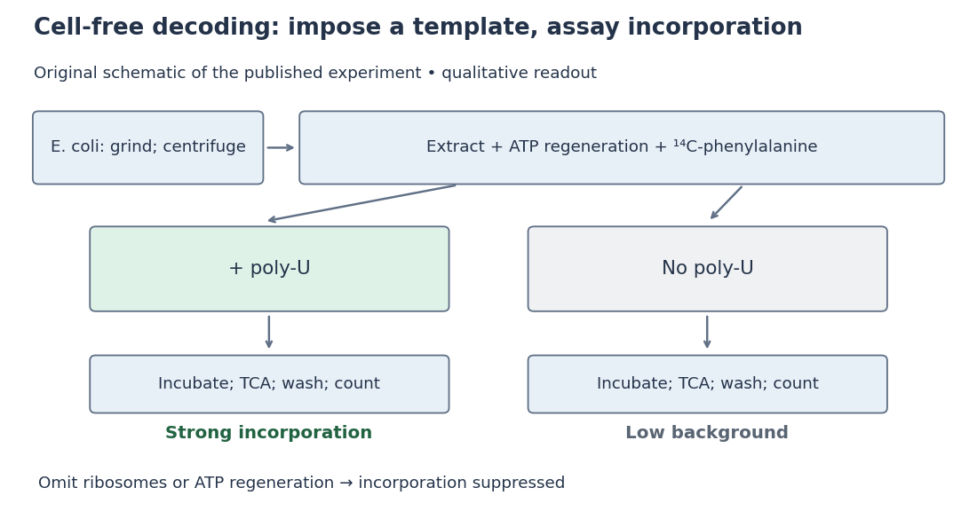
Nirenberg and Matthaei experiment: preparation, comparison and readout
. Original schematic; readout summarizes qualitative published observations. Details.The inference connects an imposed RNA template to a specific amino acid product. Yet repeated U alone cannot distinguish one-, two- or three-letter coding units: every possible group still contains only U. Neither triplet length nor the complete genetic code follows.[ref]
The dependence of cell-free protein synthesis in E. coli upon naturally occurring or synthetic polyribonucleotides by Marshall Warren Nirenberg and J. Heinrich Matthaei. p. 1601, Discussion and Note added in proof: coding-unit length not established
Cell-free translation uses isolated ribosomes and soluble components to make polypeptide chains from messenger RNA outside an intact biological cell.
Articles by others on the same topic

In biology, translation is a crucial process in the expression of genes, where the genetic code carried by messenger RNA (mRNA) is translated into a polypeptide chain, forming proteins. This process occurs in the ribosomes, which can be found in the cytoplasm or on the endoplasmic reticulum in eukaryotic cells.


