Mitosis allocates copied chromosomes to daughter cell nuclei. The spindle apparatus aligns sister chromatids before separation in anaphase.
The copying has already occurred during S phase, before mitosis. Nuclear chromosome segregation and cytokinesis, the partition of the cytoplasm, are distinct processes: two daughter cell nuclei can form inside one undivided biological cell.[ref]
The Cell Cycle. Biology 2e by Mary Ann Clark and others; §10.2, Interphase and The Mitotic Phase; Figure 10.5: replication precedes nuclear division, which can occur without cytokinesis
The original cartoon follows a hypothetical diploid animal biological cell with . Its two homologous chromosome pairs are and ; upper- and lower-case letters identify homologues, not dominant and recessive alleles. In prophase and metaphase, each replicated chromosome contains two sister chromatids: four replicated chromosomes contain eight chromatids, rather than eight replicated chromosomes. The X shape depicts two associated copies, not the shape of every chromosome throughout the cell cycle.
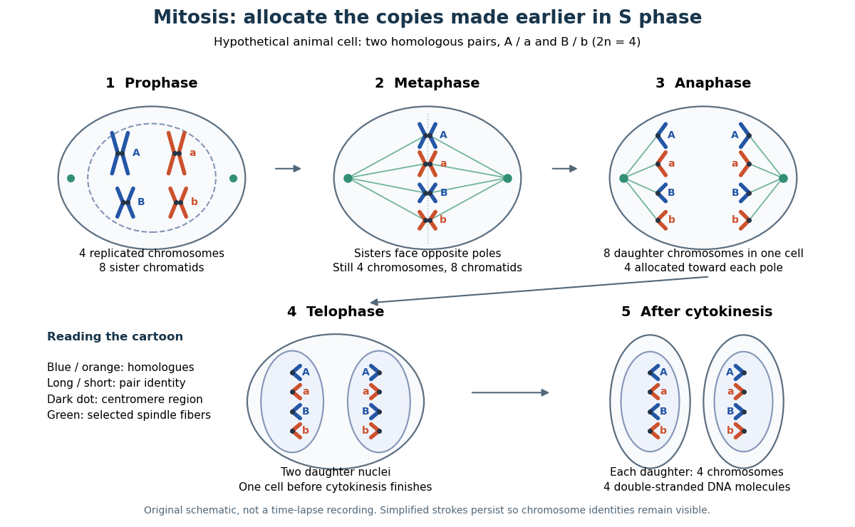

This open mitosis cartoon shows nuclear envelope breakdown and reassembly. Closed mitosis in some yeasts retains the nuclear envelope while daughter chromosomes separate inside it; the cartoon is therefore not a universal cellular architecture.[ref]
The Cell: A Molecular Approach, 2nd edition by Geoffrey M. Cooper. Chapter 8, The Nucleus during Mitosis, Dissolution of the Nuclear Envelope; Figures 8.29–8.30: open and closed mitosis
In open mitosis, nuclear envelope breakdown permits spindle apparatus microtubules to reach kinetochores at centromere regions.
In metaphase, replicated chromosomes align near the spindle apparatus equator, with their two sister chromatids connected through kinetochores toward opposite poles.
In mitotic anaphase, sister chromatids separate into daughter chromosomes moving toward opposite poles.
Release of cohesin-mediated association permits the sister centromere regions to separate. Each former chromatid is then counted as an independent daughter chromosome. This changes the counting convention without making additional DNA copies. For the example, the whole undivided biological cell transiently contains eight daughter chromosomes, four moving toward each pole. Movement toward poles and separation of the poles both contribute to partition in the illustrated animal setting.[ref]
The Cell Cycle. Biology 2e by Mary Ann Clark and others; §10.2, Karyokinesis, Figure 10.6 and Anaphase: release of cohesion, daughter chromosomes and spindle elongation
In open mitosis, telophase includes reassembly of nuclear envelopes around the separated chromosome groups and relaxation of their condensation.[ref]
Count the same hypothetical diploid animal biological cell in three ways: chromosomes, double-stranded DNA molecules, and nuclear DNA amount. Assume one continuous double-stranded DNA molecule per unreplicated chromosome or replicated chromatid, complete S phase, and no losses or mutations. Exclude mitochondrion DNA. Here is the size of one haploid set, and is its unreplicated nuclear DNA amount, as defined in cell-cycle DNA content. A double-stranded DNA molecule counts as one, despite containing two strands.
| Stage and compartment | Chromosomes | Double-stranded DNA molecules | Nuclear DNA amount |
|---|---|---|---|
| One G1 phase parent | 4 unreplicated | 4 | |
| One prophase or metaphase biological cell | 4 replicated | 8 in 4 pairs of sister chromatids | |
| Whole undivided anaphase biological cell | 8 separated daughter chromosomes | 8 | total |
| One anaphase pole's allocation | 4 daughter chromosomes | 4 | allocated |
| Each daughter after mitosis and cytokinesis | 4 unreplicated | 4 | each |
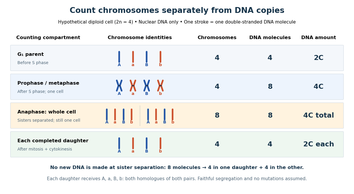

The allocation can be checked copy by copy. DNA replication turns into , into , and similarly for . One daughter receives and the other . Each therefore inherits the same four chromosome identities and the same gene and allele content as the unreplicated parent, under the stated assumptions. Across both daughters there are DNA molecules and : partition conserves the material present after S phase.
The transient whole-cell count of eight does not mean that each daughter's ploidy has doubled. Each completed daughter is diploid, containing both homologues of each homologous chromosome pair. This differs from separating homologous chromosomes during meiosis I. Faithful mitosis preserves a nuclear genome through growth and renewal; equal inherited genes need not imply equal gene expression or equal cell fate. Mutations or chromosome segregation errors can break the ideal inheritance pattern.
The spindle checkpoint can delay anaphase while kinetochore attachment is incomplete. This couples progression to the state of the spindle apparatus, rather than requiring every biological cell to separate its sister chromatids after a fixed waiting time. Alignment in a picture alone is not a demonstration that this control is satisfied.[ref]
Control of the Cell Cycle. Biology 2e, §10.3, Regulation at Internal Checkpoints, Figure 10.11 and The M Checkpoint
A checkpoint component participating in kinetochore-associated signalling and mitotic checkpoint complexes.
A checkpoint regulator involved in attachment surveillance and inhibitory complexes controlling anaphase-promoting complex–Cdc20.
A checkpoint component participating in inhibition of Cdc20 as part of the mitotic checkpoint complex.
A protein assembly that inhibits productive anaphase-promoting complex–Cdc20 activity while kinetochore-associated checkpoint signalling remains active.
In 1995, Conly L. Rieder, Richard W. Cole, Alexey Khodjakov and Greenfield Sluder tested whether an unattached kinetochore supplies the inhibitory signal delaying anaphase. Living PtK1 biological cells on glass coverslips were mounted in Rose chambers at –. An inverted microscope with a objective recorded differential interference contrast microscopy video. A pulsed laser focused through that objective destroyed selected chromosome regions. The readout was chromosome movement and anaphase onset before and after surgery.[ref]
The checkpoint delaying anaphase in response to chromosome monoorientation is mediated by an inhibitory signal produced by unattached kinetochores by Conly L. Rieder and others. pp. 941–942, Abstract and Materials and Methods: Cell Culture; Laser Microsurgery and Video-enhanced Light Microscopy
Destroying the last unattached kinetochore permitted anaphase without that chromosome aligning. Cutting chromosome arms served as a damage control: progression still awaited attachment of the last naturally one-pole-attached chromosome. Correlative electron microscopy checked the absence of the targeted kinetochore. Together these results located an inhibitory activity in or near the unattached kinetochore, rather than establishing a requirement for alignment itself.[ref]
The checkpoint delaying anaphase in response to chromosome monoorientation is mediated by an inhibitory signal produced by unattached kinetochores by Conly L. Rieder and others. pp. 943–947, arm-cutting control, Table I, Figures 1, 5 and 6; destruction of the last unattached kinetochore and correlative electron microscopy
This intervention localized control; it did not identify every signaling molecule or guarantee faithful inheritance after damage. The cultured PtK1 preparation differs from the hypothetical cartoon. Neither the cartoon nor this experiment establishes identical control in every species.
Cytokinesis partitions the cytoplasm. Without cytokinesis, two cell nuclei can share one biological cell. [ref]
Cytokinesis makes physically separate daughter biological cells by partitioning their cytoplasm and completing their cell membrane boundaries. Mitosis divides nuclear material. These processes can overlap during M phase, but completion of one does not imply completion of the other.[ref]
The Cell Cycle. Biology 2e by Mary Ann Clark and others; §10.2, The Mitotic Phase and Cytokinesis: nuclear division versus physical daughter-cell separation
For an original compartment-counting example, consider a hypothetical diploid biological cell after two daughter cell nuclei have formed. Let be the number of chromosomes in one haploid set, and let be that set's unreplicated nuclear DNA amount, as in cell-cycle DNA content. Assume faithful mitosis, no nuclear DNA replication during the partition, and no loss of nuclear DNA; exclude mitochondrion DNA.
| Counting boundary | Before cytokinesis | After cytokinesis |
|---|---|---|
| Biological cells | 1 | 2 |
| Cell nuclei in each biological cell | 2 | 1 |
| Each cell nucleus | chromosomes, | chromosomes, |
| Nuclear DNA per biological cell | ||
| Nuclear DNA across all descendants |
The check is to follow the two existing cell nuclei: initially one boundary encloses both; finally each daughter boundary encloses one. The total remains two cell nuclei, twenty chromosomes and of nuclear DNA. Thus the amount per biological cell halves because the counting boundary changes, without any new DNA replication or change of nuclear ploidy. Doubling the number of biological cells need not double their combined material.
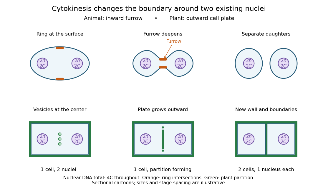

Coordinating chromosome segregation with cytoplasm partition lets descendants inherit nuclear information inside separate working compartments. Equal nuclear DNA allocations do not require equal daughter sizes or equal allocations of every organelle. The geometry of cytokinesis connects an internal allocation to the creation of two independently bounded biological cells.
The final membrane-separation event that resolves the intercellular bridge between daughter cells, distinct from earlier furrow ingression.
A structure within the late intercellular bridge that organises events associated with final cell separation.
An actomyosin ring is a contractile assembly containing actin filaments and myosin II. In typical animal cytokinesis, it forms in the cell cortex beneath the cell membrane. Its contraction draws the boundary inward to produce a cleavage furrow; an inward groove is not yet complete separation of the daughters.[ref]
The Events of M Phase. The Cell: A Molecular Approach, 2nd edition, Chapter 14, The Events of M Phase, Cytokinesis and Figure 14.30: actin–myosin II ring beneath the plasma membrane
A cleavage furrow is the inward indentation of the cell membrane during cytokinesis. It is the visible deformation; the actomyosin ring is machinery beneath that boundary.[ref]
The Cell Cycle. Biology 2e by Mary Ann Clark and others; §10.2, Cytokinesis, animal-cell paragraph and Figure 10.9
In 1961, Raymond Rappaport gently removed membrane and jelly from sand dollar ova with a constricted pipette. After fertilization, he used an inverted optical microscope and rack-and-pinion manipulator to press a needle-mounted glass sphere through an egg onto the chamber floor, producing a torus-shaped biological cell. Visible spindle apparatus positions and cleavage furrow progression were the readout.[ref]
Experiments concerning the cleavage stimulus in sand dollar eggs by Raymond Rappaport. Experiments concerning the cleavage stimulus in sand dollar eggs, pp. 81–82, Materials and Methods: egg preparation, inverted microscope and sphere manipulation
First cytokinesis opened one side into a horseshoe containing two cell nuclei. During the next division, daughters separated from its free ends; an additional cleavage furrow crossed between mitotic asters of different spindle apparatuses. Control eggs supplied the timing comparison. This region lacked intervening chromosomes or a previous nearby spindle apparatus. This supported mitotic aster-associated positioning, without identifying the signal or establishing universal necessity.[ref]
Experiments concerning the cleavage stimulus in sand dollar eggs by Raymond Rappaport. pp. 81–82, introductory experiment summary, Results §1 and Figure 1 legend: horseshoe, second division, control comparison and furrowing between separate mitotic figures
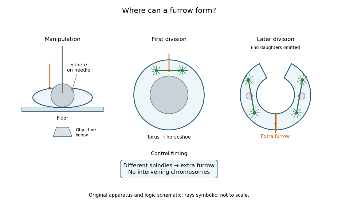
Manipulation and inference in the toroidal sand dollar egg experiment
. Original apparatus and logic schematic; mitotic aster rays are symbolic. Details.
A cell plate is an internal partition assembled during plant cytokinesis by fusion of transport vesicles. It grows outward to join the parental cell membrane, providing daughter boundaries and material for a new plant cell wall.[ref]
The Events of M Phase. The Cell: A Molecular Approach, 2nd edition by Geoffrey M. Cooper; Chapter 14, The Events of M Phase, Cytokinesis and Figure 14.31: vesicle fusion, outward expansion and joining the parental plasma membrane
Articles by others on the same topic
There are currently no matching articles.