RNA couples iron availability to the production of proteins that acquire, store, export and use iron. The clearest mammalian mechanism uses structured iron-responsive elements in untranslated regions and iron-regulated binding proteins. The RNA element encodes a regulatory address and its position determines the response; the main sensing machinery is protein-based, so this system should not be described as an autonomous iron-binding RNA riboswitch.
An iron-responsive element is a stem-loop with conserved sequence and structural features recognized by iron regulatory proteins. At low available iron, RNA-binding IRP1 and IRP2 are active. IRP1 is bifunctional: an iron–sulfur-cluster-containing form acts as cytosolic aconitase, whereas loss of the functional cluster permits high-affinity RNA binding. Biochemical disassembly of its cluster connected these two activities directly.[ref]
Cellular regulation of the iron-responsive element binding protein: disassembly of the cubane iron-sulfur cluster results in high-affinity RNA binding by David J. Haile and others. Cluster state and RNA-binding activity
IRP2 does not use the same aconitase switch. Under iron- and oxygen-replete conditions, the FBXL5 ubiquitin-ligase pathway promotes IRP2 degradation; instability of the iron-sensitive FBXL5 regulator during iron depletion helps preserve IRP2.[ref]An E3 ligase possessing an iron-responsive hemerythrin domain is a regulator of iron homeostasis by Ameen A. Salahudeen and others. FBXL5 sensing, stability and IRP2 degradation
Redox and oxygen conditions can therefore modify the relation between total cellular iron and RNA-binding activity.The canonical 5′ example is ferritin mRNA. Binding of an IRP near the cap prevents efficient recruitment of the translation-initiation machinery, lowering ferritin synthesis when iron is scarce. This avoids committing a limited resource to storage. When iron is plentiful, reduced IRP occupancy releases repression, allowing more ferritin to store iron and limit its reactive pool. Deletion analysis and transfer of a synthetic ferritin element to another reporter established a cis-acting signal sufficient to confer iron-responsive biological translation.[ref]
Identification of the iron-responsive element for the translational regulation of human ferritin mRNA by Matthias W. Hentze and others. Ferritin leader deletion and synthetic-element transfer
This response can change biological translation before a major change in transcript abundance occurs.At the 3′ end of transferrin receptor 1 mRNA, multiple IREs participate in a stability-control region. IRP binding during iron shortage protects the message from destabilization, increasing receptor production and iron uptake. At higher iron, loss of protection permits more rapid decay and reduces uptake. John L. Casey and colleagues showed that the transferrin-receptor 3′ region conferred iron-regulated RNA abundance and that isolated elements placed in a 5′ reporter context instead conferred translational control.[ref]
Iron-responsive elements: regulatory RNA sequences that control mRNA levels and translation by John L. Casey and others. Context-dependent reporter regulation by ferritin-like stem-loops
Thus similar recognition chemistry supports opposite outputs through different transcript positions and associated machinery.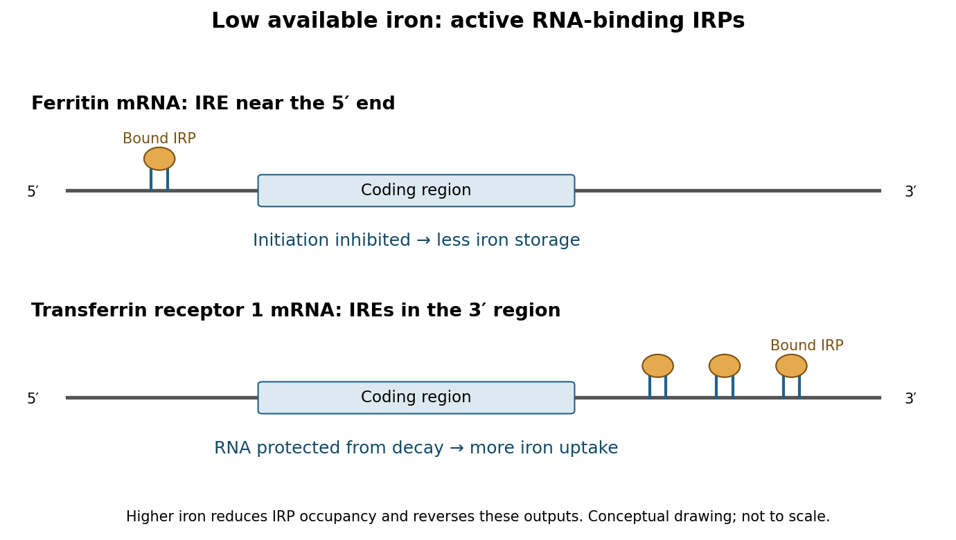
Low iron produces coordinated but opposite outputs from 5′ and 3′ iron-responsive elements
. Details. 
The network also adjusts use and release. A 5′ IRE in erythroid ALAS2 mRNA couples synthesis of a haem-pathway enzyme to iron availability, helping limit production of porphyrin precursors when metal incorporation is restricted.[ref]
Identification of a novel iron-responsive element in murine and human erythroid delta-aminolevulinic acid synthase mRNA by Thomas Dandekar and others. Erythroid ALAS leader element and iron-dependent translation
IRE-containing ferroportin transcripts can reduce iron export through translational repression during deficiency.[ref]Ferroportin IRE regulation study by Athina Lymboussaki and others. IRE deletion reporters distinguish protein-output regulation from RNA abundance
These are transcript-specific rules, not universal predictions from a gene name: alternative transcripts can lack an IRE, and ferroportin is also regulated at the protein level by systemic signals. The central RNA logic coordinates uptake with conservation, rather than simply maximizing the expression of every iron-related protein.Regulation beyond the mammalian IRE system illustrates a broader eukaryotic principle. In budding yeast, iron deficiency induces the RNA-binding regulator Cth2, which recognizes AU-rich elements in selected 3′ regions and accelerates degradation of messages encoding iron-consuming metabolic functions. This remodels resource allocation rather than protecting an uptake message. Sergi Puig and colleagues combined expression and RNA-turnover analysis with RNA-element recognition to establish targeted degradation during deficiency.[ref]
Coordinated remodeling of cellular metabolism during iron deficiency through targeted mRNA degradation by Sergi Puig and others. Cth2-dependent turnover of iron-utilization transcripts
It is therefore misleading to generalize that iron shortage always stabilizes iron-related RNA: the direction depends on the transcript and its physiological role.To establish these mechanisms experimentally, compare iron chelation with iron restoration, measuring available iron, IRP–RNA binding, transcript half-life and newly synthesized protein separately. Match reporter transcription and mutate recognition features without inadvertently changing general RNA stability. For a stem-loop, a compensatory paired mutation helps distinguish loss of structure from loss of a specific sequence contact. Electrophoretic binding assays with purified protein, cellular crosslinking and rescue with functional versus binding-defective IRP variants connect recognition to regulation. RNA-abundance measurements alone cannot establish translational repression, and chelators can cause broad stress, so unrelated control reporters and viability measurements are needed. The decisive principle is position-dependent RNA control: low iron can repress storage and export biological translation while preserving uptake RNA and reducing expendable iron-consuming programmes.
Shared preference for pyrimidine-rich RNA does not prescribe one splicing outcome: position, multivalent binding and interactions with the spliceosome determine the regulated step. Sex-lethal acts in the Drosophila sex-determination programme, whereas PTBP1 regulates many mammalian precursor RNAs in a cell-type-dependent manner. Their effects must be interpreted against competing splice sites and the proteins normally recognizing them.
During early spliceosome assembly, U1 snRNP recognizes a 5′ splice site, while factors including U2AF help recognize a polypyrimidine tract and 3′ splice-site region and promote assembly around the branch point. Alternative sites compete within this context. Binding of another RNA-binding protein can occlude an essential interaction, organize a repressive complex or favour use of a competing site; recognition need not be determined solely by an isolated site's consensus strength.
A particularly clear SXL mechanism operates on precursor RNA encoding Transformer protein. In the absence of SXL, U2AF recognizes a strong default 3′ site, producing an unproductive transcript. In female biological cells, SXL binds the pyrimidine-rich region at that site and competes with U2AF. The weaker alternative female site can then be used, producing functional Transformer protein. Reconstituted splicing experiments showed the antagonism, and adding a U2AF effector domain to SXL converted its activity from repression toward activation.[ref]
The protein Sex-lethal antagonizes the splicing factor U2AF to regulate alternative splicing of transformer pre-mRNA by Juan Valcárcel and others. In vitro splice-site switch and effector-domain fusion
The domain-fusion result is important: related RNA recognition can direct different outcomes because the proteins recruit different downstream activities. SXL need not directly activate the female acceptor to shift usage toward it; repression of the stronger competitor is sufficient in this substrate.SXL also maintains its own productive expression through exclusion of a male-specific exon containing a premature stop. This provides positive feedback in female biological cells. It is not adequately described as another instance of simple U2AF displacement. Work on the SXL autoregulatory substrate identified a specialized arrangement of AG dinucleotides and a pyrimidine tract: U2AF and SPF45 recognize different features, and interaction with SXL inhibits SPF45-dependent progression at the second catalytic step.[ref]
Splicing regulation at the second catalytic step by Sex-lethal involves 3′ splice site recognition by SPF45 by María José Lallena and others. Autoregulatory exon 3 and second-step inhibition
SXL can consequently alter site selection after early assembly has occurred. Accumulation of a first-step intermediate in an appropriate biochemical assay distinguishes such a block from failure to recruit the spliceosome initially. The same regulator can use different mechanisms on different RNAs.PTBP1 has four RNA recognition motifs and commonly binds CU-rich sequences at more than one position. Structural work showed distinct RNA-recognition arrangements and an organization of its third and fourth domains compatible with bridging separated RNA segments.[ref]
Structure of PTB bound to RNA: specific binding and implications for splicing regulation by Florian C. Oberstrass and others. RNA-bound domain structures and implications for recognition
Förster resonance energy transfer, NMR and splicing assays subsequently supported RNA looping by these domains: contacts on either side of a segment can organize an RNA geometry that disfavors exon use.[ref]RNA looping by PTB: evidence using FRET and NMR spectroscopy for a role in splicing repression by Rajan Lamichhane and others. Looping measurements and functional tests
These observations support a physical repression mechanism, but a short model RNA loop does not prove that every endogenous PTBP1-regulated exon is looped out.Other PTBP1 arrangements can interfere with early assembly or communication between sites without directly replacing U2AF at the same tract. Binding near or within an alternative exon can impair its definition, whereas in another arrangement PTBP1 can favour exon inclusion, including by suppressing a competing event. Genome-wide perturbation and positional analyses revealed both activation and repression associated with different binding contexts.[ref]
Position-dependent alternative splicing activity revealed by global profiling of alternative splicing events regulated by PTB by Miriam Llorian and others. Global splicing responses and position-dependent activity
Thus neither “all pyrimidine binders are repressors” nor “PTBP1 always causes skipping” is a valid general rule. protein abundance, RNA architecture, neighbouring factors and related PTBP family members change the effective regulatory complex.To discriminate mechanisms, map direct contacts by crosslinking and immunoprecipitation, measure exon-junction usage after acute depletion, and rescue with wild-type or selected RNA-binding/interface mutants. Use a minigene splicing reporter to mutate individual tracts, change their spacing or move them relative to a regulated exon, then confirm important effects at the endogenous locus. Assays of U2AF or U1 snRNP association and native spliceosomal assembly can reveal an early block; time-resolved splicing-intermediate analysis can reveal a late one. Control reporter transcription and precursor abundance, and account for nonsense-mediated decay, because an unstable mature isoform can conceal the splice choice that generated it. Mutants must retain expression and the intended remaining contacts. SXL supplies examples of both competition and late catalytic control; PTBP1 illustrates multivalent, context-dependent organization of splice-site recognition. RNA affinity is only one part of specificity.
I would test three separable claims: that a distinct assembly exists, that particular mRNAs are recruited or retained there, and that this recruitment causally alters their biological translation. Establishing one claim does not establish the others. In particular, a fluorescent punctum need not be a phase-separated liquid, and enrichment of untranslated RNA need not mean that the assembly caused repression.
Begin by tagging a candidate resident at its endogenous locus, verifying that the tagged protein retains its normal abundance and function. Identify additional resident proteins and RNAs with independent probes, assess ultrastructure or cell membrane exclusion where necessary, and follow assembly and dissolution over physiological time courses. Measure concentration dependence, partitioning and exchange rather than classifying the structure from roundness alone. fluorescence recovery after photobleaching reports mobility and exchange, but recovery can occur in non-liquid assemblies; fusion and rapid recovery are supporting observations rather than decisive tests. Avoid interpreting dissolution by 1,6-hexanediol as specific evidence, because it can perturb other cellular structures and processes. If phase separation is proposed, purified-component reconstitution should establish appropriate concentration, salt and RNA dependences, including sequence and valency controls and comparison with estimated cellular concentrations.
To identify recruited mRNAs, combine granule enrichment with RNA sequencing, controlling nonspecific recovery and input transcript abundance. Validate candidates by single-molecule fluorescence in situ hybridization using endogenous RNAs, negative-control RNAs and probes spanning different transcript regions. Three-dimensional segmentation and randomized spatial controls help distinguish genuine enrichment from chance overlap in a crowded cytoplasm. A useful RNA partition coefficient is , estimated from molecule numbers and accessible volumes rather than uncorrected total intensity. A small assembly can have high concentration but contain only a small fraction of the cellular transcript, so report both enrichment and the fraction of molecules recruited.
Live imaging using an MS2- or PP7-tagged RNA can measure entry, exit, dwell time and transport relative to the assembly. Compare the labelled transcript with the untagged endogenous distribution: repeated hairpins, coat-protein binding and fluorescent-protein expression can alter processing or mobility. Track whole particles in three dimensions where possible to separate actual residence from transient passage through an imaging plane. A localization element identified in a candidate 3′ region can be deleted or transferred to a neutral reporter, with matched RNA abundance and integrity, to test sequence-dependent targeting. crosslinking and immunoprecipitation of the proposed recruitment factor then tests direct contact rather than mere colocalization.
biological translation should be measured on the same RNA molecules wherever possible. A reporter bearing RNA-label sites and an N-terminal repeated peptide tag, such as SunTag, makes an mRNA visible separately from its nascent chains. A bright nascent-chain focus at an RNA can report ongoing biological translation, while completed proteins diffuse away. Puromycin-mediated release and initiation-block/run-off controls validate the translation-dependent signal; tag maturation, antibody-fragment availability and altered peptide behaviour need controls. Fixed-cell protein abundance or a ribosomal-protein stain is inadequate: either can be present without active biological translation at the identified RNA.
This caution has experimental force. Single-molecule work by Daniel Mateju and colleagues showed that some stress-granule-associated mRNAs undergo biological translation, including complete biological translation cycles, rather than being obligatorily silent.[ref]
Single-Molecule Imaging Reveals Translation of mRNAs Localized to Stress Granules by Daniel Mateju and others. Simultaneous RNA tracking and nascent-chain imaging
For the proposed new assembly, compare the probability and rate of biological translation before entry, during residence and after exit, with matched cytoplasmic molecules of the same transcript. If loss of biological translation consistently precedes recruitment, the assembly may sort previously repressed RNA. If recruitment precedes a reproducible biological translation change, that supports a different direction of action but still requires perturbation to exclude a common upstream cause.Causality requires a perturbation that separates recruitment or condensation from other functions. Acutely deplete an assembly scaffold and rescue with wild-type protein or a separation-of-function variant that retains RNA binding and normal abundance but fails to assemble, or conversely assembles but cannot recruit the chosen RNA. Verify those properties directly. Studies of G3BP1/G3BP2-deficient biological cells showed that stress-granule assembly requirements depend on the initiating stress, motivating matched tests of the upstream stress response rather than assuming one knockout removes every granule type.[ref]
G3BP–Caprin1–USP10 complexes mediate stress granule condensation and associate with 40S subunits by Nancy Kedersha and others. Double-deficient cells, rescue and stress-specific assembly
Measure global biological translation, stress signalling, RNA stability and biological cell viability alongside the local reporter. Loss of a granule accompanied by wholesale loss of biological translation machinery is not a specific test of granule function.An orthogonal manipulation could inducibly recruit the same RNA to the assembly or release it, using inert recruitment controls and a non-condensing scaffold. Artificial clustering can itself obstruct biological translation, so this establishes conditional sufficiency only. Reversible manipulation and rescue, together with endogenous cis-element edits, provide stronger evidence. A convincing result connects a defined RNA-recruitment mechanism to a reversible biological translation change while preserving RNA abundance and upstream cellular state; phase behaviour and regulatory function should be concluded separately.
chromatin has organization across overlapping length scales, and each assay measures a particular aspect rather than a universal “openness”. Local DNA exposure, histone occupancy, chemical state, long-range proximity and position within the nucleus are related but distinct. A strong experimental design chooses the relevant scale and combines a genomic assay with an independent structural or functional measurement.
At the molecular scale, DNA wraps around a histone octamer to form a nucleosome core, with roughly 147 base pairs protected; linker DNA connects neighbouring units, and linker histones can affect their organization. The core contains two copies each of H2A, H2B, H3 and H4. Crystallography established the detailed DNA–histone arrangement using a defined nucleosome core particle.[ref]
Crystal structure of the nucleosome core particle at 2.8 Å resolution by Karolin Luger and others. Atomic structure of the reconstituted core particle
Such structures explain local contacts, but a purified core does not specify the arrangement of an entire chromosome in a living nucleus. histone variants, tail modifications and ATP-dependent remodelling change stability and access without requiring DNA-sequence changes.For local accessibility, ATAC-seq uses transposase-mediated insertion of sequencing adapters into accessible DNA. It maps accessible promoters and regulatory elements, and its fragment-length distribution can contain signatures of nucleosome-free regions and neighbouring nucleosomes. The original assay combined rapid accessibility mapping with information about nucleosome organization.[ref]
Transposition of native chromatin for fast and sensitive epigenomic profiling of open chromatin, DNA-binding proteins and nucleosome position by Jason D. Buenrostro and others. ATAC-seq development and accessible-region profiling
DNase I hypersensitivity provides a related cleavage-based measure. Both have sequence bias and depend on preparation and enzyme exposure; a footprint can reflect enzyme preferences or protected binding, and is not a direct identification of the occupying transcription factor. Compare biological replicates, enzyme-dose controls, appropriate background and biological cell composition. Accessibility does not prove that an element regulates a particular gene.nucleosome mapping by MNase-seq addresses occupancy and spacing more directly: partial micrococcal-nuclease digestion preferentially removes exposed linker DNA, and sequencing protected fragments maps nucleosome-associated regions. Digestion titration is essential because over-digestion can preferentially lose fragile nucleosomes and create apparent depletion; MNase also has sequence preferences and can recover other protected particles. Compare fragment sizes, histone enrichment and an independent accessibility assay. Chemical mapping or defined in vitro reconstitution can refine particular nucleosome positions, while single-molecule measurements can expose heterogeneity hidden in a population average.
Chemical states require different assays. histone acetylation and methylation at particular residues can correlate with distinct regulatory states; they are not interchangeable with physical accessibility. chromatin immunoprecipitation followed by sequencing, or targeted CUT&RUN, maps association of a histone modification or protein with genomic regions. antibody specificity, histone abundance, input/background and calibrated normalization matter, especially when the modification changes globally. In CUT&RUN, an antibody recruits a nuclease to release nearby DNA, reducing some backgrounds associated with bulk chromatin fragmentation.[ref]
An efficient targeted nuclease strategy for high-resolution mapping of DNA binding sites by Peter J. Skene and Steven Henikoff. Principle and validation of targeted chromatin cleavage
DNA methylation can be measured by appropriately controlled bisulfite-based or direct-read approaches; standard bisulfite measurements do not automatically distinguish every modified cytosine species. These assays locate a chemical mark, not the geometric folding of the fibre.At the level of nucleosome arrays, regular compact fibres can form in some reconstituted or specialized conditions. A universal hierarchy in which every interphase chromosome passes through a uniform 30-nm fibre is not supported. electron tomography and related imaging can examine the distribution and packing of chromatin directly. ChromEMT combined DNA-selective staining with electron tomography and revealed irregular chromatin chains and variable compaction in biological cells.[ref]
ChromEMT: Visualizing 3D chromatin structure and compaction in interphase and mitotic cells by Horng D. Ou and others. In situ chromatin ultrastructure and compaction
Fixation, staining, sectioning and limited sampled volumes must be considered; cryogenic and super-resolution approaches provide complementary views. Dense staining also need not imply a distinct biochemical chromatin state.Longer-range folding produces loops and domains of enhanced contact. chromosome conformation capture crosslinks, digests and ligates spatially associated fragments; targeted variants test selected loci, whereas Hi-C samples contacts genome-wide. Contact matrices reveal local domains and broad A/B compartmentalization. High-resolution in situ Hi-C mapped chromatin loops and their association with CTCF orientation and cohesin.[ref]
A 3D map of the human genome at kilobase resolution reveals principles of chromatin looping by Suhas S. P. Rao and others. In situ contact maps, loop anchors and domain structure
A topologically associating domain is a region with preferential internal contacts under a stated analysis, not necessarily a stable, sharply bounded object in every biological cell. Contact frequency is neither an absolute distance nor proof that two sites directly interact. Restriction-site distribution, crosslinking, ligation, coverage and bin size affect resolution and interpretation.Perturbation can distinguish mechanisms underlying similar-looking domains. Acute cohesin loss eliminated loop domains while compartment organization persisted, showing that local loop organization and large-scale compartmental segregation are separable.[ref]
Cohesin loss eliminates all loop domains by Suhas S. P. Rao and others. Acute depletion, loss of loops and persistence of compartments
Imaging by DNA fluorescence in situ hybridization can measure distance distributions between chosen loci and test heterogeneity; live labels track dynamics but can perturb loci. Agreement between contact enrichment and a shift in distance distributions is stronger than either assay alone, although the observables still differ.At nuclear scale, chromosomes occupy territories and regions associate preferentially with the nuclear lamina or nucleolus. DNA imaging maps territories and radial positions, while lamina-association assays such as DamID map sequences near a labelled nuclear compartment over an integration interval. The original DamID approach tethered a DNA adenine methyltransferase to a chromatin protein to record nearby genomic regions.[ref]
Identification of in vivo DNA targets of chromatin proteins using tethered Dam methyltransferase by Bas van Steensel and Steven Henikoff. Targeted methylation as a proximity record
Such maps record proximity and history rather than a single fixed structure. Mitotic condensation is a distinct organization with additional constraints and chromosome-shaping machinery, not simply maximally closed interphase chromatin. The appropriate pairing is exposure assays for local access, protected-fragment assays for nucleosomes, protein/mark profiling for composition, contact assays for genomic proximity and imaging for spatial organization, with perturbations to connect organization to function.ribosome profiling turned initiation from a small set of reporter-based examples into a genome-wide, condition-dependent map, while specialized variants extended observation from translating ribosomes to start-site selection and scanning. The central distinction is that ordinary footprints locate occupied ribosomes; they do not directly label every initiation event or measure the movement of a scanning small subunit.
In a typical experiment, rapidly preserve biological translation complexes, digest exposed RNA, isolate ribosome-protected fragments and sequence them alongside matched total RNA. Assign each footprint to a ribosomal position using length-specific offsets calibrated on established coding regions. Enrichment over coding sequences and three-nucleotide periodicity support active elongation. The initial yeast work demonstrated genome-wide biological translation measurements and widespread regulation not inferable from mRNA abundance alone.[ref]
Genome-wide analysis in vivo of translation with nucleotide resolution using ribosome profiling by Nicholas T. Ingolia and others. Footprint method and translational responses in yeast
Counts normalized to transcript abundance provide an operational translation-efficiency measure,For treatment comparisons, a count model testing the interaction between assay type and treatment is preferable to unstable ratios at low counts. More occupancy can result from increased initiation or slower passage, so changes in elongation, premature termination and global normalization must be considered before assigning a TE change specifically to initiation.A major advance was recognition of pervasive translated upstream open reading frames and alternative starts. Mammalian stem-cell profiling found many leader-region biological translation events, including near-cognate starts, and alternative coding products.[ref]
Ribosome profiling of mouse embryonic stem cells reveals the complexity and dynamics of mammalian proteomes by Nicholas T. Ingolia and others. Mammalian footprints, leader translation and alternative initiation
Initiation upstream of an annotated start can add an N-terminal extension; downstream in-frame initiation can remove targeting or regulatory sequences, while another frame creates a different product. This exposed a coding and regulatory landscape missed by annotation based only on a single longest open reading frame. A footprint-supported short ORF is not automatically a stable functional microprotein: start-codon mutation, peptide evidence and phenotype are separate tests.Ordinary elongation profiles often make the exact starting site ambiguous. Brief harringtonine treatment permits much ongoing elongation to run off while newly initiating ribosomes accumulate near starts, generating start-associated peaks. lactimidomycin-based initiation profiling preferentially enriches early ribosomes and, when compared with an elongation-inhibitor profile, improves discrimination of initiation sites. Sooncheol Lee and colleagues used this strategy to map alternative mammalian start sites at high resolution.[ref]
Global mapping of translation initiation sites in mammalian cells at single-nucleotide resolution by Sooncheol Lee and others. Parallel initiation- and elongation-inhibitor profiling
These approaches reveal that the first AUG is not invariably chosen: local sequence context, near-cognate codons and competing upstream sites influence selection. However, drug uptake, exposure time and sequence-dependent trapping can change the observed distribution. A high peak can reflect capture efficiency or dwell time rather than a proportionate initiation flux, so compare time courses, untreated profiles and reporters with mutated candidate starts.The regulatory significance of upstream biological translation is particularly clear during stress. In the GCN4 leader, biological translation of a short upstream frame can be followed by retention of a scanning small subunit. Reacquisition of the eIF2–GTP–initiator-tRNA ternary complex determines whether it reinitiates at an inhibitory upstream frame or proceeds to the main coding region. Amino-acid limitation lowers ternary-complex availability and changes that competition. Profiling measures occupancy on upstream and main regions simultaneously, testing redistribution across endogenous transcripts rather than relying only on reporter output. The reinitiation mechanism predates profiling; the advance is transcriptome-wide evidence for its deployment and condition dependence. Similarly, changes in cap-dependent recruitment can be mapped by perturbing mTORC1 signalling: profiling identified selectively regulated transcript programmes rather than a uniform effect on every mRNA.[ref]
The translational landscape of mTOR signalling steers cancer initiation and metastasis by Andrew C. Hsieh and others. mTOR perturbation and selective translational programmes
An important limit is that standard 80S footprints do not directly demonstrate scanning. translation complex profiling addresses this by preserving and separately profiling small-subunit-associated and 80S complexes. In yeast, Stuart K. Archer and colleagues mapped small-subunit occupancy along leaders and around initiation and recycling transitions, providing evidence for otherwise difficult-to-trap intermediates.[ref]
Dynamics of ribosome scanning and recycling revealed by translation complex profiling by Stuart K. Archer and others. Small-subunit and ribosome profiling across the translation cycle
This makes different predictions from a model that treats every leader footprint as elongation. Nevertheless, the measurement is a distribution of captured complexes, not a movie of one subunit; crosslinking and footprint-length choices can affect which states are recovered.Validation is therefore integral to the mechanistic conclusions. Use rapid, consistent harvesting, biological replicates, multiple footprint lengths and matched RNA-seq; inspect rRNA contamination, mapping ambiguity and frame periodicity. Test start-site edits at the endogenous locus or in reporters preserving the relevant leader. Measure the predicted N termini or protein isoforms where possible, and use run-off or independent synthesis measurements to separate initiation changes from altered elongation. A structured RNA or another protected ribonucleoprotein can also produce a short sequencing fragment, especially in a very short proposed ORF. biological cell mixtures and transcript-isoform changes can create apparent alternative initiation, so transcript architecture must be resolved. Profiling has revealed the extent and plasticity of initiation-site choice and upstream biological translation; perturbations and specialized capture methods are needed to turn an occupancy map into an initiation mechanism.
Articles by others on the same topic
There are currently no matching articles.