Topics (384k) Articles (398k) Users (410) Discussions (242) Comments (383) Files (13k) New article

Medical-test reference from the United States National Library of Medicine. The opening definition and measurement description are used here; individual results require clinical context.
A reference range is an interval derived from measurements in a specified reference population under specified analytical conditions. It gives context for a laboratory result; it is not automatically a boundary separating disease from health. Different methods, ages and reference populations can lead to different intervals. [ref]
How to Understand Your Lab Results by MedlinePlus. Reference ranges: laboratory differences, healthy people outside the interval and illness within it
For an illustrative central 95% interval, about 5% of measurements from the defining population lie outside by construction. If 20 independent tests each have a 5% outside probability, the probability of at least one outside result is . Independence is an assumption, but the calculation shows why many tests increase incidental flags. A clinical decision threshold can instead be chosen for a particular diagnostic or treatment purpose.
Dysphagia means difficulty swallowing. It can involve preparation and transfer of material in the mouth, passage through the pharynx or movement down the oesophagus. These stages require coordinated muscle activity, sensation and neural control. Dysphagia names a functional difficulty rather than one unique disease or anatomical lesion. [ref]
Dysphagia by NIDCD. Fact sheet, What is dysphagia?, How do we swallow? and How does dysphagia occur?
Swallowing also protects the airway: food transport and temporary closure of the breathing passage must be coordinated. A delay in initiating the response and a mechanical obstruction can both disrupt swallowing through different mechanisms. Difficulty moving a bolus, pain during swallowing and entry into the airway are related observations but should not be treated as identical definitions.
Palpation is examination by touch with the hands or fingers. It uses physical contact to assess features such as an organ's apparent size, position, consistency and tenderness. The observed feature and the resulting clinical diagnosis are separate: feeling a moving or resistant region does not by itself identify the complete underlying anatomy or disease.[ref]
The force applied during palpation is part of the measurement. In an original simplified indentation example, suppose a probe produces of displacement under a load. The secant stiffness for that observation isA second specimen requiring at the same displacement has twice this apparent stiffness. That ratio is not automatically a ratio of intrinsic elastic modulus values: contact geometry, specimen thickness, support and deformation rate can alter the force-displacement curve. Touch supplies a mechanical observation whose interpretation requires an appropriate model.
A clinical measurement study makes the distinction concrete. S. D. Eilen and colleagues compared precordial palpation with two-dimensional echocardiography in 41 patients. They compared the location and palpable diameter of the apical impulse with left ventricle end-diastolic volume. A lateral impulse location was sensitive but not specific for enlargement. Within the reported subgroup without ventricular hypertrophy, an impulse diameter above in the left-lateral position had reported diagnostic sensitivity and diagnostic specificity . Those are results for that study's condition and subgroup, rather than universal performance figures for touch.[ref]
Accuracy of precordial palpation for detecting increased left ventricular volume by S. D. Eilen and others. Abstract, Annals of Internal Medicine99 (1983), pp.628–630: cohort, location result and subgroup diameter result
A small probability example explains why both attributes matter. In a stipulated set with 20 enlarged and 80 non-enlarged hearts, a rule detecting 18 enlarged hearts but also flagging 24 non-enlarged hearts has diagnostic sensitivity and diagnostic specificity . Only of its positive results identify enlargement. These invented counts are not a reconstruction of the 41-patient study. They show why a palpable feature must be tested against an independent reference and interpreted with the population being examined.
Repeating a specified observation can assess reproducibility, while comparison with echocardiography assesses a different question: correspondence with the chosen anatomical reference. Holding body position and observation method consistent helps distinguish those sources of variation. A reliable description of what was felt is necessary evidence, but reproducibility alone does not establish the causal explanation.
Pulse oximetry estimates arterial oxygen saturation noninvasively from wavelength-dependent optical signals that vary with the arterial pulse. A typical sensor combines red and infrared light-emitting diodes with a photodetector. The pulsatile component helps distinguish changing arterial blood contribution from relatively steady tissue and blood contributions; it is not a direct measurement of arterial oxygen partial pressure.
A primary instrument-design note implements 660 nm and 940 nm illumination, a shared photodetector and alternating channel acquisition. It separates pulsatile and steady components before estimating saturation.[ref]
Pulse Oximeter Fundamentals and Design by Santiago Lopez. Sections 2.2 and 3.1.2, printed pp. 3–5 and 7, optical channels and acquisition
These wavelengths and electronics describe that implementation rather than every possible sensor.Writing pulsatile and steady signal magnitudes as AC and DC, a common normalized optical quantity isFor original channel values AC, DC, AC and DC, this ratio is 0.5. Halving both red-channel magnitudes leaves the ratio unchanged. This illustrates intensity normalization; it does not determine a saturation without a validated relation between and saturation.
That relation is experimentally checked against an independent reference. The FDA's technical guidance describes comparing oximeter readings with simultaneous co-oximetry of arterial samples during controlled inspired-oxygen changes in volunteers, and reporting an accuracy statistic.[ref]
Pulse Oximeters: Premarket Notification Submissions by the FDA. Clinical Performance, SpO2 Accuracy: reference arterial sampling, inspired mixtures and paired readings
Such comparisons test the whole sensor–algorithm measurement chain, rather than only the arithmetic of a two-channel ratio.One summary is the root mean square differenceFor invented paired errors percentage points, percentage points even though mean error is zero. A small mean bias does not guarantee small individual errors.
Motion, weak pulsatile signals and additional absorbing hemoglobin species can invalidate assumptions behind a simple estimate. Pulse oximetry does not measure pH, carbon-dioxide tension or hemoglobin concentration. A stable displayed percentage therefore cannot establish every aspect of ventilation, oxygen content or gas exchange.
Arterial blood gas analysis applies blood gas analysis to a sample taken from an artery, providing a time-specific observation of systemic arterial gas tensions and acid-base state. Arterial oxygen partial pressure concerns oxygen tension, while arterial carbon-dioxide partial pressure and pH provide different information. The arterial qualifier is a sampling condition, not merely a label attached to any blood result.[ref]
Arterial Blood Gas (ABG) Test by MedlinePlus. What is an arterial blood gas test? measured quantities
The measurement chain includes obtaining an identified sample, preserving its gas composition and analyzing it with a checked instrument. Air contamination, fluid contamination and analysis delay can change the specimen after it leaves the circulation. A reproducible analyzer reading cannot repair a specimen that no longer represents the intended physiological state.[ref]
Equal oxygen tension need not mean equal arterial oxygen content. An original illustrative carrier model writeswhere is hemoglobin concentration, its oxygen-bound fraction and the carrier capacity per gram. Choose mL/g, mL/(dL·mmHg), and mmHg. Samples with and g/dL then contain about 20.00 and 10.15 mL O/dL, respectively. They have identical stipulated tension and saturation but very different oxygen content. The constants define the illustration; these are not patient results or diagnostic thresholds.
Interpretation also depends on the conditions when the sample was obtained. Inspired oxygen fraction, ventilator settings when applicable and sampling time help distinguish a changed respiratory state from a changed measurement condition. A later pulse oximetry reading is neither a simultaneous sample nor a measurement of carbon-dioxide tension.
Arterial blood gas analysis supplies evidence about gas transport and acid-base balance, but no individual number alone identifies the entire cause of an abnormal result. Measured co-oximetry and calculated oxygen saturation should also be distinguished when unusual hemoglobin species matter.[ref]
An inverted pendulum has its center of mass above its pivot. For a point mass on a massless rod of length , let measure departure from the upward vertical. The gravitational potential energy is , and the equation of motion is .
For small , linearization gives exponentially growing and decaying solutions with rate : the upright equilibrium is unstable. A moving support or an applied torque can stabilize it through feedback control. Balancing a rod on a hand makes this visible: the hand continually moves the pivot to counter the growing departure.
Statics studies mechanical equilibrium: configurations in which the relevant forces and torques balance. For a rigid body at rest in an inertial frame, the conditions are and , with all torques calculated about the same point .
A horizontal beam carrying a downward load illustrates why both equations are necessary. Vertical force balance fixes the sum of the support reactions, while torque balance determines how the load is divided between supports. Equilibrium alone does not imply dynamical stability: a perfectly upright inverted pendulum satisfies force and torque balance but falls after a sufficiently small disturbance.
A portal system carries blood through two capillary beds in sequence before returning it to the heart. The connecting vessel sends material collected from the first bed to the second without first mixing it through the whole systemic circulation. In the hepatic example, the portal vein takes blood from digestive organs to the liver; the hypophyseal portal system connects the hypothalamus with the anterior pituitary.[ref]
Anatomy and Physiology 2e by J. Gordon Betts and others. sections17.3 and20.5, hypothalamic–pituitary connection and hepatic portal circulation
A simple transport calculation explains the advantage for chemical signaling. If a source adds a substance at rate to a steady blood flow , with negligible removal along the connection, its added concentration is . In a hypothetical example, and give . Mixing the same input into instead gives . These are explanatory flow rates, not measured human values.
Direct sampling makes the portal route experimentally accessible. In a 1982 study, Iain J. Clarke and James T. Cummins measured GnRH in portal plasma and LH in peripheral plasma of conscious ewes while maintaining pituitary function. The paired pulses connected a local hypothalamic signal with the downstream pituitary response.[ref]
The systemic circulation carries blood from the left ventricle through body tissues and back to the right atrium. Its main arterial outflow is the aorta, and major venous return channels include the superior vena cava and inferior vena cava.[ref] The separate pulmonary circulation then passes through the lungs before blood returns to the left heart.
The two circuits operate in series, while many systemic tissue beds receive arterial supply in parallel. In an original fixed-resistance model, beds sharing pressure difference haveFor a stipulated mmHg and two resistances 30 and , flows are 3 and 1.5 litres per minute, totaling 4.5. The equivalent resistance is 20 in those resistance units. Adding flows is appropriate here; adding parallel resistances is not.
Real vascular resistance changes with vessel geometry and physiology. Some routes also contain successive exchange beds, as in a portal system. The parallel-bed calculation is therefore a deliberately reduced model, not a complete vessel map.
Conservation of mass constrains sustained output from the right and left heart. If their flows differ, a vascular compartment must gain or lose blood. For the pulmonary pool,A hypothetical imbalance of adds 0.2 litres in one minute if maintained. Long-term matching of flows is a consequence of finite storage, rather than a rule that both instantaneous waveforms must be identical.
For a whole-body oxygen balance, systemic cardiac output must be paired with compatible arterial and mixed venous oxygen content. A regional venous sample answers a regional question; it does not become whole-body return simply because it was taken from a large vein.
The coronary circulation supplies and drains the heart's own tissue. Coronary arteries branch from the aorta to supply the myocardium; cardiac veins return blood toward the right atrium.[ref] Blood moving through a ventricular cavity is not the same flow as blood perfusing the surrounding muscle.
Much coronary venous return passes through the coronary sinus, while anterior cardiac veins provide a direct route to the right atrium.[ref] Consequently a coronary-sinus sample represents its drainage territory rather than automatically every part of the heart. Anatomical pathways must match the region whose blood flow and oxygen uptake are being estimated.
An original tissue-balance example uses regional flow , arterial oxygen content and matching venous oxygen content in the same units. Then regional coronary oxygen uptake isThe arterial delivery rate is in those units. Delivery and uptake differ because oxygen remains in the returning blood. The values are stipulated, not normal-reference limits.
A primary 2007 experiment measured coronary volumetric flow using continuous saline thermodilution, an infusion catheter and a pressure/temperature guidewire, with comparisons in dogs and patients.[ref]
Direct volumetric blood flow measurement in coronary arteries by thermodilution by Wilbert Aarnoudse and others. Abstract, Methods
This provides a concrete way to investigate the flow term; it does not by itself measure the oxygen-content difference.The arterial map also varies among hearts. A radiopaque-injection study classified 178 of 200 examined hearts as right dominant by the origin of the posterior interventricular artery.[ref]
Functional dominance of the right coronary artery: incidence in the human heart by C. E. Nerantzis and others. Abstract
Dominance names an anatomical branching criterion, not a claim that one artery supplies the entire heart.A motile cilium is a eukaryotic cilium that actively bends or rotates to move the cell or the surrounding fluid. Its internal axoneme contains organized microtubules and motor machinery. In familiar bending cilia, dynein uses energy from ATP to drive relative sliding of neighboring microtubule doublets; constraints on that sliding convert motor activity into a change of shape.[ref]
Molecular Biology of the Cell, 4th edition by Bruce Alberts and others. Chapter 16, Cilia and Flagella: axonemal motors, sliding and bending.
Many motile cilia have a 9+2 arrangement: nine outer doublets around two central singlets. The nine counts doublets, rather than nine individual outer tubules. Motility should not be defined solely by this pattern, since some motile cilia have other arrangements. Conversely, a ciliary-looking projection is not necessarily motile.
A classic experiment separated the force-producing mechanism from the intact structure's constraints. K. E. Summers and I. R. Gibbons isolated sea-urchin sperm axonemes and briefly treated them with trypsin. Adding ATP then caused active separation into tubules and groups of tubules. The experiment supported sliding between structural elements as a source of motion, rather than requiring each tubule to shorten like a contracting thread.[ref]
Adenosine triphosphate-induced sliding of tubules in trypsin-treated flagella of sea-urchin sperm by K. E. Summers and I. R. Gibbons. Abstract: retained initial 9+2 architecture and ATP-induced disintegration after limited digestion.
The long beating projection of a eukaryotic sperm cell and short motile cilia share this general mechanical framework. A bacterial flagellum has a different construction and motor. Comparing their functions alone would hide the difference between bending a microtubule assembly and rotating a bacterial filament.
An axoneme is the organized microtubule-based core of a eukaryotic cilium or flagellum. It supplies a structural framework and, in motile forms, carries motor and linking components that turn relative microtubule sliding into movement. The axoneme is an internal assembly, not the entire membrane-covered projection.[ref]
Molecular Biology of the Cell, 4th edition by Bruce Alberts and others. Chapter 16, Cilia and Flagella: microtubule organization and sliding motors.
The familiar 9+2 arrangement contains nine outer doublets surrounding two central singlets. It describes spatial organization, not eleven identical independent tubules: each outer doublet has two associated tubules. Other axonemal arrangements occur, so the term does not require every cilium to have a central pair or to be motile.
An important mechanical test used isolated sea-urchin sperm axonemes. After brief treatment with trypsin, the preparations initially retained the characteristic organization, but addition of ATP caused active separation into individual tubules and groups. K. E. Summers and I. R. Gibbons interpreted these observations as evidence for sliding between the structural elements.[ref]
Adenosine triphosphate-induced sliding of tubules in trypsin-treated flagella of sea-urchin sperm by K. E. Summers and I. R. Gibbons. 1971 abstract: preserved starting architecture and ATP-induced active disintegration after digestion.
The experiment separates two roles that can otherwise be conflated. Motors such as dynein generate relative motion, while structural constraints normally keep sliding components in an organized assembly. Weakening the constraints reveals sliding as separation; preserving them permits bending. The disintegration experiment does not imply that a normally beating motile cilium repeatedly falls apart and rebuilds itself.
A sister chromosome is one of the copies produced by replicating a particular chromosome. The term emphasizes shared replication origin, in contrast to homologous chromosomes, which may carry different parental alleles. Its exact counting convention must be stated: an ordinary replicated eukaryotic chromosome contains two sister chromatids, while chromosome copies produced by an additional genome-doubling cycle can participate as separate meiotic partners.
This distinction matters in some parthenogenesis mechanisms. A 2010 whiptail-lizard study found an extra premeiotic chromosome doubling and used fluorescence in situ hybridization probes to distinguish homologs. The paired meiotic partners were sister copies produced by the first of two premeiotic replication cycles. Observations of pairing structures and chiasmata showed that the system did not simply omit meiotic recombination.[ref]
Sister chromosome pairing maintains heterozygosity in parthenogenetic lizards by Aracely A. Lutes and others. Abstract and Figures1–4: doubled complement and homolog-distinguishing probes
For a simplified one-locus example, label two homologs and . An extra doubling supplies copies . Pairing an copy with its sister and an copy with its sister permits the meiotic products to retain both allele types after reduction. Pairing with instead presents a different segregation and recombination problem. This explains why identifying which chromosomes pair is necessary; observing a doubled DNA amount alone does not establish the mechanism.
Sister copies are initially expected to carry the same inherited sequence, apart from replication changes. This does not make them two different parental homologs, nor imply that every parthenogenesis pathway preserves heterozygosity by the same process.
Pinned article: Introduction to the OurBigBook Project
Welcome to the OurBigBook Project! Our goal is to create the perfect publishing platform for STEM subjects, and get university-level students to write the best free STEM tutorials ever.
Everyone is welcome to create an account and play with the site: ourbigbook.com/go/register. We belive that students themselves can write amazing tutorials, but teachers are welcome too. You can write about anything you want, it doesn't have to be STEM or even educational. Silly test content is very welcome and you won't be penalized in any way. Just keep it legal!
Intro to OurBigBook
. Source. We have two killer features:
- topics: topics group articles by different users with the same title, e.g. here is the topic for the "Fundamental Theorem of Calculus" ourbigbook.com/go/topic/fundamental-theorem-of-calculusArticles of different users are sorted by upvote within each article page. This feature is a bit like:
- a Wikipedia where each user can have their own version of each article
- a Q&A website like Stack Overflow, where multiple people can give their views on a given topic, and the best ones are sorted by upvote. Except you don't need to wait for someone to ask first, and any topic goes, no matter how narrow or broad
This feature makes it possible for readers to find better explanations of any topic created by other writers. And it allows writers to create an explanation in a place that readers might actually find it.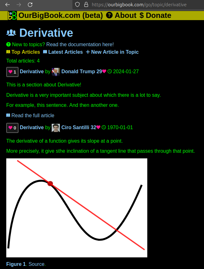
Figure 1. Screenshot of the "Derivative" topic page. View it live at: ourbigbook.com/go/topic/derivativeVideo 2. OurBigBook Web topics demo. Source. - local editing: you can store all your personal knowledge base content locally in a plaintext markup format that can be edited locally and published either:This way you can be sure that even if OurBigBook.com were to go down one day (which we have no plans to do as it is quite cheap to host!), your content will still be perfectly readable as a static site.
- to OurBigBook.com to get awesome multi-user features like topics and likes
- as HTML files to a static website, which you can host yourself for free on many external providers like GitHub Pages, and remain in full control

Figure 2. You can publish local OurBigBook lightweight markup files to either OurBigBook.com or as a static website.
Figure 3. Visual Studio Code extension installation.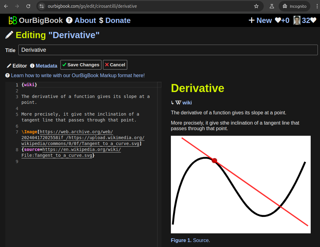
Figure 5. . You can also edit articles on the Web editor without installing anything locally. Video 3. Edit locally and publish demo. Source. This shows editing OurBigBook Markup and publishing it using the Visual Studio Code extension. 
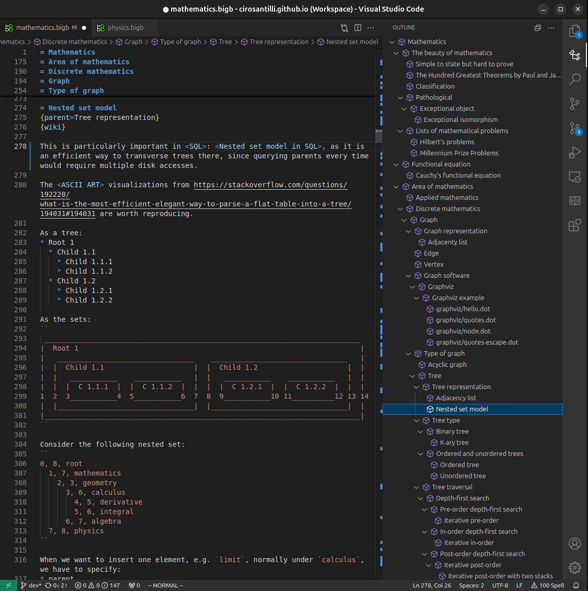
- Infinitely deep tables of contents:

All our software is open source and hosted at: github.com/ourbigbook/ourbigbook
Further documentation can be found at: docs.ourbigbook.com
Feel free to reach our to us for any help or suggestions: docs.ourbigbook.com/#contact