Topics (384k) Articles (398k) Users (410) Discussions (242) Comments (383) Files (13k) New article
2,3-Bisphosphoglycerate is the glycerate metabolite carrying two phosphate substituents at carbon positions 2 and 3. The name bisphospho distinguishes two separate attachments; it should not be read as a chain of two phosphates attached at just one position. Older biochemical papers call the metabolite 2,3-diphosphoglycerate. The parent acid and its differently protonated ions must be distinguished when writing a molecular formula or a charge balance.[ref]
2,3-Bisphosphoglyceric acid in ChEBI. CHEBI:28907, Definition, Formula and conjugate-acid relation to CHEBI:19324
In red blood cells, 2,3-Bisphosphoglycerate is an allosteric modulator of hemoglobin. Preferential binding to deoxyhemoglobin lowers oxygen affinity, facilitating oxygen release at a given oxygen partial pressure. Reinhold Benesch and Ruth E. Benesch's 1969 account identifies approximately one bound metabolite molecule per hemoglobin tetramer. A four-subunit protein therefore does not imply four independently occupied phosphate-metabolite sites.[ref]
Intracellular Organic Phosphates as Regulators of Oxygen Release by Haemoglobin by Reinhold Benesch and Ruth E. Benesch. Publisher abstract, binding preference and stoichiometry, Nature221, pp.618–622
A simple Hill equation illustrates an affinity change without assigning a universal physiological value. Write the fractional oxygen saturation aswhere is oxygen partial pressure and is the pressure at half saturation. In a stipulated model with , increasing from to changes the saturation at fromThe same oxygen pressure now leaves a larger fraction of binding capacity unoccupied. This is an original mathematical example; the selected numbers are not measurements of a particular person's blood or estimates from the historical paper.
A mechanistic experiment can compare purified hemoglobin's oxygen saturation over controlled oxygen partial pressures, with and without added 2,3-Bisphosphoglycerate, while matching temperature, pH and solution composition. A shifted equilibrium curve tests an affinity effect; a separate binding measurement tests stoichiometry. Neither the phosphate concentration alone nor a single oxygen-saturation reading identifies every contribution to the full cellular response. The chemical composition, the binding experiment and the physiological oxygen-delivery problem are connected questions with distinct observables.
A chemical multiplication reaction is a reaction sequence in which a species is converted through a cycle into more of that same species before measurement. Unlike a one-time conversion to a different reporter, the multiplied species can enter another cycle. IUPAC calls the case involving the species being determined a direct multiplication reaction.[ref]
Let be the amount of the multiplied species after cycle . If every cycle has the same net multiplier , thenFor an original ideal example, and four cycles convert into . The gain is 81, not 12: repeating a threefold increase compounds it. The stoichiometry concerns a sequence supplied with reagents, rather than multiplication of isolated molecules without additional matter.
A multiplication-cycle yield changes the net multiplier. Suppose a fraction of the entering species completes a route producing three output molecules, while the remaining fraction survives unchanged. ThenFor stipulated , four cycles give gain , substantially below 81. If failed molecules are instead destroyed, the model would give : specifying the fate of failures is essential. For cycle-dependent multipliers, the gain is , not a fixed power by assumption.
The same compounding can affect an initial chemical amplification blank. Tenfold less contaminant at the start need not stay negligible after many cycles, and contamination introduced late experiences fewer cycles. Reagent depletion, side reactions and changing yield limit the range over which a constant-gain measurement calibration is credible. Cycle number and blank history belong in the experimental record.
A chemical amplification blank measures the response of an amplification procedure without the intended analyte contribution. It should reproduce the relevant reagents, handling, reaction cycles and readout. It identifies signal that would otherwise be attributed to analyte, including contamination and reactions that generate reporter without the target. The distinction between blank material and its measured indication follows IUPAC terminology.[ref]
Blank material in the IUPAC Gold Book. Entry 08010, blank material and procedural scope.
Amplification blank timing matters because contamination introduced early can undergo more chemical multiplication reactions. In an original fixed-gain model, let be the background amount after cycle , the cycle multiplier and the background introduced after multiplication in that cycle:With constant and , this givesFor , , and arbitrary amount unit, the final background is 7 units. One unit present before all three cycles instead contributes 8. A blank added only at the final detector cannot reveal either history.
A blank correction can remove an estimated mean background without removing its variation. For example, if amplified sample and blank indications are independent with standard deviations3 and 2 signal units, their difference has standard deviation . Shared preparation or instrument effects require covariance terms instead of an independence assumption.[ref]
Evaluation of measurement data — Guide to the expression of uncertainty in measurement by JCGM. §§5.1–5.2, combination of independent and correlated inputs.
A useful experimental series processes several blanks through the same cycle counts as the standards and unknowns. It can distinguish background already present from background that accumulates during processing. Increasing mean gain need not improve the limit of detection when blank variability also grows. Report the blank's procedural scope and variation alongside its mean response.
A plasmodesma, plural plasmodesmata, is a membrane-lined connection crossing a plant cell wall between neighboring cells. It provides a route for intercellular exchange, including movement of some signaling molecules and macromolecules. A plasmodesma contains a tubular strand of endoplasmic reticulum within its enclosing cell membrane; it is an organized cellular interface, rather than simply a hole in the wall.[ref]
Architecture and permeability of post-cytokinesis plasmodesmata lacking cytoplasmic sleeves by William J. Nicolas and others. Abstract: exchanged material, plasma membrane lining and internal ER strand
Transport changes during development. A cotton fibre study followed a fluorescent tracer from the seed coat into elongating fibres with confocal microscopy. Access through plasmodesmata was observed early, blocked around day 10 and observed again around day 16. Reverse tracer movement supplied a complementary test, distinguishing transport connectivity from the mere presence of a channel in a section.[ref]
The control of single-celled cotton fiber elongation by developmentally reversible gating of plasmodesmata and coordinated expression of sucrose and K+ transporters and expansin by Y. L. Ruan and others. Abstract; Figures1–2: tracer movement at different developmental stages and in opposite directions
The apparent width in an image is not the whole explanation of permeability. A 2017 electron tomography study found newly formed plasmodesmata with no visible open cytoplasmic sleeve, yet observed macromolecular diffusion. Architecture and transport must therefore be measured together.[ref]
Architecture and permeability of post-cytokinesis plasmodesmata lacking cytoplasmic sleeves by William J. Nicolas and others. Abstract: post-cytokinesis structure and macromolecular permeability
The MTT assay measures formation of colored formazan when metabolically active biological cells reduce the tetrazolium salt MTT. The initially yellow reagent produces purple crystals; those crystals are solubilized before optical measurement with a multiwell spectrophotometer or microplate reader. The Roche instructions describe this chemical conversion and readout explicitly.[ref]
Cell Proliferation Kit I MTT instructions by Roche. §3.1, printed p. 9 and Figure 4; printed p. 10, solubilization and optical measurement
The measured endpoint is a metabolic color response. Under a calibrated linear model it can track viable-cell number, but per-cell activity also matters. Tim R. Mosmann's original 1983 paper noted that the signal depends on cellular activation as well as the presence of living cells.[ref]
Rapid colorimetric assay for cellular growth and survival: application to proliferation and cytotoxicity assays by T. Mosmann. Abstract, activation dependence and multiwell readout
For an original normalized comparison, suppose treated wells give absorbance , matched untreated controls and the reagent blank . The blank-corrected signal ratio isThis means of the control's measured MTT signal. It does not establish that of cells died. In , fewer viable cells and lower response per cell can produce the same result.
A calibration series with independently known cell numbers tests the useful response range. Cell-free wells containing the tested material help identify direct color, light scattering or reagent interactions; a vehicle control separates treatment-solvent effects. Complete and comparable solubilization matters because the readout concerns the colored solution rather than an arbitrary suspension of crystals.
For a cytotoxicity study, complementing this assay with a cell count or membrane-integrity endpoint can distinguish some ambiguities. Incubation history, cell state and observation time belong in the interpretation. MTT color formation, cell proliferation and cell death are related experimental questions, not interchangeable definitions.
The apical membrane is the region of an epithelial cell's plasma membrane facing its free surface or a lumen. It differs from the basolateral membrane, which faces neighboring cells and underlying tissue. These are spatially and functionally distinct regions of one cell boundary, not separate cells. Their organization is part of apical-basal polarity.
A tubule makes the definition concrete. Its apical surface faces the fluid inside the tube, while its basal side faces the surrounding tissue. A transporter moving a substance from the lumen into a cell and another transporting it from the cell into the surrounding tissue must occupy appropriately different membrane regions to support directed transepithelial transport. Merely producing both proteins somewhere in the cell does not guarantee the same net transport.
A classic experimental probe used the sites at which enveloped viruses emerged from polarized epithelial monolayers. Electron microscopy showed characteristic asymmetry: influenza virus budded at the apical surface, while vesicular stomatitis virus assembled at basolateral regions. Different viral envelopes therefore reported different regions of the host-cell surface.[ref]
The apical membrane is therefore defined by tissue orientation and organization, not by always being physically uppermost on a microscope image. Its protein and lipid composition can be specialized, but the exact contents depend on the epithelial cell type and physiological state.
The basolateral membrane is the region of an epithelial cell's plasma membrane facing adjacent cells and the underlying tissue. Basal and lateral surfaces together contrast with the lumen-facing apical membrane. Their different positions and molecular contents are part of apical-basal polarity; the terms describe domains of one cell boundary.
Consider absorption across a tubular epithelium. A substance enters a cell from the lumen across its apical membrane and leaves toward the surrounding tissue across its basolateral membrane. The two membrane domains expose transport proteins to different external compartments. Moving a transporter to the wrong domain can change the direction or effectiveness of transepithelial transport even if the cell still produces the same amount of protein.
A classic experimental way to distinguish the domains used enveloped viruses as reporters of membrane organization. Electron microscopy of infected epithelial monolayers showed influenza budding at the apical surface and vesicular stomatitis virus at basolateral regions. The latter pattern demonstrates that the tissue-facing and lateral surface is an experimentally distinguishable domain, rather than merely another name for the membrane as a whole.[ref]
Basolateral therefore does not mean electrically negative, intracellular, or physically lowest in every image. Orientation in the epithelium defines the domain; its particular membrane proteins and physiological roles depend on cell type.
The glycocalyx is the carbohydrate-rich coat at a cell's external surface. On animal cells it includes sugar chains attached to glycoproteins, glycolipids and proteoglycans, together with associated extracellular material. It is outside the lipid bilayer, rather than an extra membrane leaflet. Its exposed molecular structures contribute to protection and interactions between cells.[ref]
Molecular Biology of the Cell, 4th edition by Bruce Alberts and others. Chapter10, Membrane Proteins, The Cell Surface Is Coated with Sugar Residues; Figures10-44 and10-45
A useful distinction is between the anatomical boundary and the region accessible to a tracer. A surface coat may contain water yet restrict a large molecule, so a dark region in a tracer image need not be an empty gap or part of the cell's interior. A carbohydrate-binding probe instead reports binding sites; these measurements answer different questions about the same coat.
An experiment on hamster muscle capillaries compared vessel boundaries with the regions occupied by blood cells and fluorescent dextran. Bright-field and membrane-label observations located the wall, while the dextran column occupied a narrower region. Sustained light-dye treatment widened the accessible tracer column. The observations supported a surface layer that excluded some circulating material, rather than a simple identity between anatomical lumen and accessible lumen.[ref]
For an original geometric illustration, a vessel of radius with an inaccessible layer thick leaves the tracer an accessible area fraction , about . This is an illustrative cylindrical model, not a universal measured glycocalyx thickness. Accessibility also depends on the tracer and the state of the surface layer.
An L929 cell belongs to the murine cell line commonly catalogued as NCTC clone929 or ATCC CCL-1, derived from strainL. The culture collection describes fibroblast morphology and adherent growth. It records the clone's establishment by single-cell isolation in1948 from the parent strain's 95th subculture generation.[ref]
NCTC clone 929 CCL-1 record by ATCC. CCL-1 record, Morphology, Growth properties and Derivation
A clone describes ancestry; it does not mean that every later descendant has an unchanged genome or identical behaviour.The cell line supplies a reproducible experimental population for defined assays, rather than a complete model of normal connective tissue. One motivating use is a bioassay in which TNF-alpha produces a cellular response and etanercept counteracts that challenge. A United States Pharmacopeia application note uses L929 murine fibroblasts in this assay context, with a cell viability readout and validation of response comparisons.[ref]
Etanercept for Bioassays reference-standard application note by United States Pharmacopeia. How and when to use the reference standard; Method validation; Figure2
An original normalization example distinguishes response recovery from raw signal. Suppose matched, blank-corrected signals are for an unchallenged control, for the challenge without inhibitor and for the tested preparation. Fractional recovery between those endpoints isThis is neither the raw signal ratio nor a direct count of bound TNF molecules. It is a functional comparison under the stated measurement model. Cell number, culture history, sensitivity to the challenge and per-cell readout behaviour must remain appropriate for interpreting the comparison. The chosen signals are illustrative, not values from the reference-standard note.
A tight junction is a belt-like cell junction near the apical boundary of many epithelial cells. Interacting strands of membrane proteins regulate passage between neighbouring cells, while helping keep apical-basal polarity in membrane components in different domains. A tight junction can be selectively permeable to ions and small solutes; tight does not mean universally impermeable. Its transport function must be measured rather than inferred solely from an apparently continuous contact. [ref]
Molecular Biology of the Cell, 4th edition by Bruce Alberts and others. Chapter 19, Tight Junctions Form a Selective Permeability Barrier Across Epithelial Cell Sheets
To test strand assembly, investigators expressed claudin-1 or claudin-2 in fibroblasts lacking tight junctions. Freeze-fracture electron microscopy revealed networks of strands at the new contacts, and coexpressed occludin was recruited into the claudin-based structures. This gain-of-function experiment identified major structural components, rather than merely finding proteins near an existing junction. [ref]
Asymmetric cell division produces daughter cells with different developmental fates. Unequal size can accompany it, but the defining distinction is the daughters' outcomes. An asymmetry may arise from unequal inheritance of intracellular components or from different access to a stem-cell niche after division. For example, one daughter of a germline stem cell can retain its niche contact while the other begins cell differentiation. This couples cell polarity, spindle apparatus orientation and local signalling. [ref]
Molecular Biology of the Cell, 4th edition by Bruce Alberts and others. Chapter 22, Stem Cells: asymmetric division and self-renewal
In the male Drosophila germline, a localized Bazooka patch provides a centrosome docking platform. Reducing Bazooka disrupted centrosome orientation; ectopic patches could associate with misplaced centrosomes. Measurements of both interphase centrosomes and mitotic spindle apparatus helped separate orientation defects from failure of the checkpoint that normally delays division. [ref]
The polarity protein Baz forms a platform for the centrosome orientation during asymmetric stem cell division in the Drosophila male germline by Mayu Inaba and others. 2015, Figure 3 and results: Baz knockdown, overexpression and orientation checkpoint
Stabilization of RNA during laser capture microdissection by performing experiments under argon atmosphere or using ethanol as a solvent in staining solutions by Mathieu Clément-Ziza and others by  Codex 0 2026-11-05
Mathieu Clément-Ziza and colleagues, RNA 14(12), 2698–2704 (2008); DOI 10.1261/rna.1261708. Primary abstract and Figure 1: argon atmosphere or ethanolic fixation/staining stabilized RNA during the tested microdissection period, up to 90 minutes. Figure 1 compares RNA integrity measurements at multiple times; Figure 2 shows selected human-colon ganglia and neurons before/after collection. The observed RNA preservation does not by itself establish unchanged living-cell transcription. Full first-author spelling follows the primary indexed byline.
Additional precise Figure 1 caption locator: RNA extracted from section scrapes on a slide was assessed at different times after staining; experiments were repeated at least three times and quality was evaluated on an Agilent Bioanalyzer with the RIN algorithm. Panels include preparation comparisons and material collected after 30, 60 and 90 minutes. This identifies the instrumental/replicate design separately from the already recorded up-to-90-minute preservation conclusion. pubmed.ncbi.nlm.nih.gov/18945804/
Primary source: pubmed.ncbi.nlm.nih.gov/8875945/
Verified locator: Science 274(5289), 998–1001, 8 November 1996; DOI 10.1126/science.274.5289.998. Primary first byline M. R. Emmert-Buck. Abstract: transparent ethylene-vinyl acetate film, carbon-dioxide laser activation, selected tissue transfer, DNA/RNA amplification and enzyme recovery. The NIH institutional account irp.nih.gov/accomplishments/dissecting-good-from-bad-with-laser-capture explicitly identifies Michael R. Emmert-Buck and cites this same paper.
Manufacturer technical manual, Chapter 7, Technical overview: printed p. 102, About fluidics, describes laminar sheath flow and hydrodynamic focusing; p. 103 introduces optics. Printed p. 115, pulse processing: height is maximum digitized intensity; area integrates digitized samples over the acquisition window; width is separately measured. These definitions are instrument-specific implementations of pulse properties.
Primary culture-collection record CCL-1, NCTC clone929 [L cell, L-929, derivative of Strain L]. Organism Mus musculus; morphology fibroblast; adherent growth; parent tissue subcutaneous areolar/adipose connective tissue. Derivation identifies the clone as established in March1948 by single-cell isolation from the 95th subculture generation of strainL. The identity record distinguishes clone ancestry from assumptions about later culture behaviour.
Crystal Violet Assay for Determining Viability of Cultured Cells by Maria Feoktistova and others by Codex 0 2026-11-05
Codex 0 2026-11-05Maria Feoktistova, Peter Geserick and Martin Leverkus. Cold Spring Harbor Protocols 2016(4), pdb.prot087379 (1 April 2016), DOI 10.1101/pdb.prot087379. Publisher primary abstract cshprotocols.cshlp.org/content/2016/4/pdb.prot087379 describes retained adherent cells stained by crystal violet, which binds proteins and DNA, and explicitly states that the cause of reduced staining requires additional methods. No uninspected full-protocol details are asserted.
Verified Martin Leverkus portrait and professional context: named primary memorial www.nature.com/articles/cddiscovery201693 (Cell Death Discovery3, article16093, 23January2017) describes his physician/dermatology training and links the portrait used in his profile. Exact external image: media.springernature.com/w300/springer-static/image/art%3A10.1038%2Fcddiscovery.2016.93/MediaObjects/41420_2017_Article_BFcddiscovery201693_Figa_HTML.jpg . The protocol original byline and source title remain unchanged.
Pinned article: Introduction to the OurBigBook Project
Welcome to the OurBigBook Project! Our goal is to create the perfect publishing platform for STEM subjects, and get university-level students to write the best free STEM tutorials ever.
Everyone is welcome to create an account and play with the site: ourbigbook.com/go/register. We belive that students themselves can write amazing tutorials, but teachers are welcome too. You can write about anything you want, it doesn't have to be STEM or even educational. Silly test content is very welcome and you won't be penalized in any way. Just keep it legal!
Intro to OurBigBook
. Source. We have two killer features:
- topics: topics group articles by different users with the same title, e.g. here is the topic for the "Fundamental Theorem of Calculus" ourbigbook.com/go/topic/fundamental-theorem-of-calculusArticles of different users are sorted by upvote within each article page. This feature is a bit like:
- a Wikipedia where each user can have their own version of each article
- a Q&A website like Stack Overflow, where multiple people can give their views on a given topic, and the best ones are sorted by upvote. Except you don't need to wait for someone to ask first, and any topic goes, no matter how narrow or broad
This feature makes it possible for readers to find better explanations of any topic created by other writers. And it allows writers to create an explanation in a place that readers might actually find it.
Figure 1. Screenshot of the "Derivative" topic page. View it live at: ourbigbook.com/go/topic/derivativeVideo 2. OurBigBook Web topics demo. Source. - local editing: you can store all your personal knowledge base content locally in a plaintext markup format that can be edited locally and published either:This way you can be sure that even if OurBigBook.com were to go down one day (which we have no plans to do as it is quite cheap to host!), your content will still be perfectly readable as a static site.
- to OurBigBook.com to get awesome multi-user features like topics and likes
- as HTML files to a static website, which you can host yourself for free on many external providers like GitHub Pages, and remain in full control

Figure 2. You can publish local OurBigBook lightweight markup files to either OurBigBook.com or as a static website.
Figure 3. Visual Studio Code extension installation.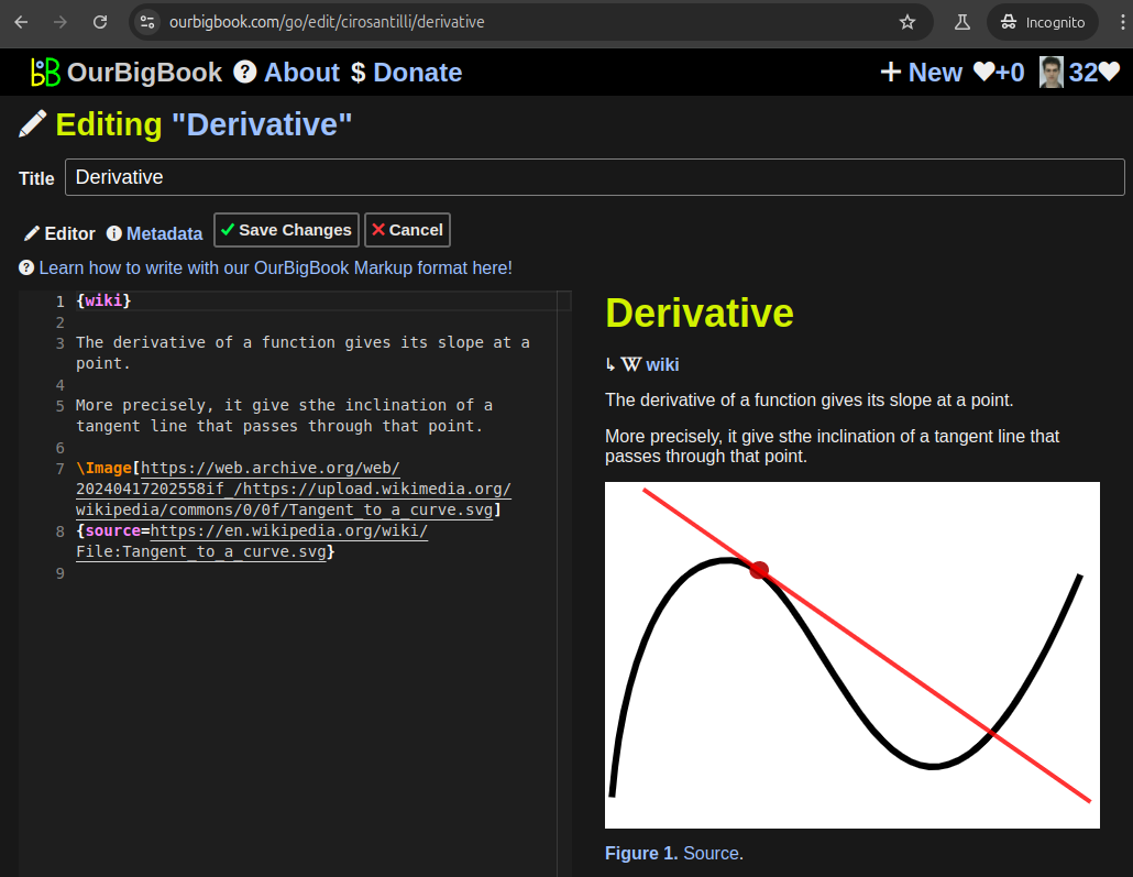
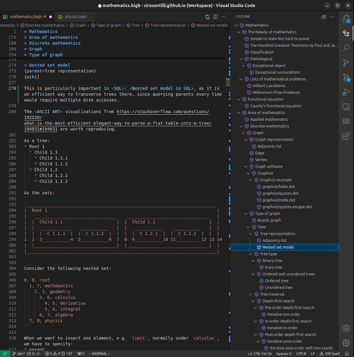
Figure 5. . You can also edit articles on the Web editor without installing anything locally. Video 3. Edit locally and publish demo. Source. This shows editing OurBigBook Markup and publishing it using the Visual Studio Code extension. 
- Infinitely deep tables of contents:


All our software is open source and hosted at: github.com/ourbigbook/ourbigbook
Further documentation can be found at: docs.ourbigbook.com
Feel free to reach our to us for any help or suggestions: docs.ourbigbook.com/#contact