Topics (384k) Articles (398k) Users (410) Discussions (242) Comments (383) Files (13k) New article

1-aminocyclopropane-1-carboxylate synthase is a pyridoxal phosphate-dependent enzyme that converts S-adenosylmethionine into 1-aminocyclopropane-1-carboxylate, commonly abbreviated ACC, with methylthioadenosine as the other principal product. ACC is a precursor in the plant pathway producing the gaseous signal ethene, usually called ethylene in plant physiology. The synthase makes the precursor; a subsequent enzyme catalyzes the ethylene-forming step.[ref]
Characterization and sequencing of the active site of 1-aminocyclopropane-1-carboxylate synthase by W. K. Yip and others. Abstract: PLP dependence and biosynthetic role
The precursor/enzyme distinction helps interpret a perturbation. Reducing synthase activity can reduce pathway output even if the downstream machinery remains able to respond. In a 1991 tomato experiment, expression of Antisense RNA against the synthase inhibited fruit ripening. Applying ethylene or propylene reversed the effect, supporting a role for the pathway's signal in triggering ripening rather than merely accompanying it.[ref]
Reversible inhibition of tomato fruit senescence by antisense RNA by P. W. Oeller and others. Abstract: antisense intervention and external-gas reversal
For an original pathway example, write substrate ACC signal. Blocking the first arrow and restoring the response by supplying the final signal places the block upstream of that response. It does not by itself prove that every intermediate remains unchanged or that this enzyme controls all developmental responses.
Heparinase is a name for a heparin-cleaving enzyme, classified as heparin lyase in the enzyme nomenclature. It breaks specified glycosidic bonds in heparin-type polysaccharides by eliminative cleavage. The reaction identity matters when using the enzyme to perturb a complex cell-surface layer.[ref]
In a 1990 experiment, researchers perfused individual capillaries in situ with enzyme-containing solution. Heparinase treatment at least doubled tube hematocrit, the fraction of the capillary's volume occupied by red blood cells, without a significant change in red-cell velocity. Vehicle alone and heat-denatured enzyme did not produce that response.[ref]
The controls connect the response to active enzymatic treatment rather than simply perfusing fluid. The observation supports a contribution of the endothelial glycocalyx to red-cell distribution. It does not mean that hematocrit and flow velocity are the same quantity, or that one enzyme experiment completely identifies every affected surface component. The substrate specificity and preparation used remain part of the experiment's interpretation.
Aromatase is an enzyme that converts androgens such as testosterone into estrogens, including estradiol. Its activity links the availability of a precursor substrate to production of a chemically different hormonal signal. The amount of enzyme, available substrate and accumulated product are separate quantities.[ref]
Improved FSH sensitisation and aromatase assay in human granulosa-lutein cells by A. Lambert and others. Abstract, androgen-to-estrogen conversion and assay design
A 2000 experiment made that distinction useful. Researchers cultured human granulosa-lutein cells, stimulated them with follicle-stimulating hormone, then washed the cells and supplied testosterone for a two-hour challenge. They measured secreted estradiol. Separating the longer stimulation period from the short substrate challenge reduced the confounding effect of progressively depleting testosterone during culture.[ref]
Improved FSH sensitisation and aromatase assay in human granulosa-lutein cells by A. Lambert and others. Abstract, preincubation, FSH stimulation, wash and substrate challenge
The experiment illustrates how an enzyme assay can distinguish regulatory induction from immediate catalytic performance. A larger accumulated product amount could otherwise reflect more active enzyme, more available substrate, or a longer reaction time. It is the controlled comparison, not the product measurement alone, that supports an interpretation of altered aromatase activity.
RPE65 is an enzyme of the visual cycle in the retinal pigment epithelium. It converts all-trans-retinyl esters into 11-cis-retinol, coupling a change of molecular geometry to processing of an ester substrate. The product participates in the pathway that replenishes the light-sensitive pigment system.[ref]
RPE65 is the isomerohydrolase in the retinoid visual cycle by Gennadiy Moiseyev and others. Abstract, substrate, product and catalytic role
A 1998 mouse experiment established a requirement for the protein: deficiency disrupted production of 11-cis vitamin-A derivatives and altered the visual response. Such a result identifies a necessary pathway component without by itself proving which reaction it catalyzes.[ref]
A 2005 study supplied more direct biochemical evidence. Expressing RPE65 in cultured cells produced the relevant activity when lecithin retinol acyltransferase was coexpressed to provide the substrate. Activity depended on the amount of expressed RPE65.[ref]
RPE65 is the isomerohydrolase in the retinoid visual cycle by Gennadiy Moiseyev and others. Abstract, recombinant expression and substrate-supplying partner
The comparison illustrates why an inactive preparation can reflect missing substrate supply as well as an absent or defective enzyme.Neutrophil elastase is a serine protease stored in granules of neutrophils. Its ability to cleave proteins can alter structures outside or within the cell, depending on where the active enzyme is located. A particularly illuminating example connects proteolysis to organization of chromatin.[ref]
In a 2010 study, activation caused neutrophil elastase to reach the cell nucleus, where it partly degraded particular histones. Experiments with isolated nuclei and purified enzyme connected that cleavage to chromatin decondensation; Western blot measurements followed the protein changes. The study also found defective neutrophil extracellular trap formation in mice lacking the enzyme in its tested infection model.[ref]
Neutrophil elastase and myeloperoxidase regulate the formation of neutrophil extracellular traps by Venizelos Papayannopoulos and others. Abstract and Fig. 1E–I, histone processing, purified enzyme and nuclear decondensation
Myeloperoxidase is an enzyme that can use hydrogen peroxide to oxidize chloride, forming hypochlorous acid. The enzyme occurs in neutrophils and other immune cells, where its oxidant chemistry contributes to antimicrobial activity. One balanced representation of the reaction is:The equation identifies consumed reactants and product; the enzyme itself is regenerated during catalysis.[ref]
A protein's catalytic reaction does not exhaust its possible biological roles. A 2010 study found that myeloperoxidase enhanced chromatin decondensation together with neutrophil elastase, with the enhancement independent of myeloperoxidase's enzymatic activity in the tested system.[ref]
Neutrophil elastase and myeloperoxidase regulate the formation of neutrophil extracellular traps by Venizelos Papayannopoulos and others. Abstract and Fig. 1H, cooperative effect on isolated nuclei
This provides a useful experimental distinction: removing a protein and selectively disabling one catalytic activity need not have identical consequences. To assign a phenotype to oxidant production, an experiment must test that chemistry rather than infer it solely from the presence or absence of myeloperoxidase.
Lecithin retinol acyltransferase is an enzyme that transfers an acyl group from phosphatidylcholine, also called lecithin, to retinol, producing a retinyl ester. The donor's position and the acceptor's alcohol group are part of the reaction definition; describing it merely as an enzyme that binds vitamin A would omit its chemical function.[ref]
Lecithin retinol acyltransferase in IUBMB Enzyme Nomenclature. EC 2.3.1.135, accepted reaction and sn-1 acyl transfer
This reaction supplies ester forms for retinoid storage and metabolism. It is distinct from changing retinyl-chain geometry, even though both operations occur in the broader visual cycle.[ref]
Lecithin retinol acyltransferase in IUBMB Enzyme Nomenclature. Comments, ester products and substrate recognition
A 2005 experiment illustrates why the distinction matters. Cultured cells expressing RPE65 required coexpression of lecithin retinol acyltransferase to provide the ester substrate for the measured production of 11-cis-retinol.[ref]
RPE65 is the isomerohydrolase in the retinoid visual cycle by Gennadiy Moiseyev and others. Abstract, coexpression requirement
Missing product can therefore reflect a failure of upstream substrate supply rather than a failure of the downstream enzyme itself. Measuring both steps helps distinguish a pathway requirement from the identity of the catalyst performing a particular transformation.In collagen synthesis, prolyl 4-hydroxylase is the enzyme that converts selected peptide-bound proline residues into Trans-4-hydroxy-L-proline. The official name procollagen-proline 4-dioxygenase emphasizes both the protein substrate and oxygen-dependent reaction. It acts on procollagen and other proteins with collagen-like domains.[ref]
The reaction couples hydroxylation of a proline residue to conversion of 2-Oxoglutarate into succinate and carbon dioxide, consuming molecular oxygen. The enzyme requires ferrous iron and vitamin C. It operates in the lumen of the endoplasmic reticulum and recognizes sequences containing , where denotes a variable residue and Gly denotes glycine.[ref]
This is a post-translational modification: the peptide chain exists before its proline side groups are hydroxylated. The modified residues support the collagen triple helix. In a simplified fully coupled assay, one mole of hydroxylated residues corresponds to one mole of succinate formed. Measuring a coproduct can therefore report reaction progress, provided appropriate controls establish that it comes from the coupled enzyme reaction rather than other sources.
HIV-1 protease is an aspartic protease of HIV-1. It catalyses peptide bond hydrolysis during viral maturation. The active enzyme is a protein dimer made from two identical subunits, each about . Thus a single isolated subunit and the assembled catalytic enzyme are different molecular objects.[ref]
Cleavage specificity depends on the amino-acid residues around the bond. In the nomenclature , the vertical line marks the cleaved bond: is on one side and on the other. The enzyme favors a hydrophobic residue, while varies and is often proline.[ref]
HIV-1 protease in IUBMB Enzyme Nomenclature. Reaction, substrate-position specificity
A short peptide assay can therefore test cleavage of one specified sequence, but its result is conditional on that substrate and the assay conditions. Measuring disappearance of the uncleaved peptide or appearance of a cleavage product addresses chemical processing directly. It differs from measuring the number of viral particles, which additionally depends on many other steps. The enzyme's role in maturation explains why inhibiting its activity can disrupt that process and why it is an antiviral drug target.[ref]
A prenyltransferase is an enzyme that transfers a prenyl group, an isoprenoid-derived carbon group, to an acceptor. The term covers multiple reactions; the acceptor can be a small molecule rather than a protein. It should therefore not be interpreted as meaning only an enzyme for protein prenylation.
One concrete reaction joins the five-carbon dimethylallyl diphosphate and five-carbon isopentenyl diphosphate to make ten-carbon geranyl diphosphate, releasing diphosphate. Carbon counting gives , while the substrate names specify the activated donors and the product structure. The official entry for this enzyme also notes poor acceptance of larger prenyl diphosphates as donors: a generic transfer label does not imply unrestricted substrate choice.[ref]
A different example links this chemistry to Vitamin K. In 2010, Kimie Nakagawa and colleagues identified UBIAD1 as an enzyme for menaquinone-4 biosynthesis. Reducing its expression suppressed conversion of deuterium-labelled vitamin-K derivatives into labelled product; expression in insect cells restored a production system, and nuclear magnetic resonance helped identify the product.[ref]
Identification of UBIAD1 as a novel human menaquinone-4 biosynthetic enzyme by Kimie Nakagawa and others. Abstract, gene suppression, expression and product identification
Combining perturbation with chemical identification provides stronger evidence for an enzyme assignment than finding a similar-looking gene sequence alone.The cholesterol side-chain cleavage enzyme, also designated CYP11A1, is a Cytochrome P450 that converts cholesterol into pregnenolone and 4-Methylpentanal. Its official accepted name is cholesterol monooxygenase (side-chain-cleaving), EC 1.14.15.6. Two hydroxylations precede cleavage between carbon positions 20 and 22.[ref]
Cholesterol side-chain cleavage enzyme in IUBMB Enzyme Nomenclature. overall reaction, steps 1a–1c and Comments
Adrenodoxin supplies electrons to the enzyme; NADPH is an upstream electron source. For the fully coupled net reaction, each pregnenolone formed requires three oxygen molecules and six reduced adrenodoxins. Thus an ideal conversion consumes oxygen and six micromoles of one-electron donor equivalents. The carbon balance is , partitioning the original steroid and its shortened side chain.[ref]
Cholesterol side-chain cleavage enzyme in IUBMB Enzyme Nomenclature. Reaction and electron-transfer pathway
In 2011, Natalia Mast and colleagues combined X-ray crystallography with solution spectroscopy. Their structure contained 22R-Hydroxycholesterol, the first intermediate, whose hydroxyl oxygen lay from the haem iron. Changing adrenodoxin conditions altered carbon monoxide access to reduced haem in a spectroscopic test.[ref]
Together, these measurements connect substrate positioning with a perturbation in solution. The proposed movement of intermediates during successive reactions is a mechanistic interpretation of the combined evidence; a single structure is a snapshot of one bound state.
Heparosan-N-sulfate-glucuronate 5-epimerase is an enzyme that converts selected D-glucuronate residues into L-iduronate within N-sulfated heparosan. This carbon-5 epimerization changes a stereocenter while preserving the number of sugar residues. The official classification is EC 5.1.3.17.[ref]
Heparosan-N-sulfate-glucuronate 5-epimerase in IUBMB Enzyme Nomenclature. Reaction and Comments
The surrounding chain is part of the substrate. Neighboring N-sulfated groups are required for recognition, and additional sulfate substitution can restrict the reaction. The enzyme should not therefore be described as simply acting on free glucuronate in any chemical setting.[ref]
Heparosan-N-sulfate-glucuronate 5-epimerase in IUBMB Enzyme Nomenclature. Comments, substrate context and exclusions
A 2019 study by Claire Debarnot and colleagues followed conversion with nuclear magnetic resonance using a carbon-13-labelled polymer. Under the reported conditions, the human enzyme approached approximately iduronate content. The researchers also compared site-directed mutagenesis variants: no activity was detected for two proposed catalytic-residue substitutions even at the stated higher enzyme concentration.[ref]
Substrate binding mode and catalytic mechanism of human heparan sulfate d-glucuronyl C5 epimerase by Claire Debarnot and others. Overall Structures of hGlce; Figure 1A and legend
For an illustrative 100 eligible sugar positions, a conversion gives 38 iduronate and 62 glucuronate positions, still 100 in total. The experiment changes chemical identity within a chain rather than extending it. The reported fraction belongs to specified conditions; it is not a universal composition of heparan sulfate.
Guanylate cyclase is an enzyme that converts GTP into cyclic guanosine monophosphate, releasing diphosphate. The official classification is EC 4.6.1.2. The phosphate retained in the product forms a cyclic linkage involving the ribose 3' and 5' positions.[ref]
Guanylate cyclase in IUBMB Enzyme Nomenclature. Reaction and systematic name
The reaction changes nucleotide connectivity rather than making the guanine-containing part from scratch. In an ideal closed assay with no competing reactions, forming of cyclic guanosine monophosphate consumes of GTP and produces of diphosphate. The enzyme's name specifies a chemical transformation; it does not identify a unique concentration or cellular rate.
Synthesis and accumulation also differ. If a phosphodiesterase simultaneously removes the cyclic product, its pool obeys a material balance,with rates expressed in compatible concentration-per-time units. For constructed rates of for both processes, the product concentration is constant even though both reactions are active.
Consequently, an assay of guanylate cyclase should distinguish product formation from net product accumulation, using defined conditions or an independent estimate of removal. A constant cyclic-nucleotide signal alone does not establish that synthesis has stopped.
Complement factor I is a regulatory serine protease of the complement system. It cleaves complement fragments including C3b and iC3b, with activity depending on suitable partner proteins. Its official enzyme classification is EC 3.4.21.45.[ref]
Complement factor I in IUBMB Enzyme Nomenclature. accepted name, Reaction and Comments
For cleavage of C3b, the substrate binds a partner such as complement factor H or complement receptor 1. Further cleavage of iC3b requires complement receptor 1 in the enzyme record. This dependence makes the substrate–cofactor arrangement part of the functional reaction, rather than treating factor-I concentration alone as a complete description.[ref]
Complement factor I in IUBMB Enzyme Nomenclature. Comments, substrate-specific cofactor requirements
A 2009 structural and functional study examined the first four domains of complement factor H. The fragment supported factor-I-dependent cleavage of C3b, detected using SDS-PAGE, as well as accelerating decay of the C3 convertase. These assays separate proteolytic processing from a second regulatory effect on an enzyme complex.[ref]
A useful control comparison therefore holds complement factor I and substrate constant while adding or omitting the required partner. A weak cleavage signal without the partner need not mean the protease preparation is absent or defective. The experiment tests the assembled reaction system.
Pyridoxal 5'-phosphate synthase, EC 1.4.3.5, is an enzyme that oxidizes pyridoxamine phosphate or pyridoxine phosphate to pyridoxal phosphate. Despite the accepted synthase name, these reactions start with an already phosphorylated vitamin. The enzyme uses flavin mononucleotide as a cofactor.[ref]
Pyridoxal 5'-phosphate synthase in IUBMB Enzyme Nomenclature. accepted name, reactions 1–2 and Comments
Both reactions consume oxygen and produce hydrogen peroxide. The pyridoxamine-phosphate reaction additionally consumes water and releases Ammonia, because its aminomethyl group becomes an aldehyde. The pyridoxine-phosphate reaction changes a hydroxymethyl group into an aldehyde without releasing that nitrogen-containing coproduct.[ref]
This difference supplies an analytical example. If an ideal mixture undergoes complete conversion of of each substrate, it produces pyridoxal phosphate and peroxide, but only ammonia. A peroxide-only readout combines contributions from both substrates; the coproduct balance contains additional information.
Those values assume only the stated coupled reactions. In an actual enzyme assay, other peroxide- or ammonia-producing activities can interfere, so chemical product identification and appropriate controls remain important. Enzyme identity, substrate identity and the chosen reporter reaction each determine what the signal means.
Chondroitin-glucuronate 5-epimerase changes D-glucuronate residues in a chondroitin chain into L-iduronate residues in a dermatan chain. Its classification is EC 5.1.3.19. The reaction alters carbon-5 stereochemistry without adding a sugar residue or changing the residue's elemental formula.[ref]
Chondroitin-glucuronate 5-epimerase in IUBMB Enzyme Nomenclature. Reaction and accepted name
This connects polymer modification to dermatan sulfate formation. It is a different enzyme assignment from heparosan-N-sulfate-glucuronate 5-epimerase, whose substrate belongs to another polysaccharide pathway. Similar names do not make the polymer substrates interchangeable.[ref]
Chondroitin-glucuronate 5-epimerase in IUBMB Enzyme Nomenclature. Comments, distinction from EC 5.1.3.17
In 1982, A. Malmström and L. Åberg reported distinct uronosyl-epimerase activities associated with dermatan- and heparan-sulfate biosynthesis in microsomal preparations from cultured fibroblasts. The comparison made substrate context experimentally relevant rather than treating all carbon-5 epimerization as one nonspecific activity.[ref]
Biosynthesis of dermatan sulphate. Assay and properties of the uronosyl C-5 epimerase by A. Malmström and L. Åberg. Abstract, two microsomal epimerase activities
For a constructed 50-position substrate segment, conversion of ten eligible glucuronate residues yields ten iduronate residues and 40 unchanged positions. The chain still contains 50 positions. A measurement sensitive only to total sugar amount or shared molecular mass cannot establish that this stereochemical conversion occurred.
A C3 convertase is an enzymatic complex of the complement system that cleaves Complement C3 into C3a and C3b. Different activation routes assemble different C3-cleaving complexes. The important shared activity is selective substrate cleavage, rather than a universal single protein carrying the name.[ref]
Assembly and lifetime matter because a catalytic complex can process many substrate molecules. In an idealized constant-rate example, active complex turning over at produces cleavage products in ten seconds, provided substrate remains abundant. Each consumed C3 molecule supplies one C3a fragment and one C3b fragment; the enzyme is not consumed on every turnover.
A 2009 study of complement factor H combined structural and functional measurements. The tested factor-H fragment accelerated decay of preassembled C3 convertase and supported complement factor I-dependent C3b cleavage in a separate assay.[ref]
Structure of complement fragment C3b-factor H and implications for host protection by complement regulators by Jin Wu and others. Figure 1d–e, convertase decay and cofactor assays
These observations separate dismantling an active assembly from cutting its protein components. Both can regulate complement activity, but they are not the same measured reaction. Comparing complex lifetime, substrate cleavage and component abundance therefore gives more mechanistic information than measuring total complement protein alone.
Guanylate kinase is an enzyme that transfers a phosphoryl group from ATP to guanosine monophosphate, producing ADP and guanosine diphosphate:Its official classification is EC 2.7.4.8. The enzyme can also use the corresponding deoxy-nucleotide acceptor and deoxy-ATP donor.[ref]
A constructed mixture containing ATP and guanosine monophosphate, initially without products, permits at most of each product through the forward reaction. At least ATP remains if no other reactions consume it. Per turnover, the phosphate inventory is conserved: .
This ceiling concerns stoichiometry. Reversible chemical equilibrium, substrate concentrations and assay conditions determine the actual conversion; the inventory alone does not specify an enzyme rate.
In 1992, M. Konrad isolated and characterized the yeast GUK1 gene encoding guanylate kinase, connecting a chemically defined activity with a molecularly identifiable catalyst.[ref]
Cloning and expression of the essential gene for guanylate kinase from yeast by M. Konrad. Abstract, reversible phosphoryl transfer and gene identification
The kinase reaction should also be distinguished from guanylate cyclase, which converts GTP to a cyclic nucleotide rather than transferring ATP's terminal phosphoryl group to GMP.Serotonin N-acetyltransferase transfers an acetyl group from acetyl-CoA to serotonin, making N-acetylserotonin and coenzyme A. The IUBMB accepted name aralkylamine N-acetyltransferase, EC 2.3.1.87, reflects its wider substrate range. This acetyl-transfer step precedes the final methyl-transfer step of melatonin synthesis.[ref]
For an idealized isolated forward reaction, acetyl-CoA and serotonin permit at most N-acetylserotonin, leaving at least serotonin. That calculation is a stoichiometric bound, not a measured rate or a prediction of complete conversion to melatonin.
In 1970, D. C. Klein and J. L. Weller measured a circadian rhythm in rat pineal gland N-acetyltransferase activity, with activity more than 15-fold higher at night than by day. The rhythm persisted in darkness and blinded animals and was suppressed by constant illumination. Their in-vitro comparisons implicated norepinephrine in activity regulation.[ref]
Geraniol synthase converts geranyl diphosphate and water into geraniol and diphosphate. The accepted IUBMB name is geranyl diphosphate diphosphatase, EC 3.1.7.11. That classification describes the overall reaction; it does not on its own identify every bond-breaking step in the mechanism.[ref]
In a 2004 study, Yoko Iijima and colleagues purified activity from sweet-basil leaf glands. The plant enzyme produced geraniol from geranyl diphosphate, and expression of the identified gene yielded a catalyst with similar activity.[ref]
Characterization of Geraniol Synthase from the Peltate Glands of Sweet Basil by Yoko Iijima and others. Abstract, purification and expression
The key mechanistic test supplied water containing oxygen-18. Label appeared in geraniol, supporting a route in which cleavage produces a geranyl cation that is captured by water. Merely observing unlabelled geraniol would not distinguish this from a route retaining the substrate's original alcohol oxygen.[ref]
An ideal isolated conversion of substrate produces two micromoles each of geraniol and diphosphate. Product amount checks stoichiometry, while isotope labelling tests atom origin. These measurements answer different questions, even when both support the same net reaction. The synthase name should not be read as implying that ATP is required by this specific conversion.
Pinned article: Introduction to the OurBigBook Project
Welcome to the OurBigBook Project! Our goal is to create the perfect publishing platform for STEM subjects, and get university-level students to write the best free STEM tutorials ever.
Everyone is welcome to create an account and play with the site: ourbigbook.com/go/register. We belive that students themselves can write amazing tutorials, but teachers are welcome too. You can write about anything you want, it doesn't have to be STEM or even educational. Silly test content is very welcome and you won't be penalized in any way. Just keep it legal!
Intro to OurBigBook
. Source. We have two killer features:
- topics: topics group articles by different users with the same title, e.g. here is the topic for the "Fundamental Theorem of Calculus" ourbigbook.com/go/topic/fundamental-theorem-of-calculusArticles of different users are sorted by upvote within each article page. This feature is a bit like:
- a Wikipedia where each user can have their own version of each article
- a Q&A website like Stack Overflow, where multiple people can give their views on a given topic, and the best ones are sorted by upvote. Except you don't need to wait for someone to ask first, and any topic goes, no matter how narrow or broad
This feature makes it possible for readers to find better explanations of any topic created by other writers. And it allows writers to create an explanation in a place that readers might actually find it.
Figure 1. Screenshot of the "Derivative" topic page. View it live at: ourbigbook.com/go/topic/derivativeVideo 2. OurBigBook Web topics demo. Source. - local editing: you can store all your personal knowledge base content locally in a plaintext markup format that can be edited locally and published either:This way you can be sure that even if OurBigBook.com were to go down one day (which we have no plans to do as it is quite cheap to host!), your content will still be perfectly readable as a static site.
- to OurBigBook.com to get awesome multi-user features like topics and likes
- as HTML files to a static website, which you can host yourself for free on many external providers like GitHub Pages, and remain in full control

Figure 2. You can publish local OurBigBook lightweight markup files to either OurBigBook.com or as a static website.
Figure 3. Visual Studio Code extension installation.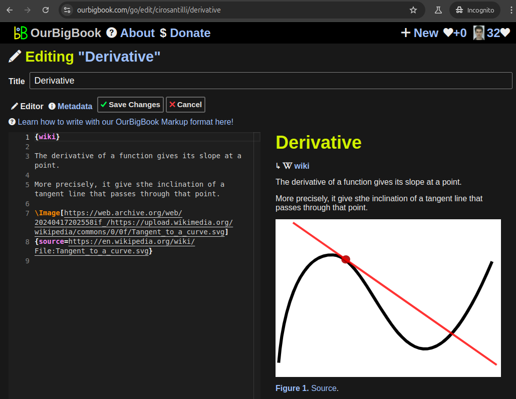
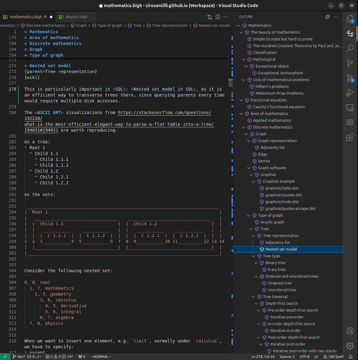
Figure 5. . You can also edit articles on the Web editor without installing anything locally. Video 3. Edit locally and publish demo. Source. This shows editing OurBigBook Markup and publishing it using the Visual Studio Code extension. 
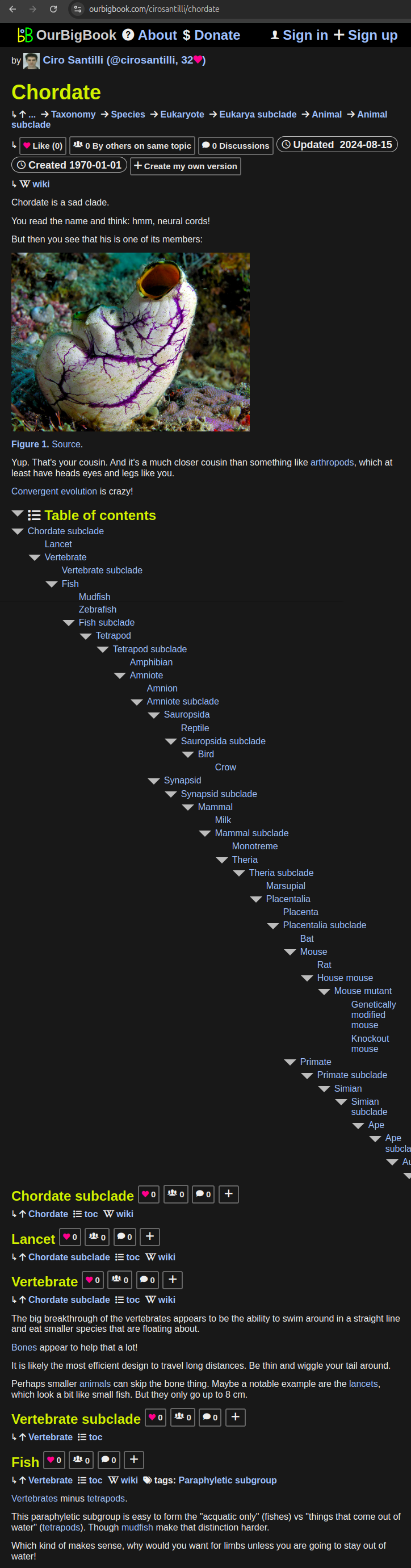
- Infinitely deep tables of contents:
All our software is open source and hosted at: github.com/ourbigbook/ourbigbook
Further documentation can be found at: docs.ourbigbook.com
Feel free to reach our to us for any help or suggestions: docs.ourbigbook.com/#contact